Reading...
![]()
Play button
![]()
Play button
![]()
Use LEFT and RIGHT arrow keys to navigate between flashcards;
Use UP and DOWN arrow keys to flip the card;
H to show hint;
A reads text to speech;
23 Cards in this Set
- Front
- Back
- 3rd side (hint)
|
Wall of the heart
Consists of three layers |
The outer layer, or epicardium (visceral layer of pericardium, a serous membrane that forms the pericardial sac).
The middle layer or myocardium The inner layer endocardium |
|
|
list
|
.
|
|
|
|
Endocardium
3ct layers |
-a simple squamous
-endothelium subendothelial loose connective tissue -Thick layer of dense connective tissue -a simple squamous -endothelium -subendothelial loose connective tissue -subendocardial layer |
|
|
|
Myocardium
4ct |
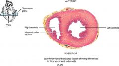
Middle layer is composed of cardiac muscle fibers
In atria myocardium is thin (low pressure) In ventricles myocardium is thick, left 3 times thicker than right. Muscles are arranged in 2 layers superficial & deep. Cardiac muscles of the atria & ventricles are completely separated by the fibrous skeleton. |
|
|
|
Myocardium
Contain 3 types of cardiocytes |
1. Contractile cardiocytes which contract to pump blood through circulation.
2. Myoendocrine cardiocytes producing atrial natriuretic factor. . Nodal cardiocytes Sinoatrial node Atrioventricular node (Purkinje’s fibers , present in subendocardium, are modified cardiocytes |
|
|
|
Atrial & Ventricular cardiac muscle
Atrial cardiac fibers |
Smaller average diameter
T-tubules are few or absent spherical granules Atrial natriuretic factor |
|
|
|
Atrial Natriuretic Factor
5ct |
Stimulates diuresis
Stimulates natriuresis Relaxes cardiovascular muscles by antagonizing the action of vasopressin and angiotensin II Prevents sodium & water reabsorption from causing hypervolemia & hypertension. ANF is released in response to the increased pressure across the atrial wall. |
|
|
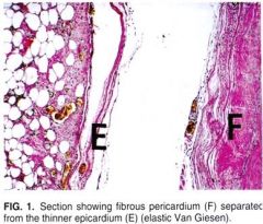
Epicardium
|
Serous covering external surface of heart / visceral
part of the pericardial sac It consists of: 3ct |
|
|
|
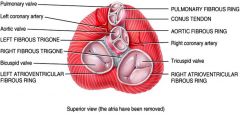
Cardiac Skeleton
|
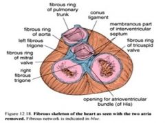
A. Dense connective tissue is concentrated between atria & ventricles and act as electric insulator
B. Anchors cardiac muscle fibers, heart valves and major arteries C. Three main parts of cardiac skeleton are a. Annuli fibrosa, encircle atrioventricular and arterial orifices (aorta & pulmonary), composed of dense irregular connective tissue, with some elastic fibers & fat cells. They provide attachment to the leaflets of the valves. |
|
|
|
Cardiac skeleton
|
Trigona fibrosa
Septum membranaceum: found in interventricular septum dense regular connective tissue which looks like aponeurosis |
|
|
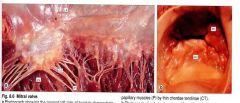
Cardiac valves
2 sets of valves |
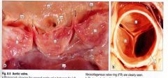
AV valves
Semilunar valves |
|
|
|
Cardiac Valves
Valves are anchored to heart musculature by = |
dense connective tissue
|
|
|
|
Cardiac Valves
dense connective tissue, which |
A. provide valves with greater tensile strength
B. extends into the core of chordae tendinae |
|
|
|
Cardiac Valves
Dense connective tissue is covered by |
areolar (loose) connective tissue,
covered on both sides by endocardium lacking subendocardial layer. |
|
|
|
Cardiac Valves
Endocardium on atrial surface is |
thicker than on ventricular & contain more
|
|
|
list items
|
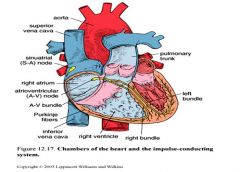
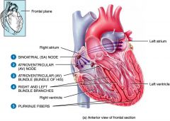
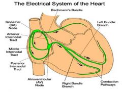
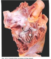
SA
Bachmans Bundle Ant-SA internodel Tract Middle Internodal Tract Posterior Internodal Tract AV Node Bundle of His Left BB RBB Conduction pathways Purkinje Fibers |
|
|
list items
Nodal fibers are 5ct |
Diffuse
Irregularly arranged Small nodal cardiocytes compared to myocardial cardiocytes More connective tissue between cardiocytes Intercalated discs not as apparent as in myocardial cardiocytes . Capillaries not as abundant as in myocardial cardiocytes. |
SA
Bachmans Bundle Ant-SA internodel Tract Middle Internodal Tract Posterior Internodal Tract AV Node Bundle of His Left BB RBB Conduction pathways Purkinje Fibers |
|
list items
|
SA
Bachmans Bundle Ant-SA internodel Tract Middle Internodal Tract Posterior Internodal Tract AV Node Bundle of His Left BB RBB Conduction pathways Purkinje Fibers |
|
|
list items
SA Bachmans Bundle Ant-SA internodel Tract Middle Internodal Tract Posterior Internodal Tract AV Node Bundle of His Left BB RBB Conduction pathways Purkinje Fibers |
.
|
|
|
Sinoatrial Node
|
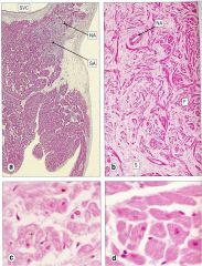
a: SA node, Low power
b: Medium power SA node showing irregular whorled network of small nodal fibers embedded in the bulky fibrocollagenous stroma. c: High magnification SA node d: Normal atrial muscle fibers |
|
|
list
|
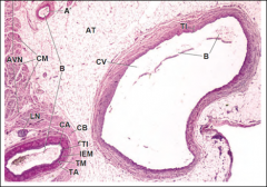
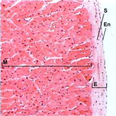
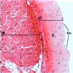
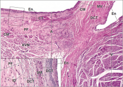
HEART. KEY:
A, atrium ; AT adipose tissue AVN atrioventricular node CM cardiac muscle; CT connective tissue DCT dense connective tissue En endothelium ID intercalated disk IEM MV mitral valve PF Purkinje fibers SM smooth muscle V, ventricle |
|
|
list
|
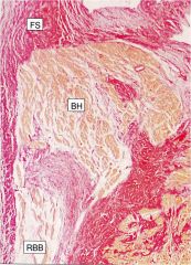
Bundle of His: BH
Right Bundle Branch: RBB |
|
|
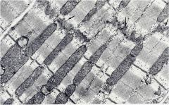
Myocardial muscle
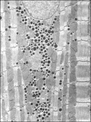
Purkinje’s fibers: describe = |
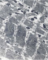
Large diameter striated muscle fibers with atypical intercalated discs.
Contain reduced no. of myofibrils located at periphery. Rich in glycogen & mitochondria Positive reaction for acetylcholinesterase |
|