![]()
![]()
![]()
Use LEFT and RIGHT arrow keys to navigate between flashcards;
Use UP and DOWN arrow keys to flip the card;
H to show hint;
A reads text to speech;
333 Cards in this Set
- Front
- Back
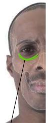
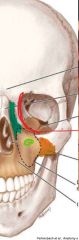
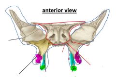
Name the bony landmark in green |
infra-orbital ridge |
|
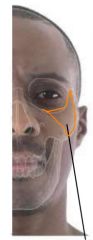
Name the bony landmark in orange |
zygomatic bone |
|

Name the bony landmark in pink |
angle of mandible |
|
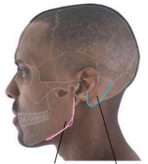
Name the bony landmark in blue |
mastoid process |
|

Name the landmark in blue |
Auricular tubercule |
|
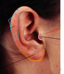
Name the landmark in pink |
tragus of ear |
|

Name the landmark in orange |
lobule of auricle |
|
|
What landmark does the Gow-Gates mandibular block target? |
tragus of ear (due to proximity to foramen ovale) |
|
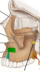
Name the maxillary process in orange |
zygomatic process of maxilla |
|

Name the maxillary process in blue |
frontal process of maxilla |
|

Name the foramen in green |
infraorbital foramen |
|
|
What landmark does the infraorbital block target? |
infraorbital foramen |
|
Name the maxillary landmark in green |
canine fossa |
|

Name the maxillary landmark in blue |
canine eminence |
|

Name the foramen in yellow |
alveolar foramen |
|

Name the maxillary landmark in orange |
maxillary tuberosity |
|
|
What landmark does the anterior superior alveolar block target? |
canine fossa (targets maxillary incisors & canines) |
|
|
What landmark does the posterior superior alveolar block target? |
alveolar foramen (targets maxillary molars) |
|
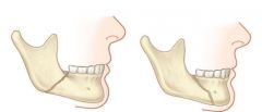
Which mandibular fracture is more favourable? Why? |
RIGHT is more favourable; heals better since anterior half can rest on the posterior half for support |
|

LATERAL VIEW Name the mandibular landmark in blue (top) |
mandibular notch |
|

LATERAL VIEW Name the mandibular landmark in purple |
condylar process |
|

LATERAL VIEW Name the mandibular landmark in pink (top) |
coronoid process |
|

LATERAL VIEW Name the mandibular landmark in green |
oblique line |
|

LATERAL VIEW Name the mandibular landmark in fuschia |
alveolar process |
|

LATERAL VIEW Name the mandibular landmark in the yellow circle |
mental foramen |
|
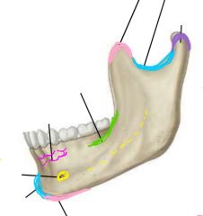
LATERAL VIEW Name the mandibular landmark in blue (bottom) |
mental protuberance (1x, on midline; ie, the chin) |
|

LATERAL VIEW Name the mandibular landmark in pink (bottom) |
mental tubercle (2x, one on each side of the mental protuberance) |
|

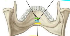
MEDIAL VIEW Name the mandibular landmark in green |
lingula (the "tongue" of the bone around a foramen) |
|

MEDIAL VIEW Name the mandibular landmark in the yellow |
mandibular foramen |
|
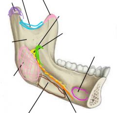
MEDIAL VIEW Name the mandibular landmark in orange |
mylohyoid line (attachment of mylohyoid muscle for floor of mouth) |
|

MEDIAL VIEW Name the mandibular landmark in the pink oval |
pterygoid fovea (for attachment of masticatory muscles) |
|

MEDIAL VIEW Name the mandibular landmark in brown |
submandibular fossa (where submandibular gland rests) |
|

MEDIAL VIEW Name the mandibular landmark in fuschia |
sublingual fossa (where sublingual gland rests) |
|
|
What landmark does the mental block target? |
mental foramen |
|
|
What landmark does the inferior alveolar block target? |
lingula, at level of mandibular foramen |
|
|
What landmark does the buccal block target? |
oblique line of mandible |
|

Name the mandibular landmark in green |
superior mental spines (attaches genio-glossus muscle) |
|

Name the mandibular landmark in blue |
inferior mental spines (attaches genio-hyoid muscle) |
|
Name the mandibular landmark in yellow |
digastric fossa |
|
|
The bony plate covering which teeth is thicker? (mandibular or maxillary) |
mandibular bony plate is thicker (harder for anaesthetic to diffuse through) |
|

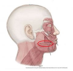
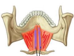
Name the neck triangle in green |
carotid triangle (2x lateral) |
|
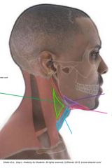
Name the neck triangle in blue |
muscular triangle (1x medial) |
|

Name the neck triangle in pink |
submental triangle (1x medial) |
|

Name the neck triangle in purple |
submandibular triangle (2x lateral) |
|
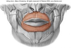
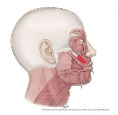
Name this muscle and its function |
orbicularis oris: forms "o" shape with mouth |
|
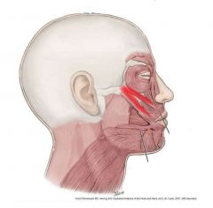
Name this muscle and its function |
risorius: for fake smile |
|
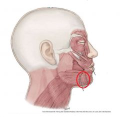
Name this muscle and its function |
depressor anguli oris: pulls angle of mouth down
|
|

Name this muscle and its function |
depressor labii inferioris: pulls lips down |
|
Name this muscle and its function |
mentalis: allows lower lip to pout |
|
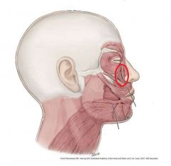
Name these muscles and their function |
levator labii superioris: pulls lips up |
|
Name this muscle and its function |
levator anguli oris: lifts corners of lips up for real smile |
|
Name these muscles and their function |
zygomaticus (major = lower, minor = upper): elevate upper lip for real smile |
|
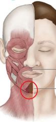
Name this muscle and its function |
buccinator: contracts cheek inwards |
|
|
What muscle is the buccinator continuous with? |
superior pharyngeal constrictor |
|
|
What structure pierces the buccinator muscle? |
parotid duct |
|

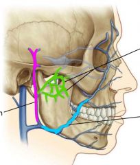
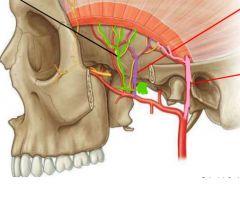
Identify the artery in fuschia |
angular artery (terminal branch of facial artery) |
|

Identify the artery in dark green |
transverse facial artery (antastomosis b/w superficial temporal a. & facial a.) |
|

Identify the arteries in light green |
superior & inferior labial arteries |
|

Identify the artery in light blue |
mental artery (coming thru mental foramen) |
|
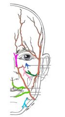
Identify the artery in dark blue |
infraorbital artery (coming thru infraorbital foramen) |
|
|
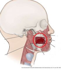
What does the "danger zone" refer to? |
facial veins without valves that enter the brain, risking spread of infection |
|

Identify the vein in fuschia |
retromandibular vein |
|
Identify the vein in blue |
facial vein |
|

Identify the veins in green |
pterygoid plexus |
|
|
Why is the pterygoid plexus a "danger zone" during anaesthesia? |
close to inferior alveolar nerve; during IA block, risk hitting vein instead, which drains to cavernous sinus of brain |
|
|
What are the 5 motor branches of the facial nerve? |
temporal, zygomatic, buccal, marginal mandibular, cervical |
|
|
Besides motor innervation, what sensory function does CN VII serve? |
conscious proprioception of the face |
|
|
What structure does the facial nerve emerge through? |
parotid gland (via stylomastoid foramen) |
|
|
What is Bell palsy? |
LMN injury to facial nerve (not forehead-sparing) |
|
|
What are the 3 sensory branches of the trigeminal nerve (CN V)? |
ophthalmic (V1), maxillary (V2), mandibular (V3) |
|

Name the 3 branches of V2 (in yellow) |
(from superior to inferior): zygomatico-temporal; zygomatico-facial; infraorbital |
|
Name the 3 branches of V3 (in blue) |
(from superior to inferior): auriculo-temporal; buccal (sensory); mental |
|

Name the nerve in blue and the teeth it innervates |
inferior alveolar nerve (branch of V3); innervates all lower teeth |
|
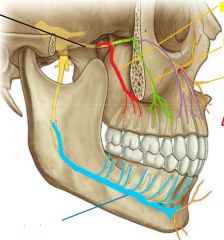
Name the nerve in red and the teeth it innervates |
posterior superior alveolar nerve (branch of V2): innervates upper molars |
|

Name the nerve in green and the teeth it innervates |
middle superior alveolar nerve (branch of V2); innervates upper premolars |
|

Name the nerve in purple and the teeth it innervates |
anterior superior alveolar nerve (branch of V2); innervates upper incisors & canines |
|
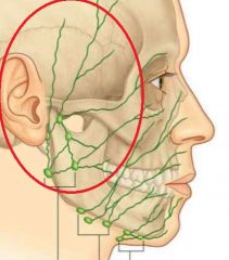
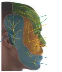
Identify the lymph nodes |
parotid lymph nodes |
|
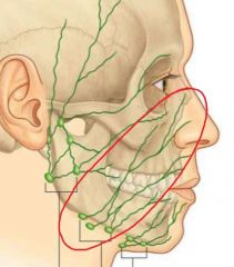
Identify the lymph nodes |
submandibular lymph nodes |
|
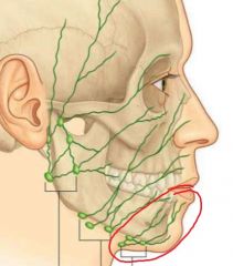
Identify the lymph nodes |
submental lymph nodes |
|

Identify the muscle in purple and its function |
genio-hyoid; hyoid elevation |
|
Identify the muscle in red and its function |
mylo-hyoid; hyoid elevation & floor of mouth |
|
Identify the muscle in yellow and its function |
digastric muscle (anterior & posterior bellies); hyoid elevation |
|

Identify the muscle in green and its function |
stylo-hyoid muscle; hyoid elevation |
|

Identify the muscle in fuschia and its function |
omo-hyoid; hyoid depression |
|

Identify the muscle in orange and its function |
thyro-hyoid; hyoid depression |
|

Identify the muscle in blue and its function |
sterno-thyroid; hyoid depression |
|
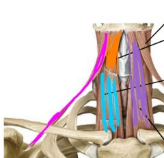
Identify the muscle in purple and its function |
sterno-hyoid; hyoid depression |
|
|
Which muscles must contract to fix the hyoid in place during mandible depression? |
all infra-hyoid muscles |
|
|
Which muscles must contract to fix the mandible in place during hyoid elevation? |
all mastication muscles |
|

Identify the artery in dark blue |
external carotid a. |
|
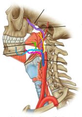
Identify the artery in dark green |
superior thyroid a. |
|

Identify the artery in fuschia |
lingual a. |
|

Identify the artery in light blue |
facial a. |
|

Identify the artery in light green |
tonsillar a. |
|

Identify the artery in yellow |
ascending pharyngeal a. |
|

Identify the artery in purple (deep to mandible) |
maxillary a. |
|
|
Which lymph nodes drain from the upper lip? |
submandiblar |
|
|
Which lymph nodes drain from the lower lip? |
submental |
|
|
Which way do the retro-auricular lymph nodes drain? |
with superficial cervical chain (along external jugular vein) |
|
|
Which way do the occipital lymph nodes drain? |
with deep cervical chain (along internal jugular vein) |
|
|
Which nodes are palpable under the sternocleidomastoid muscle? |
jugulo-digastric lymph nodes |
|
|
Where do the cervical lymph nodes ultimately drain into? |
directly into jugular trunk (not thru thoracic duct!!) |
|
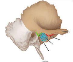
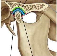
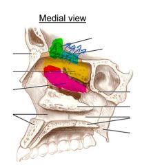
Identify the green landmark |
post-glenoid process of temporal bone |
|

Identify the blue landmark |
articular fossa of TMJ |
|

Identify the red landmark |
articular eminence of temporal bone |
|

Identify the blue structure |
capsule of TMJ |
|
Identify the green ligament |
sphenomandibular (from sphenoid to lingula of mandible, interior surface) |
|

Identify the yellow ligament |
lateral ligament of TMJ (from temporal bone to condyle) |
|

Identify the purple ligament |
stylomandibular ligament (from styloid to angle of mandible) |
|
What is the function of the fibrocartilage (in green)? |
prevent erosion of underlying bone |
|

What is the function of the articular disc (in blue)? |
prevents trauma when bones of TMJ move (ie condyle hitting articular eminence) |
|

What is the tissue in red? |
synovial membrane (secretes synovial fluid) |
|
|
What are the two directions of movement of the TMJ? |
-protrusion/retraction -depression/elevation |
|
|
Which movements of the TMJ always occur together? |
-protrusion & depression -retraction & elevation |
|
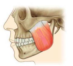
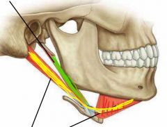
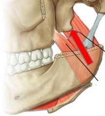
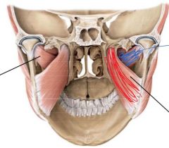
Identify this muscle and its 2 functions |
masseter; elevates and retracts mandible |
|
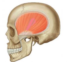
Identify this muscle and its 2 functions |
temporalis; elevates and retracts mandible |
|
|
Where on the mandible does the temporalis muscle attach? |
coronoid process of mandible |
|
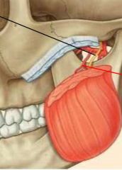
What structure must the masseteric nerve & artery pass through to reach the muscle? |
the mandibular notch |
|

Identify the pink artery and where it branches from |
superficial temporal; branch of external carotid |
|

Identify the purple artery and where it branches from |
deep temporal; branch of maxillary artery |
|
Identify the green nerve and where it branches from |
deep temporal nerve; motor branch of V3 |
|
|
What 3 nerves provide sensory innervation of the TMJ? |
deep temporal nerve; masseteric nerve; auriculo-temporal nerve |
|
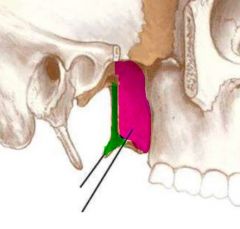
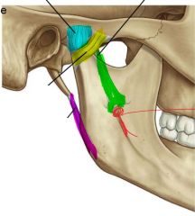
Identify the bony structure in green |
medial pterygoid plate of sphenoid bone |
|

Identify the bony structure in pink |
lateral pterygoid plate of sphenoid bone |
|
|
What is the little dangly bit of bone that comes off the medial pterygoid plate? |
the hamulus |
|
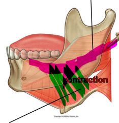
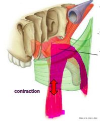
Identify this muscle and its 2 functions |
medial pterygoid muscle; retracts & elevates mandible |
|
|
Where does the medial pterygoid muscle attach? |
the MEDIAL surface of the LATERAL pterygoid plate |
|
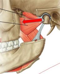
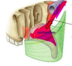
Identify this muscle and its 2 functions |
lateral pterygoid muscle; protrudes & depresses the mandible |
|
|
Where does the lateral pterygoid muscle attach? |
the LATERAL surface of the LATERAL pterygoid plate |
|
|
What happens during UNILATERAL contraction of lateral pterygoid muscle? |
contralateral deviation of mandible (since protruded & depressed from ipsilateral) |
|
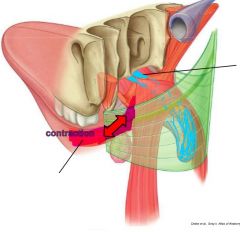
Identify the red & blue muscles |
red = medial pterygoid; blue = lateral pterygoid |
|
|
How does the auriculo-temporal nerve act as a landmark? |
it loops around the middle meningeal artery |
|
|
What is the main venous network on the lateral part of the head? |
the pterygoid plexus |
|
|
How is venous drainage of the teeth different from arterial supply? |
HAH TRICK QUESTION IT'S THE SAME THING psyyyychhh |
|
|
What function does the anterior trunk of the mandibular nerve (V3) have |
motor |
|
|
The mandibular nerve provides motor innervation via which 4 branches? |
(for all mastication muscles) superior & deep temporal nerves, lateral pterygoid nerve, masseteric nerve |
|
|
What function does the posterior trunk of the mandibular nerve (branch of V3) serve? |
sensory |
|
|
What is the ONLY sensory branch of the anterior mandibular nerve? |
buccal nerve; sensory for cheek |
|
|
The mandibular nerve provides sensory innervation via which 3 branches? |
auriculo-temporal nerve; lingual nerve; inferior alveolar nerve |
|
|
What is the ONLY motor branch of the posterior mandibular nerve? |
mylohyoid muscle |
|
|
What is the risk in anaesthesizing the mylohyoid? |
can become paralyzed, making it hard for patient to swallow if done bilaterally |
|
|
What space is posterior and superior to the space of the body of the mandible? |
pterygomandibular |
|
|
What spaces are posterior to and much larger than the pterygomandibular |
temporal & infratemporal space |
|
|
What is the risk of having these cranial spaces connected? |
risk abscess forming and spreading through to temporal space and the throat, esp if left untreated |
|
|
What is the "occlusion line" that helps shape & pattern teeth |
the curve of Spee |
|
|
What happens to the vertical dimension of the mandible with aging? |
vertical bone mass is lost due to wear & tear |
|
|
Name the indentation in the skin above the upper lip? |
philtrum |
|
|
Name the line between the lips and the surrounding skin |
vermillion border |
|
|
Name the corners of the mouth where the lips meet |
labial commissure |
|
|
Name the surface of the lip |
vermillion zone |
|
|
Name the indentation in the centre of the upper lip |
tubercle |
|
|
What are the borders of the maxillary vestibule? |
between buccal mucosa and upper teeth |
|
|
What are the borders of the mandibular vestibule? |
between buccal mucosa and the lower teeth |
|
|
What is considered the "oral cavity proper"? |
area in between the teeth |
|
|
What is the landmark behind the last mandibular molar called? |
retromolar pad |
|
|
What is the common attachment site of the buccinator and superior pharyngeal constrictor? |
pterygomandibular raphe |
|
|
What is the function of the pterygomandibular raphe? |
keeps the different functions of the two attached muscles separate so they can move independently |
|
|
Where does the pterygomandibular raphe attach to bone? |
the hamulus of the medial pterygoid plate |
|
|
What muscle makes up the bulk of the tongue? |
genio-glossus |
|
|
What is the function of the genio-glossus muscle |
protrusion of tongue |
|
|
If there is paralysis of one genio-glossus muscle, how will the tongue move? |
will move ipsilaterally, since the other side can still contract & protrude normally |
|

Name the muscle in pink and its function |
stylo-glossus; tongue retraction |
|
Name the muscle in green and its function |
hyo-glossus; flips tongue to each side ("steering wheel" of the tongue) |
|

Name the muscle in green and its function |
inferior longitudinal; shortens tongue |
|

Name the muscle in blue and its function |
superior longitudinal; shortens tongue |
|

Name the muscle in pink and its function |
vertical muscle; thins the tongue |
|
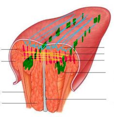
Name the muscle in yellow and its function |
transverse muscle; narrows the tongue |
|
|
What line separates the anterior 2/3 and posterior 1/3 of the tongue? |
sulcus terminalis |
|
|
What are the large papillae in a V at the posterior edge of the tongue? |
circumvallate papillae (the big-ass circular ones) |
|
|
What papillae cover the majority of the dorsal surface of the tongue? |
filiform papillae (little long skinny ones) |
|
|
What papillae cover the lateral surface of the tongue? |
fungiform papillae (short, bulbous ones) |
|
|
What embryological structure is at the centre of the sulcus terminalis? |
foramen caecum |
|
|
What is the function of the foramen caecum |
related to thyroid formation |
|
|
What causes thyroglossal duct cyst formation? |
-abnormal migration of cells OR remaining thyroglossal duct cells |
|
|
What causes black hairy tongue? |
no shedding of filiform papillae; dead layer of cells with extrinsic staining |
|
|
What causes geographic tongue? |
filiform papillae undergoing orthokeratinization, appearing more white |
|
|
What are the two main branches of the lingual artery (from the ECA)? |
deep lingual artery, sublingual artery |
|
|
What does the sublingual artery supply? (1 gland, 2 muscles) |
sublingual gland, mylohyoid, genio-hyoid |
|
|
What does the deep lingual artery supply? |
all extrinsic muscles of the tongue (genio-glossus, hyo-glossus, stylo-glossus) |
|
|
What structure does the sublingual vein run parallel to? |
hypoglossal nerve |
|
|
What is the clinical relevance of the sublingual vein? |
very superficial, so good for rapid absorption of drugs |
|
|
What is the fibrous band along the middle of the ventral surface of the tongue? |
frenulum |
|
|
What is the disorder in which there is a long frenulum preventing tongue protrusion? |
ankyloglossia |
|

What provides sensory innervation to the gingiva in yellow? |
lingual nerve
|
|

What provides sensory innervation to the gingiva in green? |
mental nerve |
|
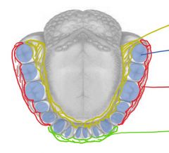
What provides sensory innervation to the gingiva in red? |
buccal nerve (V3 branch) |
|
|
What provides sensory innervation for the anterior 2/3 of the tongue? |
V3 of trigeminal |
|
|
What provides taste innervation for the anterior 2/3 of the tongue? |
chorda tympani of CN VII (facial nerve) |
|
|
What provides sensory AND taste innervation for the posterior 1/3 of the tongue? |
CN IX (glossopharyngeal) |
|
|
What provides motor innervation for the whole tongue? |
CN XII (hypoglossal) |
|
|
From which side are extrinsic tongue muscles innervated |
contralateral ONLY (so in case of UMN or LMN lesion, cannot move tongue, since no compensation by ipsilateral) |
|
|
From which side are intrinsic tongue muscles innervated |
bilateral (so can change shape of tongue if UMN lesion since compensation by other side, but cannot change shape if LMN since branches have converged by this point) |
|
|
Which direction does lymphatic drainage go in the tongue? |
bilateral; thus cannot differentiate which side infection originated based on where nodes are swollen |
|
|
Where are the openings of the sublingual glands? |
many small openings, under the sublingual folds on the floor of the oral cavity |
|
|
Where are the openings of the submandibular glands? |
one opening on each side of the frenulum (sublingual caruncles) |
|
|
Where are the openings of the parotid glands? |
on the buccal mucosa above the linea alba, posteriorly (near molars) |
|

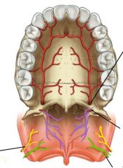
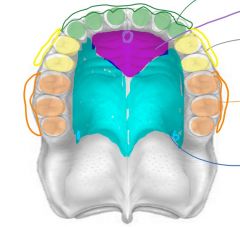
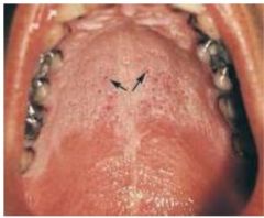
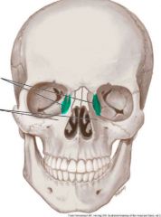
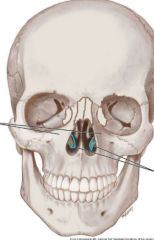
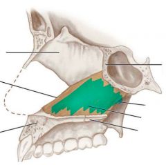
Name the blue structure (hint: at this level of tissue it is NOT a canal!) |
incisive papilla |
|
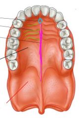
Name the green structure |
palatine rugae |
|

Name the pink structure |
median raphe |
|

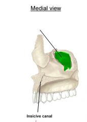
Name the structure in blue |
incisive canal |
|

Name the bone in yellow |
palatine process of maxilla |
|
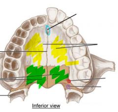
Name the bone in green |
palatine bone |
|

Name the landmark in red |
greater palatine foramen |
|

Name the landmark in purple |
lesser palatine foramen |
|
|
What area does a greater palatine block target? |
greater palatine foramen |
|
|
What area does a nasopalatine block target? |
incisive canal |
|
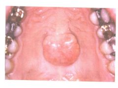
Name and define this condition |
torus palatinus; overgrowth of bone of hard palate |
|

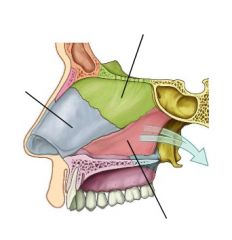
Name the muscle in blue |
buccinator |
|
Name the muscle in green |
superior pharyngeal constrictor |
|

Name the muscle in pink (at arrows), and its function |
tensor veli palatini; tenses the soft palate |
|

Name the tube structure |
pharyngotympanic tube |
|

Name the light pink structure |
palatine aponeurosis |
|
name the muscle in pink (shown contracting), and its function |
levator veli palatini; elevates the soft palate |
|
Name the muscle in pink (shown contracting) |
palatopharyngeus |
|
Name the muscle shown in pink (shown contracting) |
palatoglossus |
|

Name the structure in blue |
musculus uvulae |
|
|
What is the most anterior arch in the oral cavity and what forms it? |
palato-glossal arch, formed by palatoglossus muscle |
|
|
What is the posterior arch in the oral cavity and what forms it? |
palato-pharyngeal arch, formed by palatopharyngeus muscle |
|
|
What is the single, superior-most tonsil in Waldeyer's ring? |
pharyngeal tonsil |
|
|
What are the upper pair of tonsils in Waldeyer's ring? |
tubal tonsils |
|
|
What are the lower pair of tonsils in Waldeyer's ring? |
Palatine tonsils |
|
|
What is the single, inferior tonsil in Waldeyer's ring? |
Lingual tonsil |
|
|
Which tonsil can be affected by adenoids and obstruct nasal airways? |
pharyngeal tonsil |
|

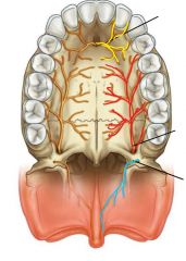
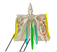
Name the artery in red & its exit point |
greater palatine artery; greater palatine foramen |
|

Name the artery in purple and its exit point |
lesser palatine artery; lesser palatine foramen |
|

Name the artery in yellow |
ascending palatine branch of facial artery |
|
Name the artery in green |
ascending pharyngeal artery |
|
|
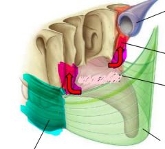
What artery does the greater palatine artery anastomose with, and where? |
sphenopalatine artery; at the incisive canal |
|

Name the nerve in yellow and its exit point |
nasopalatine nerve; incisive canal |
|
Name the nerve in red and its exit point |
greater palatine nerve; greater palatine foramen |
|

Name the nerve in blue and its exit point |
lesser palatine nerve; lesser palatine foramen |
|

What nerve innervates the orange area of the maxilla? |
posterior superior alveolar nerve |
|

What nerve innervates the yellow area of the maxilla? |
middle superior alveolar nerve |
|

What nerve innervates the green area of the maxilla? |
anterior superior alveolar nerve |
|
What nerve innervates the purple area of the maxilla? |
nasopalatine nerve |
|

What nerve innervates the blue area of the maxilla? |
greater palatine nerve |
|

Name the nerve in yellow/blue (branches into the lesser & greater palatine nerves in the oral cavity) |
Descending palatine nerve |
|
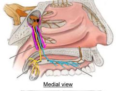
Name the landmark in purple, where nerves are travelling |
pterygopalatine canal |
|
|
What nerve innervates all muscles of the soft palate (except tensor veli palatini)? |
vagus nerve (CN X) |
|
|
What nerve innervates tensor veli palatini |
trigeminal nerve (CN V3) |
|
|
Where are palatine glands located (in relation to other layers of tissue)? |
under the keratinized outer layer of the mucosa |
|
Name this mucosa disorder and how it happens |
nicotinic stomatitis; hyperkeratinization due to heat from smoking or hot liquid |
|
What bone is shown in blue, making the medial wall of the orbit? |
lacrimal bone |
|
Name the blue structures |
inferior nasal conchae |
|
Name the landmark in green |
maxillary sinus |
|
Name the structure in blue |
inferior nasal conchae |
|
|
What is the function of the bony plates in the nasal cavity? |
close off the maxillary sinus from the nasal cavity |
|

Identify the yellow structure of the ethmoid bone |
orbital plate |
|

Identify the blue structure of the ethmoid bone |
middle nasal concha |
|
Identify the green structure of the ethmoid bone |
perpendicular plate |
|

Identify the structure circled in red on the ethmoid bone |
ethmoid air cells |
|

Identify the bony landmark in green and its function |
crista galli; attaches to falx cerebri |
|
Identify the bony landmark in blue and its function |
cribriform plate; entrance of olfactory nerve |
|

Identify the bony landmark in yellow |
superior nasal concha |
|

Identify the bony landmark in pink |
middle nasal concha |
|
|
What are the superior & middle nasal conchae projecting from? |
ethmoid bone |
|
Identify this bone |
vomer |
|
|
What does the vomer articulate with at its anterior border? |
nasal cartilage |
|
|
What does the vomer attach to at its inferior border? |
palatine bone |
|
Identify the green bone |
perpendicular plate of ethmoid bone |
|

Identify the blue structure |
septal cartilage |
|
|
What 3 structures form the nasal septum? |
septal cartilage; perpendicular plate; vomer |
|

Identify the location in blue |
maxillary sinuses |
|
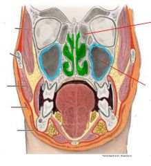
Identify the location in green |
nasal cavities |
|
|
What is the dental clinical significance of the opening from the nasal cavity to the maxillary sinus? |
inhaled air can enter the sinuses, and any resulting infections can be referred to the maxillary teeth |
|
|
Why is it important to consider the size of the maxillary sinus in Caldwell-Luc procedure (implantation)? |
large sinuses means a thin maxillary plate; may not have enough anchorage for implant, so may require filling first |
|

Identify the opening in blue (under the middle nasal concha) |
maxillary sinus opening |
|
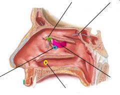
Identify the opening in green (under the middle nasal concha) |
frontal sinus opening |
|

Identify the bony landmark in pink |
bulla ethmoidalis (where air cells open into nasal cavity) |
|

Identify the opening in yellow |
naso-lacrimal duct opening |
|

Identify the opening in lavender (lol) |
sphenoid sinus opening |
|
|
What is the direction of drainage from the paranasal sinuses (except sphenoid)? |
into nasal cavity to be swallowed |
|
|
What is the clinical risk of the direction of drainage of the paranasal sinuses? |
pass Eustachian tubule; can enter and cause otitis media |
|
|
Why is maxillary sinus pain referred to the teeth? |
superior alveolar nerve (V2 branch) that innervates the teeth also goes all around the maxillary sinus |
|

Identify the artery in blue |
sphenopalatine artery (branch of ECA) |
|
What does the artery in yellow anastomose with? (hint: going thru incisive canal) |
greater palatine artery
|
|

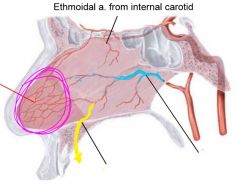
Identify the arterial plexus in pink and its clinical significance |
Kiesselbach's area; most common site of epistaxis (nose bleed) |
|

Identify the nerve in green and its function (hint: going thru incisive canal) |
nasopalatine nerve (V2 branch); sensory function |
|

Identify the nerve in blue and its function (hint: going thru cribriform plate) |
olfactory nerve (CN I); sense of smell |
|

Identify the nerve in yellow |
ethmoidal nerve (V1 branch) |
|
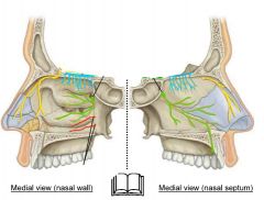
Identify the nerves in red |
nasal nerves (V2 branches) |
|
|
What are the borders of the pterygopalatine fossa? |
POSTERIOR: sphenoid bone; MEDIAL: palatine bone; ANTERIOR/LATERAL: maxillary bone |
|
|
|
roof of oral cavity |
|
|
Where does the inferior orbital fissure open? |
the orbit |
|
|
where does the sphenopalatine foramen open? |
nasal cavity |
|
|
where does the foramen rotundum open? |
cranial cavity |
|
|
where does the pterygoid canal open? |
cranial cavity |
|
|
where does the pharyngeal canal open? |
nasopharynx |
|
What structure is outlined in red? |
lesser wing of sphenoid bone |
|

What structure is outlined in blue? |
greater wing of sphenoid bone |
|

what is the structure in purple? |
lateral pterygoid plate |
|

What is the structure in blue, and the end of it in green? |
medial pterygoid plate and its hamulus |
|
|
The sphenoid wing contains the openings of which 3 canals? |
foramen rotundum & pterygoid canal (to cranial cavity); pharyngeal canal (to nasopharynx) |
|
|
The parasympathetic system has (long/short) pre-ganglionic fibres |
LONG |
|
|
the sympathetic system has (long/short) pre-ganglionic fibres |
SHORT |
|
|
Where do pre-ganglionic parasympathetic fibres synapse? |
on or near the target organ |
|
|
Where do pre-ganglionic sympathetic fibres synapse? |
on the sympathetic trunk |
|
|
the sympathetic system has (long/short) post-ganglionic fibres |
LONG (usually travel wrapped around an artery) |
|
|
the parasympathetic system has (long/short) post-ganglionic fibres |
SHORT (since ganglion is on/near the target organ) |
|
|
What gland does the otic ganglion innervate? |
parotid gland |
|
|
What is the pre-ganglionic nerve of the otic ganglion? |
lesser petrosal nerve (from CN IX) |
|
|
The post-ganglionic sympathetic fibres of the parotid gland travel with what structure to reach the gland? |
2 branches: superior temporal artery & auriculotemporal nerve |
|
|
What glands do the pterygopalatine ganglion innervate? |
palatine & lacrimal |
|
|
What is the pre-ganglionic nerve of the pterygopalatine ganglion? |
greater petrosal nerve (CN VII) |
|
|
The post-ganglionic sympathetic fibres of the palatine gland travel with what structure to reach the gland? |
internal carotid (then enter pterygoid canal) |
|
|
The submandibular ganglion innervates what 2 glands? |
submandibular & sublingual |
|
|
The post-ganglionic sympathetic fibres of the palatine gland travel with what structure to reach the gland? |
internal carotid (then enter palatine canal) |
|
|
The post-ganglionic sympathetic fibres of the submandibular gland travel with what structure to reach the gland? |
along external carotid, then facial artery |
|
|
The post-ganglionic sympathetic fibres of the sublingual gland travel with what structure to reach the gland? |
along external carotid, then lingual artery |
|
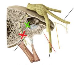
A lesion at the level of the green X would affect function of which glands? |
parasympathetic innervation of submandibular & sublingual glands (this is chorda tympani branching off of CN VII and hitchhiking with V3) |
|

A lesion at the level of the red X would affect function of which glands? |
parasympathetic innervation of submandibular, sublingual, palatine, and lacrimal glands (this is CN VII before any branching, so both the chorda tympani & greater petrosal nerve are affected, and all glands innervated by the pterygopalatine & submandibular ganglia) |
|
|
What route do sympathetic pre-ganglionic fibres follow from the spine? |
from level of T1-L2, travel up and synapse on sympathetic trunk at level of C1 |
|
|
When breathing, what is the state of the soft palate, epiglottis, and vocal chords? |
soft palate = relaxed; epiglottis = relaxed; vocal folds = open (allow air in) |
|
|
When chewing food & breathing, what is the state of the soft palate, epiglottis, and vocal chords? |
soft palate = depressed (prevent bolus swallowing); epiglottis = elevated (allow air in larynx); vocal folds = open (allow air in larynx) |
|
|
When swallowing food/liquid, what is the state of the soft palate, epiglottis, and vocal chords? |
soft palate = elevated (to allow bolus into pharynx & prevent nasal regurgitation); epiglottis = depressed (to prevent aspiration of bolus); vocal folds = closed (extra protection for larynx) |
|
|
The epiglottis is longer than the larynx and covers part of the esophagus when closed. What must be done to compensate? |
Must pull trachea forward when swallowing to keep esophagus open & uncovered |
|
|
What makes up the true vocal folds? |
the superior free edge of the crico-thyroid ligament |
|
|
What makes up the false vocal folds (aka vestibular folds)? |
the inferior free edge of the quadrangular membrane |
|
|
What is the space between the 2 sets of vocal folds, covered by a membrane? |
laryngeal ventricle |
|
|
What is the function of the thyro-arytenoid muscle? |
tenses the vocal chords |
|
|
What is the function of the oblique & transverse arytenoid muscles? |
narrow (but not totally close) the vocal chords for quiet respiration |
|
|
What is the function of the lateral crico-arytenoid muscles? |
close the vocal folds completely during swallowing |
|
|
What is the function of the posterior crico-arytenoid muscles? |
open vocal chords wide for deep breathing |
|
|
What phase of degluttition is under voluntary control? |
only the initiation segment of the oral phase |
|
|
What happens during the oral phase of degluttition? |
the tongue is squeezed against the palate to push bolus into the pharynx |
|
|
What happens during the pharyngeal phase of degluttition? |
nasal & laryngeal cavitities closed (to prevent nasal regurgitation & aspiration); upper esophageal sphincter opened (to allow entry) |
|
|
What happens during the esophageal phase of degluttition? |
contraction of esophageal muscles; peristalsis to move bolus inferiorly |
|
|
What muscles squeeze the tongue posteriorly? |
genio-glossus & styloglossus |
|
|
What muscles elevate the soft palate? |
tensor veli palatini & levator veli palatini |
|
|
What muscles elevate the larynx (via the hyoid bone)? |
supra-hyoid muscles (mylo-hyoid, genio-hyoid, stylo-hyoid, digastric) |
|
|
What muscles close the pharynx? |
the 3 constrictor muscles (superior, middle, inferior) |
|
|
What muscles carry out peristalsis of the pharynx? |
stylopharyngeus; salpingo-pharyngeus; palato-pharyngeus |
|
|
What is special about degluttition in a newborn? Why is this important? |
overlap of epiglottis & soft palate; allows continunous swallowing & breathing (ie while breastfeeding) |