Reading...
![]()
Play button
![]()
Play button
![]()
Use LEFT and RIGHT arrow keys to navigate between flashcards;
Use UP and DOWN arrow keys to flip the card;
H to show hint;
A reads text to speech;
469 Cards in this Set
- Front
- Back
- 3rd side (hint)
|
What is the Function of enzymes in catalyzing biological reactions?
|
Enzymes are biological catalysts usually composed of proteins. Catalysts are things that increase the rate of a reaction, but does not get used up during the reaction.
Structure determines function. A change in structure => a change in function. Important biological reactions catalyzed by enzymes: Metabolism DNA synthesis RNA synthesis Protein synthesis Digestion Enzymes that break down other molecules do so via reactions like hydrolysis, anything that would alter the original molecule to make it inactive. So if there is another drug or enzyme that inhibits the inhibitor, then the molecule is not broken down. For example, if an enzyme hydrolysis acetylcholine, then acetylcholine is broken down and doesn't work as a neurotransmitter. This isn't creating acetylcholine or anything. Then if a drug is inhibiting the enzyme that break down of acetylcholine, then acetylcholine is free to be the neurotransmitter and affect the target tissues. ATP is hydrolyzed to ADP. ADP is phosphorylated to ATP. |
Enzymes work at an optimal temperature and pH, typically around the physiological temperature of 36°C and a pH of 7.2. At higher temperatures the protein will denature and lose their function. They often have to interact with cofactors such as vitamins or IONS for optimal activity. Also, mutation will affect the DNA sequence coding for these proteins, leading to an altered polypeptide and often a change in spatial configuration, and therefore a change in function.
|
|
|
Explain the Reduction of activation energy.
|
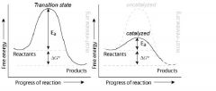
Enzymes decrease the activation energy (Ea) of a reaction by lowering the energy of the transition state.
Enzymes increase the rate of a reaction by decreasing the activation energy. Enzymes will increase the rate constant, k, for the equation rate = k[A][B]. Enzymes do NOT change the Keq of a reaction. Enzymes do not change Keq because it lowers the activation energy for BOTH forward and reverse reactions. Enzymes will make the reverse reaction go faster also. Enzymes do not change ΔG, the net change in free energy. Enzymes affect the kinetics of a reaction, but not the thermodynamics. |
|
|
|
What are Substrates and enzyme specificity?
|
Enzyme-substrate interactions occur at the enzyme's active site.
Enzyme-substrate specificity derives from structural interactions. Lock and key model: rigid active site. Substrate fits inside the rigid active site like a key. Induced fit model: flexible active site. Substrate fits inside the flexible active site, which is then induced to "grasp" the substrate in a better fit. Enzymes can be specific enough to distinguish between stereoisomers. Enzymes can be protein or RNA. Almost all enzymes in your body is made of protein. The most important RNA enzyme in your body is the ribosome. Ribosomes are made from complexes of RNAs and proteins. Ribosomes are divided into two subunits, one larger than the other. The smaller subunit binds to the mRNA, while the larger subunit binds to the tRNA and the amino acids. When a ribosome finishes reading a mRNA these two subunits split apart. Ribosomes have been classified as ribozymes. A ribozyme (from ribonucleic acid enzyme, also called RNA enzyme or catalytic RNA) is an RNA molecule possessing a well defined tertiary structure that enables it to catalyze a chemical reaction. Heat and extreme pH denatures enzymes by altering their structure. |
|
|
|
What are the 4 levels of enzyme structure?
|
Primary: this is the sequence of the protein or RNA chain.
Secondary: this is hydrogen bonding between the protein backbone. Examples include alpha helices and beta sheets (backbone H-bonding). For RNA, this is base pairing. Tertiary: this is the 3-D structure of the enzyme. This involves -R group interactions and spatial arrangement of secondary structure. Globular proteins occupy a 3-D structure and are known as a tertiary structure. Quaternary: when more than 1 chain is involved. When you hear about "dimers", "trimers", "tetramers", "oligomers", that's quaternary structure. Freezing an enzyme has no effect on its tertiary or quaternary structure; if brought back to its optimal temperature after freezing, it will still function normally. Heating, however, can cause the tertiary and quaternary structure of enzymes to become unstable, thus inactivating them. |
|
|
|
What is Feedback inhibition?
|
Controls enzyme activity.
The product of a pathway inhibits the pathway. For example, hexokinase, the first enzyme in glycolysis, is inhibited by its product glucose-6-phosphate. If something happens in a pathway where a conversion is prevented, so the product will not form, then the beginning products of the pathway (before the step that prevents the conversion) will accumulate because it is not being checked by the end product. If there is no end product, the beginning products will continue to form and accumulate through the pathway (until the pathway reaches the step that prevents the conversion). Feedback inhibition is where a certain amount of the formed product inhibits the beginning products. But if there is no product of the pathway, then the pathway isn't inhibited. |
|
|
|
What is Competitive inhibition?
|
Controls enzyme activity. An inhibitor competes with the substrate for binding to the active site. Competitive inhibition increases the amount of substrate needed to achieve maximum rate of catalysis. Competitive inhibition does NOT change the maximum possible rate of the enzyme's catalysis. You can overcome competitive inhibition by providing more substrate. Competitive inhibition is a form of enzyme inhibition where binding of the inhibitor to the enzyme prevents binding of the substrate and vice versa. In competitive inhibition, the inhibitor binds only the free enzyme, and cannot bind when the substrate is bound (in other words, it cannot bind the enzyme-substrate complex). In competitive inhibition, the maximum velocity (Vmax) of the reaction is unchanged, while the apparent affinity of the substrate to the binding site is decreased. Any given competitive inhibitor concentration can be overcome by increasing the substrate concentration in which case the substrate will outcompete the inhibitor in binding to the enzyme.
|
|
|
|
What is Non-competitive inhibition?
|
Controls enzyme activity. An inhibitor binds to an allosteric site on the enzyme to deactivate it. An allosteric site is a place on an enzyme where a molecule that is not a substrate may bind, thus changing the shape of the enzyme and influencing its ability to be active. The substrate still have access the active site, but the enzyme is no longer able to catalyze the reaction. Non-competitive inhibition decreases the maximum possible rate of the enzyme's catalysis. Non-competitive inhibition does NOT change the amount of substrate needed to achieve the maximum rate of catalysis. You can't overcome non-competitive inhibition by adding more substrate. Non-competitive inhibition is a type of enzyme inhibition that reduces the maximum rate of a chemical reaction (Vmax) without changing the apparent binding affinity of the catalyst for the substrate. Non-competitive inhibition usually applies to enzymes and differs from competitive inhibition in that the inhibitor always binds to the enzyme at a site other than the enzyme's active site (this other site is called an allosteric site). This affects the rate of the reaction catalysed by the enzyme because the presence of the inhibitor causes a change in the structure and shape of the enzyme. This change in shape means the enzyme is no longer able to bind with a substrate correctly. This reduces the concentration of 'active' enzyme resulting in a decrease in the Vmax. In this mode of inhibition, there is no competition between the inhibitor and the substrate, so increasing the concentration of the substrate still does not allow the maximum enzyme activity rate to be reached.
|
|
|
|
What is basic metabolism?
|
Metabolism consists of two parts: Catabolism and anabolism.
Catabolism is breaking stuff down for energy. Anabolism is using energy to build stuff for storage. Metabolism is the set of chemical reactions that happen in living organisms to maintain life. These processes allow organisms to grow and reproduce, maintain their structures, and respond to their environments. Metabolism is usually divided into two categories. Catabolism breaks down organic matter, for example to harvest energy in cellular respiration. Anabolism uses energy to construct components of cells such as proteins and nucleic acids. A metabolic process begins with a substrate, passes it through a unique network of interconnected chemical reactions, and releases a product. Glycolysis is an example of a metabolic process. Gycolysis converts glucose (a substrate) into pyruvate (the product) via a series of reactions. |
|
|
|
What are the Steps of aerobic metabolism (needs oxygen)?
|
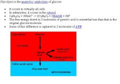
Glycolysis
Aerobic decarboxylation Krebs cycle Electron transport chain. |
|
|
|
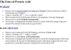
What are the Steps of anaerobic metabolism (don't need oxygen)?
|
Glycolysis
Glycolysis produces pyruvic acid as its last product. Alcohol or lactic acid fermentation. Pyruvic acid can be anaerobically converted to lactic acid in humans or broken down to ethanol and CO2 in yeast. Yeast are used to make alcoholic beverages (the bubbles is the CO2) and cause dough to rise. Fermentation occurs under anaerobic conditions and can produce either alcohol (called yeast fermentation) or lactic acid (via lactic acid fermentation). |
|
|
|
What is Aerobic metabolism of glucose ?
|
Complete oxidation of metabolite (glucose) to carbon dioxide.
~ 36 ATP produced per glucose. C6H12O6 + 6O2 => 6CO2 + 6H2O C6H12O6: this is glucose. You get it from your diet. 6O2: this is molecular oxygen that you breath in. 6CO2: this is carbon dioxide produced by aerobic decarboxylation and the Krebs cycle. Both the carbon and oxygen in this CO2 comes from the metabolite (glucose). 6H2O: this is water produced in the electron transport chain. The oxygen comes completely from the molecular oxygen that you breath in. If we were to follow the carbon in the metabolite (glucose), it will end up in carbon dioxide. If we were to follow the oxygen in the metabolite (glucose), it will end up in carbon dioxide. If we were to follow the oxygen (O2) you breath in, it will end up in water (H2O). As for the HYDROGENS, they'll either be in water, exist as protons in solution, or be transferred to some other entity. As we can see, the total reaction involves complete oxidation of the metabolite (glucose) and complete reduction of molecular oxygen. When electrons pass from the metabolite (glucose) to molecular oxygen, energy is released. The electron transport chain harnesses this energy. |
|
|
|
What is Anaerobic metabolism of glucose?
|
Partial oxidation of metabolite (glucose) to pyruvate.
2 net ATP produced per glucose. Pyruvate is then reduced to either alcohol or lactate. Bacteria reduce pyruvate to alcohol in a process called alcohol fermentation. Humans reduce pyruvate to lactate in a process called lactic acid fermentation. |
|
|
|
What are the substrates and products of glycolysis?
|
Glycolysis = convert glucose (6 carbons) to 2 molecules of pyruvate (3 carbons).
Location: cytosol. 2 net ATP made for every glucose (2 input ATP, 4 output ATP). 2 NADH made for every glucose. Occurs under both aerobic and anaerobic conditions. Glycolysis is inhibited by ATP. Glyceraldehyde 3-phosphate dehydrogenase is an enzyme that catalyzes the sixth step of glycolysis and thus serves to break down glucose for energy and carbon molecules. |
No FADH2 is produced in glycolysis. FADH2 is not produced until the Krebs cycle.
|
|
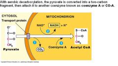
What are the substrates and products of Aerobic decarboxylation?
|
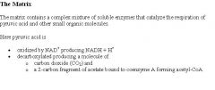
Aerobic decarboxylation = convert pyruvate (3 carbons) to an acetyl group (2 carbons). 1 NADH made for every pyruvate. Only occurs in the presence of oxygen. Acetyl group attaches to Coenzyme A to make acetyl CoA. Aerobic decarboxylation occurs in the matrix of the mitochondria. This step is also known as the link reaction, as it links glycolysis and the Krebs cycle. Sometimes it may be considered part of Krebs cycle.
Carbon dioxide is released during this step. |
|
|
|
What are the substrates and products of Anaerobic fermentation?
|
Anaerobic fermentation = redox reaction: reduce pyruvate, oxidize NADH.
1 NAD+ made for every pyruvate. Alcohol fermentation = pyruvate reduced to ethanol. Lactic acid fermentation = pyruvate reduced to lactate. The purpose of anaerobic fermentation is to regenerate NAD+, which is needed for glycolysis. |
|
|
|
Explain the Krebs cycle, substrates and products, and the general features of the pathway.
|
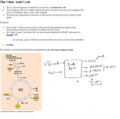
Location: matrix of mitochondria.
The final product of glycolysis are 2 pyruvates, which are each converted into acetyl coA for use in the Krebs cycle. Acetyl CoA feeds into the cycle. 3 NADH made per acetyl CoA. 1 FADH2 made per acetyl CoA. 1 ATP (GTP) made per acetyl CoA. Coenzyme A is regenerated (during the first step of the cycle). Carbon dioxide is released during the Krebs cycle as well as the aerobic decarboxylation step. Krebs cycle, TCA, Tricarboxylic acid cycle, citric acid cycle all mean the same thing. Krebs cycle is Inhibited by ATP and NADH. |
Citrate is an intermediate of the Krebs cycle - it is the first molecule formed; another term for the Krebs cycle is the Citric Acid cycle - citric named from the citrate. The Krebs cycle only forms 2 ATP directly, from the 2 acetyl coAs, all other ATPs that are formed are produced by oxidative phosphorylation.
|
|
|
What is the difference in ATP produced for prokaryotes and eukaryotes?
|

in glycolysis, a total of four molecules of ATP are produced, but two are used up in other steps in the process. Additional ATP is produced during the Krebs Cycle and the Electron Transport Chain, resulting in a grand total of 40 ATP molecules produced from the breakdown of one molecule of glucose via cellular respiration. Since two of those are used up during glycolysis, in prokaryotes a net total of 38 molecules of ATP are produced by cellular respiration. Most prokaryotes have very simple cells which lack several types of organelles present in eukaryotes, and therefore the Krebs Cycle and the Electron Transport Chain occur in the cytoplasm and/or using chemicals embedded in the cell membrane. In contrast, eukaryotes have more complex cells with more specialized organelles to perform given functions. In eukaryotes, the Krebs Cycle and Electron Transport Chain occur within the mitochondria, and thus the pyruvic acid resulting from glycolysis must be sent into the mitochondria for these reactions to occur. However, to move one molecule of pyruvic acid (remember each molecule of glucose turns into two pyruvic acid molecules) from the cytoplasm into a mitochondrion “costs” the cell one molecule of ATP (therefore two ATPs for a whole glucose), thus a net total of 36 ATP molecules per molecule of glucose is produced in eukaryotes as compared to only two in fermentation. The overall reaction for cellular respiration is C6H12O6 + 6O2 yields 6CO2 + 6H2O (+ energy for the cell to use for other things). Biology textbooks often state that 38 ATP molecules can be made per oxidised glucose molecule during cellular respiration (2 from glycolysis, 2 from the Krebs cycle, and about 34 from the electron transport system). However, this maximum yield is never quite reached due to losses (leaky membranes) as well as the cost of moving pyruvate and ADP into the mitochondrial matrix and current estimates range around 29 to 30 ATP per glucose.
|
|
|
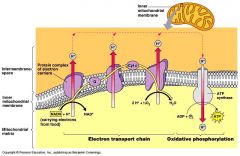
Explain the Electron transport chain and oxidative phosphorylation, substrates and products, and the general features of the pathway.
|
Location: the cristae (inner membrane of mitochondria). 1) Input NADH. 2) Proton gradient. The electron transport chain (ETC) is essentially a series of redox reactions that forms an electron gradient, where NADH gets oxidized to NAD+ and O2 gets reduced to H2O. The series of redox reactions consists of electrons passing from NADH to coenzyme flavin mononucleotide (FMN), to Coenzyme Q, iron-sulfur complexes, and cytochromes (cytochrome b, c and aa3) before finally being used to reduce oxygen. NADH is highest in energy, while O2 is lowest in energy. When electrons are passed from NADH down a series of proteins and finally to O2, energy is released. FADH2 is lower in energy than NADH, that's why it releases less energy when it gets oxidized. FADH2 skips FMN and passes its electrons to Coenzyme Q. The energy released from these reactions of the electron gradient generates a proton gradient, which drives ATP synthase to make ATP. This is called oxidative phosphorylation. The ETC is inhibited by certain antibiotics, by cyanide, azide, and carbon monoxide. Sometimes ATP synthase is referred to as ATP synthethase.
|
The cytochrome chain is located in the inner membrane of the mitochondria (the cristae).
|
|
|
What is a proton gradient?
|
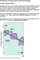
The energy released from passing electrons down the ETC is used to pump protons into the intermembrane space of the mitochondria.
H+ concentration is very high in the intermembrane space (higher than those in the matrix). Thus, this establishes an electrochemical gradient called the proton gradient. H+ wants to migrate down the proton gradient (from the intermembrane space back into the matrix), but it can only do this by going through the ATP synthase. Like a water mill, ATP synthase harnesses the energy of the falling protons to convert ADP into ATP. ATP synthase, is the final enzyme in the oxidative phosphorylation pathway. The enzyme uses the energy stored in a proton gradient across a membrane to drive the synthesis of ATP from ADP and phosphate (Pi). This phosphorylation reaction is an equilibrium, which can be shifted by altering the proton-motive force. In the absence of a proton-motive force, the ATP synthase reaction will run from right to left, hydrolyzing ATP and pumping protons out of the matrix across the membrane. However, when the proton-motive force is high, the reaction is forced to run in the opposite direction; it proceeds from left to right, allowing protons to flow down their concentration gradient and turning ADP into ATP. |
|
|
|
What is the enzyme complex ATP Synthase?
|
The Enzyme called ATP synthetase has two major components Fo and F1 factors. The Fi component resembles a doorknob protruding into the matrix from the inner membrane. It is attached by a stalk to the Fo part, which is embedded in the inner membrane and extends across it. The oxidative phosphorylation principle postulates that the High H+ gradient creates the protomotive force needed to link the electron transport chain to the ATP synthetase molecule. As 2H+ (two protons) pas back into the mitochondria matrix through the ATP synthetase molecule enough free energy is released to create one ATP molecule from ADP + Pi. For each 2H+ 1 ATP is created. Therefore since each NADH causes 6 H+s to be pumped out 3 ATP can be made for each NADH entering the respiratory chain. Also each FADH2 can pump 4 H+s. Therefore each FADH2 can cause 2ATP to be made.
|
|
|
|
What is Fat metabolism?
|
Location: beta-oxidation occurs in the matrix of the mitochondria. Ester hydrolysis occurs in the cytosol.
Fatty esters gets hydrolyzed into free fatty acids by lipases. Fatty acids enter the Krebs cycle 2 carbons at a time. For example, triglyceride gets hydrolyzed into 3 free fatty acids and glycerol. Fatty acids cannot cross the mitochondrial membrane unassisted, so they are activated at the COOH end by CoA (to be precise, it turns into a thioester). The fatty acid is then transported into the mitochondrial matrix. A process called beta-oxidation is where the fatty acid-CoA molecule is degraded into acetyl CoA molecules by a recurring cyclic sequence of four reactions. The final step (4th reaction) is the cleavage of the acetyl CoA from the original fatty acid chain. At the same time, CoA is added to the remaining part of the original fatty acid chain, but now it is 2 carbons shorter. This chain repeats the 4 reaction steps of beta oxidation, and gets shorter and shorter each time until the last cycle begins with a 4 carbon-CoA and finishes as two acetyl CoA molecules. β-oxidation produces acetyl CoA and also FADH2 and NADH. The acetyl CoA feeds into the Krebs cycle, and the FADH2 and NADH feed into the ETC. On a per gram basis, fats give the more energy than any other food source. Beta oxidation is the process by which fatty acids, in the form of Acyl-CoA molecules, are broken down in mitochondria to generate Acetyl-CoA, the entry molecule for the Krebs cycle. The beta oxidation of fatty acids involve three stages: 1)Activation of fatty acids in the cytosol 2) Transport of fatty acids into mitochondria 3)Beta oxidation in the mitochondrial matrix Fatty acids are oxidized by most of the tissues in the body. However, the brain, erythrocytes, and adrenal medulla cannot utilize fatty acids for energy requirements. |
Lipolysis is the hydrolysis of lipids. Metabolically it is the breakdown of triglycerides into free fatty acids within cells. When fats are broken down for energy the process is known as beta oxidation. Ketones are produced, and are found in large quantities in ketosis (a state in metabolism occurring when the liver converts fat into fatty acids and ketone bodies which can be used by the body for energy). The following hormones induce lipolysis: epinephrine, norepinephrine, glucagon, growth hormone and cortisol. This results in increased production of cAMP, which activates protein kinase A, which subsequently activate lipases found in adipose tissue. Triglycerides are transported through the blood to appropriate tissues (adipose, muscle, etc) by lipoproteins such as chylomicrons. Triglycerides present on the chylomicrons undergo lipolysis by the cellular lipases of target tissues which yields glycerol and free fatty acids. Free fatty acids released into the blood are then available for cellular uptake. Free fatty acids not immediately taken up by cells may bind to albumin for transport to surrounding tissues that require energy. Serum Albumin is the major carrier of free fatty acids in the blood. The glycerol also enters the bloodstream and is absorbed by the liver or kidney where it is converted to glycerol 3-phosphate by the enzyme glycerol kinase which will then rejoin the glycolysis and gluconeogenesis pathway. While lipolysis is triglyceride hydrolysis, the process by which triglycerides are broken down, esterification is the process by which triglycerides are formed. Esterification and lipolysis are essentially reversals of one another.
|
|
|
What is protein metabolism?
|
Proteins are broken down into amino acids by proteases. The carbon in the amino acid is converted to pyruvate or acetyl-CoA, (or other metabolical intermediates such as oxaloacetate), depending on what amino acid it is.
The nitrogen in the amino acid is converted to urea (for desert animals, birds and reptiles, it is uric acid). The carbon products from amino acid metabolisms can either feed to the Krebs cycle, or be the starting material for gluconeogenesis. Gluconeogenesis is a metabolic pathway that results in the generation of glucose from non-carbohydrate carbon substrates such as lactate, glycerol, and glucogenic amino acids. A glucogenic amino acid is an amino acid that can be converted into glucose through gluconeogenesis. |
|
|
What is the Double-helix structure?
|
The "double" in the double helix means that DNA is found in a double-stranded form - 2 single-stranded chains of DNA stuck to each other via hydrogen bonding of the base pairs.
The 2 single-strands are anti-parallel to each other. Going from 5' to 3' of one strand means going from 3' to 5' of the other strand. The "helix" in the double helix means that the entire thing is wound up in a spiral. |
|
|
What is the DNA composition?
|
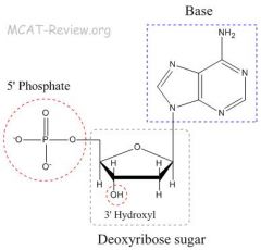
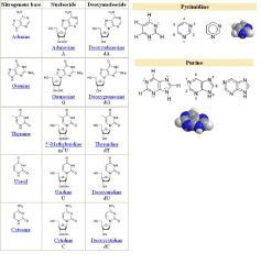
DNA composition: purine and pyrimidine bases, sugars, phosphate
dNTP is deoxyribonucleotide triphosphate and it is a generic term referring to the four deoxyribonucleotides: dATP, dCTP, dGTP and dTTP. Nucleotide = base + sugar + phosphate = Adenine, Guanine, Thymine, Cytosine. Nucleoside = base + sugar = Adenosine, Guanosine, Thymidine, Cytidine. Base can either be purines A and G (the big ones with 2 rings) or pyrimidines T and C (the small ones with 1 ring). The phosphate group gives DNA its acidity (hence the name nucleic acid). The phosphate group is phosphoric acid. DNA is a polymer. The monomer units of DNA are nucleotides, and the polymer is known as a "polynucleotide." Each nucleotide consists of a 5-carbon sugar (deoxyribose), a nitrogen containing base attached to the sugar, and a phosphate group. Deoxy means one of the hydroxyl groups is replaced by a hydrogen (which is why ribose has the extra hydroxyl group at the 2 carbon). Nucleosides consist of a nitrogenous base bound to a ribose or deoxyribose sugar. Nucleosides can be phosphorylated by specific kinases in the cell on the sugar's primary alcohol group (-CH2-OH), producing nucleotides, which are the molecular building-blocks of DNA and RNA. DNA consists of two long polymers of simple units called nucleotides, with backbones made of sugars and phosphate groups joined by ester bonds (creating phosphodiester bonds). |
|
|
|
Explain Base-pairing specificity with DNA and the concept of complementarity.
|
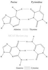
Base pairing specificity: A with T and G with C.
A forms 2 hydrogen bonds with T. G forms 3 hydrogen bonds with C. GC bonds are stronger. DNA with high GC content will be harder to break apart. Complementary strands of DNA hydrogen bond with each other. 5'-ATGC-3' will be complementary to 5'-GCAT-3' or 3'-TACG-5', but NOT 5'-TACG-3'. make sure you get the 5's and 3's right. Complementarity is a property of double-stranded nucleic acids such as DNA and RNA. Each strand is complementary to the other in that the base pairs between them are non-covalently connected via two or three hydrogen bonds. Since there is only one complementary base for any of the bases found in DNA and in RNA, one can reconstruct a complementary strand for any single strand. This is essential for DNA replication. |
In general, the hydrogen bonds are weak and the weak hydrogen bonds enable the helices to separate easily to facilitate DNA replication. It's just that the GC bonds are stronger than the AT bonds, but overall, hydrogen bonding is a weak bond (though stronger than london dispersion forces).
|
|
|
How does the DNA function in transmission of genetic information?
|
Because of the complementary nature of base pairing, DNA can transmit genetic information through replication. Deoxyribonucleic acid (DNA) is a nucleic acid that contains the genetic instructions used in the development and functioning of all known living organisms and some viruses. The main role of DNA molecules is the long-term storage of information. DNA is often compared to a set of blueprints or a recipe, or a code, since it contains the instructions needed to construct other components of cells, such as proteins and RNA molecules. The DNA segments that carry this genetic information are called genes, but other DNA sequences have structural purposes, or are involved in regulating the use of this genetic information.
If one wants to inhibit activation of a protein (such as a protein that stimulates cell death), you would want to prevent the degredation of the regulatory molecule for the protein. Although some regulatory molecules induce transcription, some molecules also repress transcription. In general, all genes need regulatory molecules - the reason why cancer goes unchecked is that its cells do not have regulatory molecules. Regulation of gene expression (or gene regulation) includes the processes that cells and viruses use to turn the information in genes into gene products. Although a functional gene product may be an RNA or a protein, the majority of known mechanisms regulate protein coding genes. Any step of the gene's expression may be modulated, from DNA-RNA transcription to the post-translational modification of a protein. Gene regulation is essential for viruses, prokaryotes and eukaryotes as it increases the versatility and adaptability of an organism by allowing the cell to express protein when needed. Furthermore, gene regulation drives the processes of cellular differentiation and morphogenesis, leading to the creation of different cell types in multicellular organisms where the different types of cells may possess different gene expression profiles though they all possess the same genome sequence. Gene regulation is necessary, so you never want to degrade the regulatory molecule. |
|
|
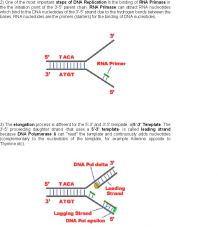
What is the first step of the Mechanism of DNA replication?
|

Mechanism of replication: separation of strands, specific coupling of free nucleic acids, DNA polymerase, primer required. First, the double stranded DNA must separate, or unwind. To do this: Helicase unwinds the DNA at the replication fork. It is what breaks the hydrogen bonds of the strands. As helicase unwinds DNA at the replication fork, the DNA ahead is forced to rotate. This process results in a build-up of twists in the DNA ahead. This tension usually leads to regions of DNA that become overwounded. DNA gyrase (a topoisomerase enzyme) is responsible for creating a small cut in the phosphate backbone of the double-stranded DNA and allowing the overwounded DNA to unwind and relax. Gyrase prevents an accumulation of twists by making temporary nicks in the double-stranded DNA. Where the DNA is nicked, the helix can unwind around the connected strand. The nick will be sealed later by another enzyme called ligase. Single-strand binding protein (SSB) is responsible for keeping the DNA unwound after the helicase. SSBs stabilize single-stranded DNA by binding to it and preventing the DNA from reclosing as replication continues. DNA gyrase can add supercoils to bacterial DNA and cause it to be more compact.
|
|
|

What is the second step of the Mechanism of DNA replication?
|
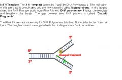
Next, you start making DNA that is complementary to the newly unwound/separated DNA. Note, all biological DNA synthesis occurs from the 5' to the 3' end. Primase gets this started by laying down a short RNA primer on the unwound DNA. Primer is required for DNA replication. The primer is made of RNA, but is complementary to the DNA sequence. Later, this RNA is replaced with DNA. There are multiple primers, 1 for the leading strand at the point where DNA polymerase replicates toward the replication fork and in multiple points on the lagging strang for the DNA polymerase to move from 5' to 3' away from the replication fork. The primer is not started at the replication fork, it is started away from the replication fork, where the DNA polymerase starts to replicate toward the replication fork. DNA polymerase then takes over starts and makes DNA that is complementary to the unwound DNA. DNA polymerase can only add deoxyribonucleotides to the 3' OH end of the RNA primer. DNA synthesis occurs on both strands of the unwound DNA. The synthesis that procedes in the direction of the replication fork is the leading strand. The synthesis that procedes in the opposite direction to the replication fork is the lagging strand. The lagging strand contains Okazaki fragments.
|
|
|
|
What are origins of replication?
|
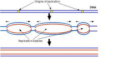
For a cell to divide, it must first replicate its DNA. This process is initiated at particular points within the DNA, known as "origins", which are targeted by proteins that separate the two strands and initiate DNA synthesis. Origins contain DNA sequences recognized by replication initiator proteins. Initiator proteins recruit other proteins to separate the DNA strands at the origin, forming a bubble. Origins tend to be "AT-rich" (rich in adenine and thymine bases) to assist this process, because A-T base pairs have two hydrogen bonds (rather than the three formed in a C-G pair)—strands rich in these nucleotides are generally easier to separate due the positive relationship between the number of hydrogen bonds and the difficulty of breaking these bonds. Once strands are separated, RNA primers are created on the template strands. More specifically, the leading strand receives one RNA primer per active origin of replication while the lagging strand receives several; these several fragments of RNA primers found on the lagging strand of DNA are called Okazaki fragments. RNase removes the RNA fragments used to initiate replication by DNA Polymerase, and another DNA Polymerase enters to fill the gaps. When this is complete, a single nick on the leading strand and several nicks on the lagging strand can be found. Ligase works to fill these nicks in, thus completing the newly replicated DNA molecule. The overall direction of replication is towards the replication fork. The overall direction of lagging-strand growth is toward the replication fork (because the strands are replicated by backing up further to the replication fork) and the leading strand is continously replicated toward the replication fork.
|
|
|
What is the third step of the Mechanism of DNA replication?
|
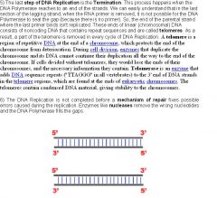
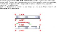
Finally, RNA primers are replaced with DNA by a special DNA polymerase. The Okazaki fragments in the lagging strands are then stitched together by DNA ligase. DNA ligase forms a phosphodiester bond between the 3' OH bond and the 5' phosphate bond. DNA synthesis is bidirectional: 2 replication forks form and proceeds in opposite directions (like an expanding bubble). Biological DNA synthesis always proceeds from the 5' end to the 3' end, for both the leading and the lagging strand. DNA polymerase has proof-reading activity, which means it corrects any mistakes (mutations) it makes. This is mismatch repair - during DNA replication, DNA polymerase proofreads the DNA as soon as it is added to the strand searching for mismatches. When found, the polymerase removes the mismatched nucleotide, adds a correct nucleotide, and continues synthesizing DNA.Replication occurs once every cell generation, during the S phase. (Cell division may occur twice in meiosis, but replication still occurs once only). It's important that cells only start replication once per generation, so that the daughter cells don't end up with extra copies of the genome. Meiosis is different than mitosis in terms of the cell division number. During mitosis, chromosomes are duplicated once, and cell divides once, therefore each daughter cell has equal chromosome number which is also equal to the mother cell’s. During meiosis, chromosomes are also duplicated, cell division occurs twice consecutively, leading the half of the chromosome number in 4 daughter cells. This process is used for generating germ line cells, the gametes. When gametes from male and female parents meet, they form normal diploid chromosome number again. However, replication only occurs in the S phase (not the M phase where meiosis and mitosis occurs). Sex cells go through meiosis, somatic cells (cells of the body) go through mitosis.
|
|
|
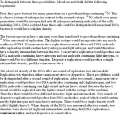
What is the Semiconservative nature of replication?
|
Newly synthesized DNA contains one old strand and one new strand.
Meselson and Stahl proved this by experiment: Basically, they used heavy (15N) DNA as the old (pre-replication) DNA, and used light (14N) nucleotides for the synthesis of new DNA. They can tell the difference between heavy and light DNA by centrifugation. What they found was that when heavy DNA undergoes one round of replication in light nucleotides, the DNA made is of intermediate weight. After the second round of replication, the DNA is split between intermediate and light weight. If DNA replication were completely conservative, only heavy and light DNA would be seen, and nothing in between. This was not the case. If DNA replication were dispersive, everything would be of intermediate weight. Again, this was not the case because after the second round of replication, light DNA was seen. |
|
|
|
How does DNA Repair during replication?
|
DNA polymerase has proof-reading activity (also called 3' → 5' exonuclease activity). If a wrong nucleotide gets incorporated, the polymerase will "back-up" and replace it with the correct one. Error correction is a property of some, but not all, DNA polymerases. This process corrects mistakes in newly-synthesized DNA. When an incorrect base pair is recognized, DNA polymerase reverses its direction by one base pair of DNA. The 3'-5' exonuclease activity of the enzyme allows the incorrect base pair to be excised (this activity is known as proofreading). Following base excision, the polymerase can re-insert the correct base and replication can continue. The special polymerase that replaces the RNA primers with DNA in the 5' → 3' direction, and also has 3' -> 5' exonuclease activity that mediates proofreading. This allows the polymerase to clear away short stretches of incorrect nucleotides (RNA or incorrect DNA) and replace it with the right ones (DNA). This process is also called repair. So both DNA polymerases, the one for the leading strand, and the one for the lagging strand, are involved in DNA repair.
|
|
|
|
What is Mismatch repair?
|
Mismatch repair: enzymes recognize incorrectly paired base-pairs and cuts out the stretch of DNA containing the mismatch. Then polymerase re-adds the correct nucleotides in.
During mismatch repair, the repair enzyme must decide what strand of DNA to cut since DNA contains 2 strands. To do this, the enzyme cuts the DNA strand that do not have methylations. The original (old) DNA has methylations, but the newly synthesized DNA do not have them until shortly after replication. Thus, there is a window of time when mismatch repair enzymes can know what strand to cut if mismatch is encountered. |
|
|
|
What is Base-excision repair?
|
Base-excision repair: a damaged base gets cut out. Then the base's sugar phosphate backbone gets cut out. And then, several more nucleotides next to the base get cut out. Finally, polymerase synthesizes the cut out nucleotides.
It is primarily responsible for removing small, non-helix distorting base lesions from the genome. BER is important for removing damaged bases that could otherwise cause mutations by mispairing or lead to breaks in DNA during replication. |
|
|
|
What is Nucleotide-excision repair?
|
Nucleotide-excision repair: damaged nucleotide(s) gets cut out and then polymerase replaces it. This is like mismatch-repair, but it's not for mismatch. It's for damages like thymine dimers, and other damages that changes normal nucleotides into abnormal nucleotides. The related nucleotide excision repair pathway repairs bulky helix-distorting lesions.
|
|
|
|
What is Nick translation for DNA repair?
|
Nick translation: this is basically 5' → 3' exonuclease activity coupled to polymerase activity. The DNA polymerase removes the RNA primers on the lagging strand and replaces the RNA with DNA. The result is a number of discontinuous DNA fragments that are "nicks" in the lagging strand. The DNA ligase then seals these nicks together and creates a continuous strand of DNA on the lagging strand. The DNA polymerase also removes the RNA primer on the leading strand and replaces it with DNA. The DNA ligase seals the 2 fragments together on the leading strand. DNA polymerase has 5' to 3' polymerase activity (to build the DNA strand), 3' to 5' exonuclease activity (to proofread the DNA strand 1 nucleotide at a time), and 5' to 3' exonuclease activity (for nick translation - replacing the RNA primers with DNA).
|
|
|
|
What is the SOS response in E. Coli for DNA?
|
SOS response in E. Coli: during replication, when there's just too much DNA damage for normal repair to handle, the SOS repair system comes along. Instead of correcting any DNA damages during replication, the polymerase replicates over the damaged DNA as if it were normal. By using the damaged DNA as a template error rates are high, but it's still better than not replicating at all.
|
|
|
|
What are sticky and blunt ends of restriction enzymes?
|
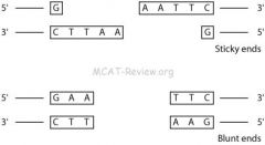
Some restriction enzymes cut to make sticky ends, which can hybridize. Some restriction enzymes cut to make blunt ends, which cannot hybridize. To cut the DNA, a restriction enzyme makes two incisions, once through each sugar-phosphate backbone (i.e. each strand) of the DNA double helix. Isolated restriction enzymes are used to manipulate DNA for different scientific applications. They are used to assist insertion of genes into plasmid vectors during gene cloning and protein expression experiments. Sticky ends are ends that overlap one another and can be put together like a puzzle. If the restriction enzyme cuts straight across the double helix, it produces blunt ends. If they cut in an offset fashion, the ends of the cut have an overhanging piece of single-stranded DNA. These are called "sticky ends" because they are able to form base pairs with any DNA molecule that contains the complementary sticky end. Any other source of DNA treated with the same enzyme will produce such molecules. Mixed together, these molecules can join with each other by the base pairing between their sticky ends. The union can be made permanent by another enzyme, DNA ligase, that forms covalent bonds along the backbone of each strand. The result is a molecule of recombinant DNA (rDNA). The ability to produce recombinant DNA molecules has not only revolutionized the study of genetics, but has laid the foundation for much of the biotechnology industry. The availability of human insulin (for diabetics), human factor VIII (for males with hemophilia A), and other proteins used in human therapy all were made possible by recombinant DNA.
|
|
|
|
Explain what palindromes are for restriction enzymes.
|
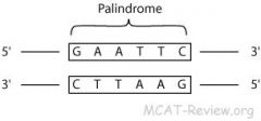
Restriction enzymes (also called restriction endonucleases) cut double stranded DNA at palindrome sequences. The resulting fragments are called restriction fragments and the site that it cuts are called restriction sites. If you read from 5' → 3' of one strand, then read from 5' → 3' of the other strand, and they are the same, then the section of the double stranded DNA that you just read is a palindrome sequence. Since the DNA is formed by two paired strands of nucleotides, and the nucleotides always pair in the same way (Adenine (A) with Thymine (T), Cytosine (C) with Guanine (G)), a (single-stranded) sequence of DNA is said to be a palindrome if it is equal to its complementary sequence read backwards. For example, the sequence ACCTAGGT is palindromic because its complement is TGGATCCA, which is equal to the original sequence in reverse complement. Restriction enzymes recognize a specific sequence of nucleotides and produce a double-stranded cut in the DNA. While recognition sequences vary between 4 and 8 nucleotides, many of them are palindromic. Endonucleases are enzymes that cleave the phosphodiester bond within a polynucleotide chain, in contrast to exonucleases, which cleave phosphodiester bonds at the end of a polynucleotide chain. Restriction endonucleases (Restriction enzymes) cleave DNA at specific sites. These enzymes are often used in genetic engineering to make recombinant DNA for introduction into bacterial, plant, or animal cells.
|
|
|
|
What is recombinant DNA?
|
Recombinant DNA is a form of DNA that does not exist naturally, which is created by combining DNA sequences that would not normally occur together. In terms of genetic modification, recombinant DNA (rDNA) is introduced through the addition of relevant DNA into an existing organismal DNA, such as the plasmids of bacteria, to code for or alter different traits for a specific purpose, such as antibiotic resistance. It differs from genetic recombination, in that it does not occur through processes within the cell, but is engineered. A recombinant protein is a protein that is derived from recombinant DNA.
Recombinant DNA technology was made possible by the discovery, isolation and application of restriction endonucleases (restriction enzymes). |
|
|
|
What is Hybridization?
|

Hybridization, also called annealing, is where DNA strands base pair with each other. In Southern blotting, DNA probes is used to hybridize onto DNA fragments containing a target sequence. In gene cloning, hybridization refers to the process where sticky ends from a restriction fragment of a gene base pairs with the same sticky ends on a plasmid. If we heat up a tube of DNA dissolved in water, the energy of the heat can pull the two strands of DNA apart (there's a critical temperature called the Tm at which this happens). This process is called 'denaturation'; when we've 'denatured' the DNA, we have heated it to separate the strands. The two strands still have the same nucleotide sequences, however, so they are still complementry. If we cool the tube again, then in the course of the normal, random molecular motion they'll eventually bump into each other ... and stick tightly, reforming double-stranded DNA. This process is called 'annealing' or 'hybridization', and it is very specific; only complementary strands will come together if it is done right. This process is used in many crime labs to identify specific strands of DNA in a mixture.
|
|
|
|
What is gene cloning?
|
The plasmid need to have a restriction site because you need to open it up for the insertion of your gene. The plasmid need to have an origin of replication because you want to clone your gene, which is inside your plasmid. The plasmid could have an antibiotic resistant gene and this would let it you kill competing, useless bacteria that doesn't have the the plasmid. When you add an antibiotic, only the bacteria with the antibiotic resistant plasmid will live. Plasmids replicate independently of the genomic DNA of the bacteria. Molecular cloning refers to the procedure of isolating a defined DNA sequence and obtaining multiple copies of it in vivo. Cloning is frequently employed to amplify (make exact copies) DNA fragments containing genes, but it can be used to amplify any DNA sequence such as promoters, non-coding sequences, chemically synthesised oligonucleotides and randomly fragmented DNA. Cloning is used in a wide array of biological experiments and technological applications such as large scale protein production. The DNA containing the target gene(s) is split into fragments using restriction enzymes. These fragments are then inserted into cloning vectors, such as bacterial plasmids or bacteriophages, which transfer the recombinant DNA to suitable host cells, such as the bacterium E. coli. Alternatively, complementary DNA is inserted into the vectors, or ‘naked’ DNA fragments can be taken up directly by a host bacterium from its medium (this is less efficient than vector transfer). Inside the host cell the recombinant DNA undergoes replication; thus, a bacterial host will give rise to a colony of cells containing the cloned target gene. Various screening methods may be used to identify such colonies, enabling them to be selected and cultured. Gene cloning facilitates DNA sequencing; it also enables large quantities of a desired protein product to be produced. Human insulin, for example, is now produced by bacteria containing the cloned insulin gene.
|
|
|
|
What is PCR?
|
Denaturation: heat (90 °C) to separate double stranded DNA template.
Annealing: cool reaction in order for primers to anneal to the now single stranded DNA template. Excess amount of primers, so some are left over after complete re-annealing of the template strands. Elongation: use heat stable polymerase to extend the primers. At this step the DNA polymerase synthesizes a new DNA strand complementary to the DNA template strand. The extension time depends both on the DNA polymerase used and on the length of the DNA fragment to be amplified. Under optimum conditions, i.e., if there are no limitations due to limiting substrates or reagents, at each extension step, the amount of DNA target is doubled, leading to exponential (geometric) amplification of the specific DNA fragment. Repeat steps 1 to 3 for n cycles. The resulting amplification of the original DNA template after n cycles is 2^n. In molecular biology, the polymerase chain reaction (PCR) is a technique to amplify a single or few copies of a piece of DNA across several orders of magnitude, generating thousands to millions of copies of a particular DNA sequence. The method relies on thermal cycling, consisting of cycles of repeated heating and cooling of the reaction for DNA melting and enzymatic replication of the DNA. Primers (short DNA fragments) containing sequences complementary to the target region along with a DNA polymerase (after which the method is named) are key components to enable selective and repeated amplification. As PCR progresses, the DNA generated is itself used as a template for replication, setting in motion a chain reaction in which the DNA template is exponentially amplified. PCR can be extensively modified to perform a wide array of genetic manipulations. In most cases of natural DNA replication, the primer for DNA synthesis and replication is a short strand of RNA. However, in the lab, PCR requires DNA primers. These are short nucleic acid polymers that are are hybridized to a target DNA, which the polymerase then extends and creates the complementary strand. |
|
|
|
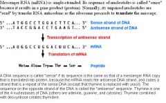
What is the typical information flow of the genetic code?
|
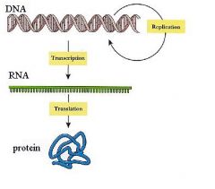
Typical information flow (DNA → RNA → protein)
DNA: resides in the nucleus. It codes information in genes. Transcription: Inside the nucleus, the DNA genes get transcribed into RNA (messenger RNAs or mRNAs). RNA: The mRNAs get transported out of the nucleus into the cytoplasm. mRNAs are working copies of the gene. Translation: ribosomes read off the mRNAs to make proteins. Protein: synthesized by ribosomes. They are the end product of what's encoded in the genes and they perform all the functions in the cell. |
|
|
|
Describe the Codon–anticodon relationship and the degenerate code.
|
Codon: The mRNA is a sequence of nucleotides, but it codes for a sequence of amino acids. To do this, every 3 nucleotide codes for an amino acid. These triplets of nucleotides are called codons. A single mRNA contains many codons.
Codons are continuous, non-overlapping and degenerate. In physics, two or more different quantum states are said to be degenerate if they are all at the same energy level. Non-degenerate means there are differences in energy in physics (electrons). Continuous because one codon follows right after another. There're no nucleotides in between. Non-overlapping because the 3 nucleotides that consist of one codon never serve as part of another codon. Degenerate because more than one codons code for a given amino acid. The degeneracy of the genetic code is where more than 1 codon on the mRNA codes for a single amino acid. The wobble phenomenon is where more than 1 anticodon can code for a codon. However, each tRNA can only be attached to one amino acid. Because the genetic code contains multiple codons that specify the same amino acid, tRNA molecules bearing different anticodons may also carry the same amino acid.Anticodon: the 3 bases on the "tip" of the tRNA. A single tRNA contains a single anticodon at the "tip" and the corresponding amino acid at the"tail". Anticodons are complementary to their corresponding codon. The codon-anticodon relationship: During translation, codons pair with anticodons so that the correct amino acids can be linked to a given codon. |
The degeneracy of the genetic codes describes how the genetic code is not precise nor specific. More than 1 codon can code for each of the different amino acids. For example, both UAU and UAC code for tyrosine. Since there are 3 bases in a codon, there are 64 possible codons and only 20 amino acids. Obviously, there will be some repeating of amino acids among the codons.
|
|
|
What are Missense and nonsense codons?
|
Missense codon: mutated codon that results in a different amino acid.
Nonsense codon: mutated codon that results in something other than an amino acid. For example, a stop codon. |
|
|
|
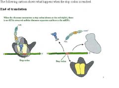
What are Initiation and termination codons (function, codon sequences)?
|
The start codon is typically AUG.
The three stop codons are UAG, UAA. and UGA. Initiation codon: signals the start of translation. Lies just downstream of the Shine Dalgarno sequence. Termination codon: signals the end of translation. Unlike other codons, tRNA are not involved. Instead a protein called "release factor" comes along and terminates translation. |
|
|
What is the mRNA composition and structure?
|
mRNA composition and structure: MRNA has RNA nucleotides, 5' cap and a poly-A tail. mRNA stands for messenger RNA. It's the product of transcription and the template for translation. The 5' cap is a modified guanine nucleotide linked in a special way to the mRNA. This protects the 5' end from exonuclease degradation. Exonucleases are enzymes that work by cleaving nucleotides one at a time from the end of a polynucleotide chain. The poly-A tail protects the 3' end of the mRNA from exonuclease degradation. Eukaryotic mRNA order of structure: 5' cap - nucleotides - 3' polyA. A 5' cap is a modified guanine nucleotide that has been added to the "front" or 5' end of a eukaryotic messenger RNA shortly after the start of transcription. Its presence is critical for recognition by the ribosome for translation and protection from exonucleases. The poly(A) tail is added after the transcription termination, and a specific cleavage of the mRNA occurs. Polyadenlyation of prokaryotic mRNAs has also been detected, but the poly(A) tails are much shorter than those in eukaryotes. Prokaryotes do not have 5' capping. Polyadenylation is the covalent linkage of a stretch of RNA which only has adenine bases to a messenger RNA molecule. The poly(A) tail and the protein bound to it aid in protecting mRNA from degradation by exonucleases. Polyadenylation is also important for transcription termination, export of the mRNA from the nucleus, and translation. The process of polyadenylation begins as the transcription of a gene finishes. The 3'-most segment of the newly-made RNA is first cleaved off by proteins; these proteins then synthesize the poly(A) tail at the RNA's 3' end.
|
|
|
What is the tRNA composition and structure (e.g., RNA nucleotides)?
|
Both tRNA (transfer RNA) and rRNA (ribosomal RNA) are products of transcription. However, they do not serve as the template of translation. tRNA is responsible for bringing in the correct amino acid during translation. At the 3' end of the tRNA, the amino acid is attached to the 3'OH via an ester linkage. tRNA structure: clover leaf structure with anticodon at the tip, and the amino acid at the 3' tail. tRNA is made of nucleotides, many of which is modified for structural and functional reasons. Transfer RNA (abbreviated tRNA) is a small RNA molecule (usually about 74-95 nucleotides) that transfers a specific active amino acid to a growing polypeptide chain at the ribosomal site of protein synthesis during translation. It has a 3' terminal site for amino acid attachment. This covalent linkage is catalyzed by an aminoacyl tRNA synthetase. It also contains a three base region called the anticodon that can base pair to the corresponding three base codon region on mRNA. Each type of tRNA molecule can be attached to only one type of amino acid, but because the genetic code contains multiple codons that specify the same amino acid, tRNA molecules bearing different anticodons may also carry the same amino acid. The structure of tRNA can be decomposed into its primary structure, its secondary structure (usually visualized as the cloverleaf structure), and its tertiary structure (all tRNAs have a similar L-shaped 3D structure that allows them to fit into the P and A sites of the ribosome). An anticodon is a unit made up of three nucleotides that correspond to the three bases of the codon on the mRNA. Each tRNA contains a specific anticodon triplet sequence that can base-pair to one or more codons for an amino acid. For example, the codon for lysine is AAA; the anticodon of a lysine tRNA might be UUU. Some anticodons can pair with more than one codon due to a phenomenon known as wobble base pairing. To provide a one-to-one correspondence between tRNA molecules and codons that specify amino acids, 61 types of tRNA molecules would be required per cell. However, many cells contain fewer than 61 types of tRNAs because the wobble base is capable of binding to several, though not necessarily all, of the codons that specify a particular amino acid. A minimum of 31 tRNA are required to translate, unambiguously, all 61 sense codons of the standard genetic code.
|
|
|
|
What is the initiation process of transcription?
|
RNA polymerase binds to the promoter (TATA box) of the double stranded DNA (closed complex). The double stranded DNA template opens up (open complex). In eukaryotes, RNA polymerase, and therefore the initiation of transcription, requires the presence of a core promoter sequence in the DNA. Promoters are regions of DNA which promote transcription. Core promoters are sequences within the promoter which are essential for transcription initiation. RNA polymerase is able to bind to core promoters in the presence of various specific transcription factors. The most common type of core promoter in eukaryotes is a short DNA sequence known as a TATA box. One transcription factor, DNA helicase, has helicase activity and so is involved in the separating of opposing strands of double-stranded DNA to provide access to a single-stranded DNA template. However, only a low, or basal, rate of transcription is driven by the preinitiation complex alone. Other proteins known as activators and repressors, along with any associated coactivators or corepressors, are responsible for modulating transcription rate.
|
|
|
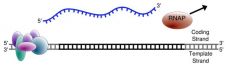
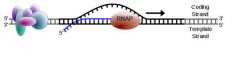
What is transcription?
|
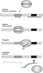
Mechanism of transcription (RNA polymerase, promoters, primer not required). Transcription, or RNA synthesis, is the process of creating an equivalent RNA copy of a sequence of DNA. Both RNA and DNA are nucleic acids, which use base pairs of nucleotides as a complementary language that can be converted back and forth from DNA to RNA in the presence of the correct enzymes. During transcription, a DNA sequence is read by RNA polymerase, which produces a complementary, antiparallel (side by side and opposite of DNA) RNA strand. As opposed to DNA replication, transcription results in an RNA complement that includes uracil (U) in all instances where thymine (T) would have occurred in a DNA complement. Transcription is the first step leading to gene expression. The stretch of DNA transcribed into an RNA molecule is called a transcription unit and encodes at least one gene. If the gene transcribed encodes for a protein, the result of transcription is messenger RNA (mRNA), which will then be used to create that protein via the process of translation. Alternatively, the transcribed gene may encode for either ribosomal RNA (rRNA) or transfer RNA (tRNA), other components of the protein-assembly process, or other ribozymes.
Uracil is the nucleotide only found in RNA and thymine is the nucleotide only found in DNA. |
|
|
|
What is rRNA composition and structure (e.g., RNA nucleotides)?
|
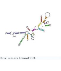
Both tRNA (transfer RNA) and rRNA (ribosomal RNA) are products of transcription. However, they do not serve as the template of translation. rRNA and protein makes up the ribosome, which is the enzyme responsible for translation. Prokaryotes have 70S ribosomes, each consisting of a small (30S) and a large (50S) subunit. Eukaryotes have 80S ribosomes, each consisting of a small (40S) and large (60S) subunit. rRNA is made of nucleotides, many of which is modified for structural and functional reasons. rRNA is highly structured because it contains the active site for catalysis. The rRNA of the large ribosomal subunit is responsible for catalyzing peptide bond formation, and can do this even without ribosomal proteins. The ribosome is composed of two subunits, named for how rapidly they sediment when subject to centrifugation. mRNA is sandwiched between the small and large subunits and the ribosome catalyzes the formation of a peptide bond between the 2 amino acids that are contained in the rRNA. Note that the S units of the subunits cannot simply be added because they represent measures of sedimentation rate rather than of mass. The sedimentation rate of each subunit is affected by its shape, as well as by its mass.
|
|
|
|
Explain the mechanism of the lac operon.
|
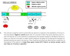
The lac operon controls the production of B-galactosidase, an enzyme that catalyzes the hydrolysis (break-down with water) of lactose into glucose and galactose. This is an inducible operon, meaning gene expression in B-galactosidase is stimulated by the presence of co-inducer, lactose. There are two regulatory mechanisms that are used to turn on lac operon. 1) The presence of lactose as a co-inducer because B-galactosidase is not needed unless lactose needs to be broken down. 2) Low levels of glucose. Because lactose breaks down into glucose and galactose, low levels of glucose signals the cell for more lactose to be broken down. Both conditions must be met for the lac operon to turn on. A co-inducer is a molecule that interacts with a repressor to free the operon from restraints on its transcription into mRNA. The repressor protein is under allosteric control - when not bound to lactose, the repressor protein can bind to the operator. When lactose is present, an isomer of lactose, allolactose, will also be present in small amounts. Allolactose binds to the allosteric site and changes the conformation of the repressor protein so that it is no longer capable of binding to the operator. Three functional genes: 1) lacZ produces B-galactosidase. This enzyme hydrolyzes the bond between the two sugars, glucose and galactose 2) lacY produces permease. This enzyme spans the cell membrane and brings lactose into the cell from the outside environment. The membrane is otherwise essentially impermeable to lactose. 3) lacA produces B-galactosidase transacetylase. The function of this enzyme is still not known.
|
The lac operon is a set of control and structural genes in E. Coli that allow the digestion of lactose. There are 3 structural genes controlled by an operator found on another part of the genome.
|
|
|
How is gene regulation controlled in prokaryotes?
|
Gene regulation is controlled by operons. Operons are a form of transcriptional control. An operon consists of the structural gene (or genes) which actually code for specific proteins and the controlling elements associated with the control of those genes. An operon typically contains several genes, all under the same control mechanism. Eukaryotes do not have operons because each of their genes is controlled by more than one regulator. Operons control genes that are based on similar functions, called mRNA transcription units. These units enable easy regulation of the genes by a simple on/off switch of the genes. The operon consists of a promoter, an operator, the structural genes, a termination sequence, and a repressor gene. The operator is the "on/off" switch - binding site for the repressor protein. The promotor gene aids in RNA polymerase binding. The repressor gene for the production of a diffusible repressor protein that either binds to the operator represses it or doesn't bind to it and allows transcription to occur. Termination sequences marks the end of gene transcription and are different from termination codons of translation (called stop codons or nonsense codons). Silencers are the repressors for prokaryotes. Enhancers are rarely found in prokaryotes because they distant regions of DNA. Eukaryotes are able to loop their promoter to the enhancer to initiate transcription. Activators are proteins that bind to the promoter region of the operon and cause transcription to occur.
|
|
|
|
How is translation regulated in prokaryotes?
|
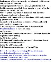
Prokaryotes also do transcription and translation. Since transcription, translation and RNA degradation in prokaryotes are coupled, regulation mainly acts at transcription level. In a few cases, a translational control can be made through : (1) Different degradation rates of mRNAS, (2) different efficiencies of translation initiation in different genes, (3) different efficiencies of translation rate due to different conformation of the mRNA (existence of secondary structures which slow down ribosome movement). Gene expression is regulated in transcription more than during translation. The regulation of gene expression in prokaryotes provides the best survival opportunities to the organism by rapid and synchronized switch of gene transcription. Prokaryotes synthesize proteins while the mRNA is being transcripted. Basically, as soon as the mRNA is created (transcribed by the RNA polymerase), the ribosome starts translating the mRNA and protein is made. The reason prokaryotes do transcription-translation coupling is because their mRNA does not have to exit a nucleus before being translated, the way the eukaryotes's mRNA has to exit the nucleus before being translated.
|
|
|
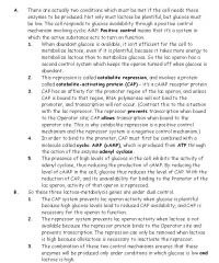
How is the lac operon controlled by glucose levels and lactose levels?
|
|
|
|
|
What happens to the lac operon if lactose is not present?
|
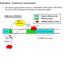
When glucose is plentiful and/or lactose is low, the desired situation is for these genes to be repressed (inactive). The repressor gene codes for the production of a diffusible repressor protein, which is present in low numbers in the cell at all times. The repressor has an affinity for the operator of the lac operon, and unless something happens to intervene, the repressor molecule will bind to the O site and block the movement of an RNA polymerase from the promoter to the structural genes, thus preventing transcription of those genes.
|
|
|
|
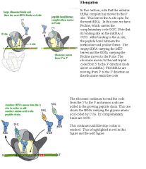
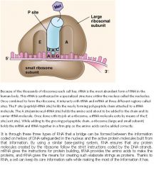
What happens at the site of the ribosome for initiation of translation?
|
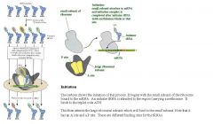
The code is actually translated on structures that are also made in the nucleus, called Ribosomes. These ribosomes provide the structural site where the mRNA sits. The amino acids for the proteins are carried to the site by "transfer RNAs,". In the cartoon to the left, these are shown as blue molecules. Each transfer RNA (tRNA) has a nucleotide triplet which binds to the complementary sequence on the mRNA (see the three letters at the bottom of each molecule).
The tRNA carries the amino acid at its opposite end. One can trace and detect binding of a particular tRNA-amino acid complex to the mRNA by labeling that amino acid. It will bind to its tRNA. In the case to the left, Phenylalanine is bound to the tRNA which carries the complementary base code AAA (adenine-adenine-adenine). This triplet code would bind to the complementary sequence on mRNA UUU (uracil X3). The mRNA is shown as a green arrow. This cartoon shows the selective binding site on the mRNA which is attached in the ribosome. It also shows the tRNA carrying the Phenylalanine bound at the site In this particular assay which uses a polyuracil mRNA, only phenylalanine-bearing tRNA is bound and detected on the filter. |
|
|
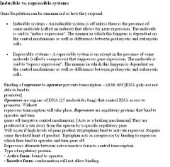
How is gene expression controlled by the trp gene in prokaryotes?
|
The trp operon is a repressible operon, meaning that gene expression of the operon is repressed by the presence of the co-repressor, tryptophan. IV. The tryptophan (trp) operon in E. coli is a negative control repressible system. It’s negative control because the system produces a repressor which functions to turn off the operon. It’s repressible because, unlike the lac operon, repression occurs when a critical substance is abundant in the cell. (For the trp operon, that critical substance is the amino acid tryptophan; for the lac operon the critical substance is lactose, and its presence removes repression.) Tryptophan is a co-repressor because it needs to bind to the inactive repressor to activate it and cause it to bind to the operator, preventing transcription. This contrasts to the lactose which is a co-inducer.
|
|
|
|
What happens to the lac operon if lactose is present (and glucose is not)?
|
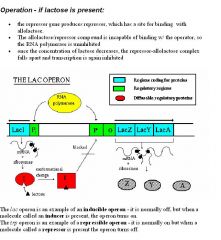
When lactose is plentiful (and glucose is not) the repression needs to be removed so the genes can be activated. The trigger for this activation needs to be lactose. Besides being attracted to the operator sequence of the operon, the repressor protein also has an affinity for a slightly unusual form of lactose called allolactose, which will be present as a small percentage of the lactose available whenever there’s lactose around. When the repressor binds to allolactose, the configuration (three-dimensional shape) of the repressor is altered, and it is no longer able to bind to the operator. It falls off, and there is nothing to prevent RNA polymerase from reading through the operator to the structural genes, and the three enzymes will thus be made. The lactose is considered a co-inducer because by it binding to the repressor, the repressor is unable to bind to the operator. 8. As the cell metabolizes the available lactose, eventually the concentration of lactose will fall far enough that there won’t be enough around to maintain the complexes with the available repressor proteins, the repressor will return to its former configuration, and will once again bind to the operator and repress the structural genes.
|
|
|
|
What happens during termination of transcription?
|
Chain termination: there are 2 ways that transcription can terminate in prokaryotes. 1) Intrinsic termination: specific sequences called a termination site creates a G-C rich hairpin loop structure on the RNA that causes the RNA to slip off the template. 2) Rho (ρ) dependent termination: a protein called the ρ factor travels along the synthesized RNA and bumps off the polymerase. Transcription termination in eukaryotes is less understood but involves cleavage of the new transcript by protein factors followed by template-independent addition of As at its new 3' end, in a process called polyadenylation (poly A tail). When transcription is complete, the transcript is released from the polymerase and, shortly thereafter, the polymerase is released from the DNA. The addition of the poly-A tail stabilizes the RNA and allows it to be exported out of the nucleus.
|
|
|
|
What is the route from the DNA code to the protein?
|
Before cell division, the DNA in our chromosomes replicates so each daughter cell has an identical set of chromosome. In addition, the DNA is responsible for coding for all proteins. Each amino acid is designated by one or more set of triplet nucleotides. The code is produced from one strand of the DNA by a process called "transcription". This produces mRNA which then is sent out of the nucleus where the message is translated into proteins. This can be done in the cytoplasm on clusters of ribosomes, called "polyribosomes". Or it can be done on the membranes of the rough endoplasmic reticulum. The cartoon to the left shows the basic sequence of transcription and translational events.
|
|
|
|
What is the process of elongation for translation?
|
|
|
|
|
What is the elongation process of transcription?
|
Nucleoside triphosphates (AUGCs) adds corresponding to the DNA template. No primer is required. Nucleosides are ribose-containing nucleotides or ribonucleotides, and are the monomers of RNA. RNA elongates as the RNA polymerase moves down the DNA template. RNA is made from the 5' to 3' direction. One strand of DNA, the template strand (or noncoding strand), is used as a template for RNA synthesis. As transcription proceeds, RNA polymerase traverses the template strand and uses base pairing complementarity with the DNA template to create an RNA copy. Although RNA polymerase traverses the template strand from 3' → 5', the coding (non-template) strand and newly-formed RNA can also be used as reference points, so transcription can be described as occurring 5' → 3'. This produces an RNA molecule from 5' → 3', an exact copy of the coding strand (except that thymines are replaced with uracils, and the nucleotides are composed of a ribose (5-carbon) sugar where DNA has deoxyribose (one less oxygen atom) in its sugar-phosphate backbone). Unlike DNA replication, mRNA transcription can involve multiple RNA polymerases on a single DNA template and multiple rounds of transcription (amplification of particular mRNA), so many mRNA molecules can be rapidly produced from a single copy of a gene. Elongation also involves a proofreading mechanism that can replace incorrectly incorporated bases. In eukaryotes, this may correspond with short pauses during transcription that allow appropriate RNA editing factors to bind.
|
|
|
|
What is termination for translation?
|
|
|
|
What are the Roles of mRNA, tRNA, and rRNA?
|
mRNA (messenger RNA): contains codons that code for the peptide sequence. tRNA (transfer RNA): contains the anticodon on the "tip" and the corresponding amino acid on the "tail". tRNA link the correct amino acid to its corresponding mRNA codon through codon-anticodon interaction. rRNA (ribosomal RNA): forms the ribosome. Catalyzes the formation of the peptide bond. RNA base-pairing specificity is where the DNA sequence is the same as the RNA copy. Ribosomal ribonucleic acid (rRNA) is the central component of the ribosome, the protein manufacturing machinery of all living cells. The function of the rRNA is to provide a mechanism for decoding mRNA into amino acids and to interact with the tRNAs during translation by providing peptidyl transferase activity.The tRNA then brings the necessary amino acids corresponding to the appropriate mRNA codon. Transfer RNA (abbreviated tRNA) is a small RNA molecule (usually about 74-95 nucleotides) that transfers a specific active amino acid to a growing polypeptide chain at the ribosomal site of protein synthesis during translation. It has a 3' terminal site for amino acid attachment. This covalent linkage is catalyzed by an aminoacyl tRNA synthetase. It also contains a three base region called the anticodon that can base pair to the corresponding three base codon region on mRNA. Each type of tRNA molecule can be attached to only one type of amino acid, but because the genetic code contains multiple codons that specify the same amino acid, tRNA molecules bearing different anticodons may also carry the same amino acid.
|
tRNA is a smaller type of RNA, which functions as a carrier of amino acid molecules. Like mRNA, tRNA is coded for by DNA, but unlike mRNA, it is a comparatively short ribonucleotide polymer instead of a long filament. Although it is largely single-stranded, there are SHORT DOUBLE-stranded segments in tRNA where the nucleotide chain LOOPS back upon itself. mRNA is the template for protein synthesis and has a poly-A tail, which plays a role in the degeneration of mRNA. rRNA is produced in the region of the nucleus known as the nucleolus.
|
|
|
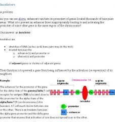
What are insulators?
|
|
|
|
|
What is the Role and structure of ribosomes?
|
Ribosome is the enzyme that catalyzes protein synthesis. Ribosome has 2 subunits - the large and the small. The large subunit is responsible for the peptidyl transfer reaction. The small subunit is responsible for the recognizing mRNA and binds to the Shine-Dalgarno sequence on the mRNA in prokaryotes. In eukaryotes, this sequence is called the Kozak sequence. Both the large and small subunits are needed for translation to occur and they come together in a hamburger fashion that sandwitches the mRNA and tRNAs in between. The ribosome is composed of two subunits, named for how rapidly they sediment when subject to centrifugation. mRNA is sandwiched between the small and large subunits and the ribosome catalyzes the formation of a peptide bond between the 2 amino acids that are contained in the rRNA. The ribosome also has 3 binding sites called A, P, and E. The A site in the ribosome binds to an aminoacyl-tRNA (a tRNA bound to an amino acid). The amino (NH2) group of the aminoacyl-tRNA, which contains the new amino acid, attacks the ester linkage of peptidyl-tRNA (contained within the P site), which contains the last amino acid of the growing chain, forming a new peptide bond. This reaction is catalyzed by peptidyl transferase. The tRNA that was holding on the last amino acid is moved to the E site, and what used to be the aminoacyl-tRNA is the peptidyl-tRNA. A single mRNA can be translated simultaneously by multiple ribosomes.
|
|
|
|
What is the peptidyl transfer reaction and what is the Shine-Dalgarno sequence?
|
The Peptidyl transferase is the primary enzymatic function of the ribosome which forms peptide links between adjacent amino acids using tRNAs during the translation process of protein biosynthesis. Peptidyl transferase activity is carried out by the ribosome. The Shine-Dalgarno sequence is a ribosomal binding site in the mRNA generally located 8 basepairs upstream of the start codon AUG. The Shine-Dalgarno sequence exists only in prokaryotes. This sequence helps recruit the ribosome to the mRNA to initiate protein synthesis by aligning it with the start codon. The eukaryotic equivalent of the Shine-Dalgarno sequence is called the Kozak sequence.
|
|
|
|
What is the mechanism of translation initiation?
|
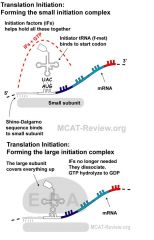
Chain Initiation: To begin translation, you need to form the initiation complex. The initiation complex is basically an assembly of everything needed to begin translation. This includes mRNA, initiator tRNA (fmet), and the ribosome (initiation factors, and GTP aids in the formation of the initiation complex). The initiation complex forms around the initiation codon (AUG), which is just down stream of the Shine-Dalgarno sequence. The Shine-Dalgarno sequence is the "promoter" equivalent of translation and is only found in eukaryotes. The sequence is called the Kozak sequence in eukaryotes. Both of these sequences recruits the small unit of the ribosome to the mRNA to initiate translation by aligning it with the start codon. Translation starts at the AUG start codon, coding for methionine. As a consequence, methionine is incorporated into the N-terminal position of all proteins in eukaryotes during translation, although it is usually removed during post-translational modification.
Initiation of translation involves the assembly of the components of the translation system which are: the two ribosomal subunits (small and large), the mRNA to be translated, the first (formyl) aminoacyl tRNA (the tRNA charged with the first amino acid), GTP (as a source of energy), and three initiation factors (IF1, IF2, and IF3) which help the assembly of the initiation complex. |
Protein synthesis (translation) requires energy.
|
|
|
What is the mechanism of translation termination?
|
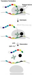
Chain termination: When a stop codon is encountered, proteins called release factors, bound to GTP, come in and blocks the A site. The peptide chain gets cleaved from the tRNA in the P site. Peptide chain falls off, and then the whole translation complex falls apart.
Amino acid activation: enzymes called aminoacyl-tRNA synthetases attach the correct amino acids to their corresponding tRNAs. ATP required. Termination occurs when one of the three termination codons moves into the A site. These codons are not recognized by any tRNAs. Instead, they are recognized by proteins called release factors, namely RF1 (recognizing the UAA and UAG stop codons) or RF2 (recognizing the UAA and UGA stop codons). These factors trigger the hydrolysis of the ester bond in peptidyl-tRNA and the release of the newly synthesized protein from the ribosome. A third release factor RF-3 catalyzes the release of RF-1 and RF-2 at the end of the termination process. |
|
|
|
What are polysomes?
|
Translation is carried out by more than one ribosome simultaneously. Because of the relatively large size of ribosomes, they can only attach to sites on mRNA 35 nucleotides apart. The complex of one mRNA and a number of ribosomes is called a polysome or polyribosome.
|
|
|
|
What is the role of antibiotics on translation?
|
Several antibiotics exert their action by targeting the translation process in bacteria. They exploit the differences between prokaryotic and eukaryotic translation mechanisms to selectively inhibit protein synthesis in bacteria without affecting the host.
|
|
|
|
What are chromosomal proteins?
|
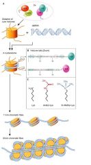
Histones: responsible for the compact packing and winding of chromosomal DNA. DNA winds itself around histone octamers. Nonhistone chromosomal proteins: all the other proteins are lumped together in this group. Responsible for various roles, such regulatory and enzymatic. A histone octamer is an octamer of the histones found at the center of a nucleosome core particle. It consists of 2 copies of each of the four core histone proteins (H2A, H2B, H3 and H4). The octamer assembles when a tetramer, containing two copies of both H3 and H4, complexes with two H2A/H2B dimers. These histones of the histone octamer all contain N-terminal tails that emanate from their central histone folds. First, eukaryotic genes are contained within a complex of DNA and protein called chromatin. The protein component constitutes about half the weight of chromatin and is composed largely of nucleosomes. The nucleosome is the basic unit of chromatin structure. It is composed of an octamer of histone proteins (two molecules each of histones H2A-H2B and histones H3-H4) wrapped with two loops containing approximately 140 base pairs of DNA. DNA is negatively charged, because of the presence of the phosphate groups (the oxygens have a negative charge), and DNA is acidic (COOH of nucleic acid). Therefore, histones wrapped around in DNA to make chromatin is basic and positively charged, for electrostatic interactions to occur.
|
|
|
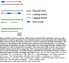
What are Telomeres and centromeres?
|
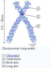
Telomere: the 2 ends of the chromosome. A telomere is a region of repetitive DNA at the end of a chromosome, which protects the end of the chromosome from deterioration. Centromere: a region on the chromosome, can be at the center or close to one of the ends. After replication, sister chromatids are attached at the centromere. During mitosis, spindle fibers are attached at the centromere and pulls the sister chromatids apart. A centromere is a region of DNA typically found near the middle of a chromosome where two identical sister chromatids come in contact. It is involved in cell division as the point of mitotic spindle.
The ends of chromosomes have DNA sequences called telomeres. DNA polymerase, the enzyme responsible for DNA replication cannot not fully synthesize the 3' end of linear DNA. At the end of a linear chromosome, DNA polymerase can synthesize the leading strand until the end of the chromosome. Before a cell can divide, it must produce a new copy of each of its chromosomes, and it does this during a specific part of interphase called the DNA-synthesis phase, or S phase, of the cell-division cycle; the part of interphase preceding S phase is called Gap 1, or G1, and the part following S phase is called Gap 2, or G2. By the end of the S phase each chromosome has been replicated to produce two complete copies, which remain joined together at their centromeres until the M phase that soon follows. Chromosome duplication requires both the replication of the long DNA molecule in each chromosome and the assembly of a new set of chromosomal proteins onto the DNA to form chromatin. |
|
|
|
What is the significance of telomeres for cellular aging and cancer?
|
|
|
|
|
What are chromosomes and what is the difference between chromosome and chromatin?
|
A chromosome is an organized structure of DNA and protein that is found in cells. It is a single piece of coiled DNA containing many genes, regulatory elements and other nucleotide sequences. Chromosomes also contain DNA-bound proteins, which serve to package the DNA and control its functions. Chromosomes vary widely between different organisms. The DNA molecule may be circular or linear, and can be composed of 10,000 to 1,000,000,000 nucleotides in a long chain. Typically eukaryotic cells (cells with nuclei) have large linear chromosomes and prokaryotic cells (cells without defined nuclei) have smaller circular chromosomes, although there are many exceptions to this rule. Furthermore, cells may contain more than one type of chromosome; for example, mitochondria in most eukaryotes and chloroplasts in plants have their own small chromosomes. In eukaryotes, nuclear chromosomes are packaged by proteins into a condensed structure called chromatin. This allows the very long DNA molecules to fit into the cell nucleus. The structure of chromosomes and chromatin varies through the cell cycle. Chromosomes are the essential unit for cellular division and must be replicated, divided, and passed successfully to their daughter cells so as to ensure the genetic diversity and survival of their progeny. Chromosomes may exist as either duplicated or unduplicated—unduplicated chromosomes are single linear strands, whereas duplicated chromosomes (copied during synthesis phase) contain two copies joined by a centromere. Compaction of the duplicated chromosomes during mitosis and meiosis results in the classic four-arm structure (pictured to the right). Chromosomal recombination plays a vital role in genetic diversity. Chromatin is basically coils of DNA (Deoxyribonucleicacid) bound to protein called histone. Chromosomes during the cycle of Interphase (G1, S, and G2), take form of chromatin. Chromatin condenses during cell divison (mitosis and meiosis) to form the Chromosomes. Chromosomes are long strands of DNA tightly wrapped around histone 'beads' which help to pack it down to fit within the cell. The cells of your body are absolutely tiny, so the tight structure of them also aids in making mitosis simpler for the cell.
|
|
|
What is the Transcription regulation of eukaryotes?
|
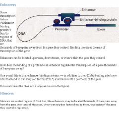
Transcription factors (protein) bind to enhancers or silencers (DNA) to affect transcription. Enhancers increase transcription when bound, while silencers decrease it. The main difference in eukaryotes that sets them apart from prokaryotes is that enhancers/silencers can be very far away from the actual promoter, and can be upstream or downstream. The DNA must must loop back on itself so that the transcription factor bound to enhancer/silencer can actually make contact with the promoter. Intermediate proteins are involved in the process.
Eukaryotes do not have bacterial transcription regulation mechanisms such as the operon. Prokaryotes are "simple," single celled organisms, so they have "simple" systems where the genes are grouped together based on similar functions into functional units of transcriptional control called operons. MANY GENES UNDER ONE CONTROL!!! There is one single on/off switch for the genes that are grouped into similar functions. However, eukaryotes are much more complex. Every cell (except gametes) have the same DNA, with the same information. Almost all eukaryotic genes must be shut off in order to allow for cell normal function (a liver cell cannot have genes for lung cells running, for example). Usually, every gene has more than one gene regulator (all of which must be on for the gene to function). Eukaryotes can't have operons because each gene has to have more than one regulator. Operons are found in prokaryotes and regulate more than 1 gene. |
|
|
|
Explain what it means for Cancer to be a failure of normal cellular controls.
|
Failure of normal cellular controls:
Cancer cells continue to grow and divide in situations normal cells would not. Cancer cells fail to respond to cellular controls and signals that would halt this growth in normal cells. Cancer cells avoid apoptosis (self-destruction) that normal cells undergo when extensive DNA damage is present. Cancer cells stimulate angiogenesis (cause new blood vessels to grow to nourish the cancer cell). Cancer cells are immortal while normal cells die after a number of divisions. Cancer cells can metastasize - break off and then grow in another location. |
|
|
|
What are oncogenes?
|
Oncogenes: genes that cause cancer when activated. The product of many oncogenes are involved in speeding up cell division. Before an oncogene is activated, it is a harmless proto-oncogene. Something occurs that changes the proto-oncogene to an oncogene. The classic exampe of oncogene is the src.
|
|
|
|
How are tRNA and rRNA processed?
|
The primary transcripts produced from most rRNA genes and from tRNA genes, like pre-mRNAs, are extensively processed to yield the mature, functional forms of these RNAs. rRNAs are encoded by a single type of pre-rRNA transcription unit. Transcription by RNA polymerase yields a primary transcript (pre-rRNA), which is processed into the mature rRNAs found in cytoplasmic ribosomes. Discovery of pre-rRNA processing was the first indication that mature cytoplasmic RNAs are derived from larger precursor RNAs synthesized in the nucleolus. Both the synthesis and processing of pre-mRNA occurs in the nucleus. Mature cytosolic tRNAs are produced from larger precursors (pre-tRNAs) synthesized by RNA polymerase in the nucleoplasm. Mature tRNAs also contain numerous modified bases that are not present in tRNA primary transcripts. Cleavage and base modification occur during processing of all pre-tRNAs. Some pre-tRNAs contain one or more introns that are spliced out during processing. Processing of pre-tRNA, like mRNA processing, occurs in the nucleus. The mature tRNAs then are transported to the cytoplasm through nuclear pore complexes. Most likely, mature tRNAs in the nucleus, like mature mRNAs and rRNAs, are bound by specific proteins that facilitate their transport through nuclear pores. Once in the cytoplasm, tRNAs are passed between aminoacyl-tRNA synthetases, elongation factors, and ribosomes during protein synthesis. Thus tRNAs generally are associated with proteins and spend little time free in the cell, as is also the case for mRNAs and rRNAs. Like pre-mRNAs, the primary transcripts produced from pre-rRNA and tRNA genes undergo extensive processing. tRNAs and rRNAs modifications: some normal nucleotides are modified to control the structure of these RNAs.
|
|
|
|
What are tumor suppressor genes?
|
Tumor suppressors: if the oncogene is the "bad" gene, tumor suppressors are the "good" genes. The product of many tumor suppressors are involved in slowing down or controlling cell division. If something happens that cause the tumor suppressor to no longer function, then the cell becomes cancerous. The classic example of tumor suppressor is the p53.
DNA must be replicated before cell division can occur (the DNA synthesis phase). Replication is called out by DNA polymerases that both copy and proofread DNA for damage and/or errors that may be present. If and when DNA damage is discovered, the cell will immediately activate its tumor suppressor genes that HALT THE CELL CYCLE in an attempt to repair the genetic lesion. If the genetic lesion is too extensive, an intracellular auto-death pathway, known as apoptosis, will be activated and cell self-digestion would follow. The attempt to carry out apoptosis may fail, perhaps because of a preexisting defect in the repair mechanism, which would increase the possibility for a malignancy to develop. |
So if a healthy cell is exposed to ultraviolet light which damages the cell's DNA, the cell would stall the cell cycle with its tumor suppressor genes to repair the damaged DNA.
|
|
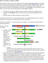
What is Posttranscriptional control and the basic concept of splicing (introns, exons)?
|
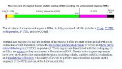
mRNAs modifications: 1) RNA splicing: sequences called introns are cut out, sequences called exons are kept and and spliced (joined) together. 2) Alternate splicing: different ways of cutting up and RNA and rejoining the exons pieces make different final RNA products. 3) 5' capping and 3' poly-A tail: these help to protect the RNA from degradation so they can last longer. After the correct modifications, RNA is transported out of the nucleus where they can function in translation. After some time, RNA is degraded. The rate and timing of RNA degradation can be controlled by the cell. All the primary transcripts produced in the nucleus must undergo processing steps to produce functional RNA molecules for export to the cytosol. Approximately 80 percent of the total RNA in rapidly growing mammalian cells is rRNA, and 15 percent is tRNA; protein-coding mRNA thus constitutes only a small portion of the total RNA. The cutting and splicing of mRNA must be done with great precision. If even one nucleotide is left over from an intron or one is removed from an exon, the reading frame from that point on will be shifted, producing new codons specifying a totally different sequence of amino acids from that point to the end of the molecule (which often ends prematurely anyway when the shifted reading frame generates a STOP codon). The removal of introns and splicing of exons is catalyzed by spliceosomes. A spliceosome is a complex of specialized RNA and protein subunits that removes introns from a transcribed pre-mRNA segment.
|
|
|
|
What are the General characteristics of fungi?
|
Fungi are made of hyphae filaments. Most fungi grow as tubular filaments called hyphae. An interwoven mass of hyphae is called a mycelium. It is through the mycelium that a fungus absorbs nutrients from its environment. The tubular walls of hyphae are often strengthened with chitin, which surrounds, supports, and protects the cells that compose a hypha.. Parasitic hyphae = haustoria. Some forms of parasitic fungi have a portion of their hyphae modified to form haustoria that are able to penetrate the tissues of a host organism. A mass of hyphae is called mycelium. Have cell wall made of chitin. All fungi are heterotrophs. They are either parasites, saprobes, or symbiotic. Some live as saprophytes (saprobes), getting their nourishment from the surroundings (often having first digested it by secreting enzymes). They perform a crucial role in nature by decomposing dead organisms are releasing their nutrients for reuse by the living. Some live in a mutually beneficial symbiotic relationship with another organism, often a plant. Some fungi are parasitic, causing serious damage to their host. Lichens = fungi + algae. Algae provides food, fungi provides water and protection. Lichens (mycobiont) are fungi that live in a mutually symbiotic association (both members benefit) with an autotrophic green alga or blue-green alga (cyanobacterium), (the "photobiont"). Lichens are another such partnership for fungi to gain nutrients from another organism. The algal partner photosynthesizes and provides food for the fungus, so it can grow and spread. Mycorrhizae = fungi + plant roots. Plant provides food, fungi provides more absorption surface area. Yeast, molds, mushrooms are all fungi. Yeast and molds are not the same. Some 80% of land plants benefit from symbiotic mycorrhiza. The plant benefits by more-efficient mineral uptake. The fungus benefits by the sugars translocated to the root by the plant.
|
|
|
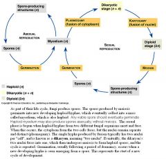
What are the General aspects of life cycle of fungi?
|
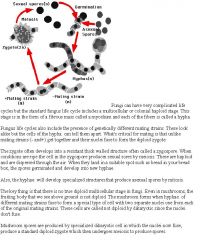
Can be sexual or asexual. Reproduces via spores or mycelial fragmentation. Many fungi can reproduce by fragmentation. Any mycelium that is fragmented or disrupted, can grow into a new colony. Many fungi are sub-cultured using this hyphal fragment technique. Most fungi have both a haploid and a diploid stage of life cycle. Fungi do not contain chlorophyll or any other means of producing their own food so they rely on other organisms for nutrition. A spore is a reproductive structure that is adapted for dispersal and surviving for extended periods of time in unfavorable conditions. A chief difference between spores and seeds as dispersal units is that spores have very little stored food resources compared with seeds. Spores are usually haploid and unicellular and are produced by meiosis in the sporangium by the sporophyte. Spore cells are usually metabolically inactive, since they are dispersed with little food reserves. Once conditions are favorable, the spore can develop into a new organism (develop into fungus via metabolism) using mitotic division, producing a multicellular gametophyte, which eventually goes on to produce gametes. Two gametes fuse to create a new sporophyte. Haploid spores produced by mitosis are used by many fungi for asexual reproduction. Spores are often classified by the structure in which meiosis and spore production occurs, for example, sporangiospores are spores produced by a sporangium. Spores form when nutrient supply is low or environmental conditions are harsh and can survive for thousands of years on minimal metabolism. However, if a question says, "all spore-forming bacteria form spores when nutrients are low" - that isn't true - don't pick "All, always, every, never - no extreme/absolute statements with science.
|
|
|
|
What are the General structural characteristics (nucleic acid and protein, enveloped and nonenveloped) of viruses?
|
Nucleic acid for viruses can be DNA or RNA. The DNA can be single stranded or double stranded and the RNA can be single or double stranded. A protein coat (called a capsid) covers the nucleic acid. Some viruses have an envelope derived from the host's cell membrane, while others lack it (nonenveloped). Enveloped viruses bud off the host's membrane. Nonenveloped viruses that penetrate the host cause the host to burst (lyse, causing the cell to die) to release more viral particles. Viruses need a host to survive. Viruses that lack envelopes (i.e., do not have nor need them) are termed non-enveloped. In non-enveloped viruses the capsids are solely responsible for attachment of the virus to the to-be-infected cell. Viruses are smaller than bacteria. Many viruses have viral envelopes covering their protein capsids. Functionally, viral envelopes are used to help viruses enter host cells. Glycoproteins on the surface of the envelope serve to identify and bind to receptor sites on the host's membrane. The viral envelope then fuses with the host's membrane, allowing the capsid and viral genome to enter and infect the host. Usually, the cell from which the virus itself buds will survive, and shed more viral particles for an extended period. The lipid bilayer envelope of these viruses is relatively sensitive to desiccation, heat and detergents, therefore these viruses are easier to sterilize than non-enveloped viruses, have limited survival outside host environments, and typically must transfer directly from host to host. For enveloped viruses, the virion particle is not complete (nor infectious) until the envelope has been acquired.
|
Viruses have capsids that cover the nucleic acid and bacteria have plasmids that have the new DNA and are inserted into the bacteria to replicate.
|
|
|
What do viruses lack?
|
Lack organelles, nucleus: Viruses don't have any organelles or a nucleus. The genetic material is simply packed inside a protein coat. Viruses are acellular. They carry out no metabolism on their own and must replicate using the host cell's metabolic machinery. In other words, viruses don't grow and divide. Instead, new viral components are synthesized and assembled within the infected host cell. The protein coat that surrounds the virus can be as rigid as a cell wall of bacteria. The key difference between bacteria and viruses is that bacteria replicate (reproduce) via fission, while viruses replicate inside of a host cell. Viruses can not replicate on their own, like bacteria.
|
|
|
|
What are the Structural aspects of typical bacteriophage?
|
Head stores genetic material.
Sheath provides a passage way for genetic material to be injected into the host bacteria. Tail fibers attach to the host bacteria. Bacteriophages are viruses that infect bacteria. |
|
|
|
What are DNA binding proteins and transcription factors?
|
DNA-binding proteins bind to DNA.
Transcription factors bind to DNA, so they have a DNA-binding domain and they are DNA-binding proteins. DNA-binding domains interact with the grooves in the double helix (major grooves and minor grooves). Advanced: common DNA-binding domains include helix-turn-helix (HTH), zinc finger, basic-region leucine zipper (bZIP). A transcription factor is a protein that binds to specific DNA sequences and thereby controls the transfer (or transcription) of genetic information from DNA to mRNA. Transcription factors perform this function alone or with other proteins in a complex, by promoting (as a transcriptional activator), or blocking (as a transcriptional repressor) the recruitment of RNA polymerase (the enzyme that performs the transcription of genetic information from DNA to RNA) to specific genes. Eukaryotic DNA have transcription factors that function as activators and repressors, while prokaryotic DNA have the operon with repressors and inducers. A defining feature of transcription factors is that they contain one or more DNA-binding domains (DBDs), which attach to specific sequences of DNA adjacent to the genes that they regulate. |
|
|
|
What is the genomic content of viruses?
|
Genomic content RNA or DNA: Viruses can contain either RNA or DNA as their genomic content. Those with RNA are called retroviruses.
|
|
|
|
What is the size of viruses relative to bacteria and eukaryotic cells?
|
Viruses are roughly 100 times smaller than bacteria, and 1000 times smaller than eukaryotic cells.
|
|
|
|
What is the Retrovirus life cycle?
|
Retrovirus life cycle: integration into host DNA. First, retrovirus enters the host. The viral reverse transcriptase then converts the viral RNA (single strand) genome into double-stranded DNA. A virally encoded enzyme called integrase adds in the viral DNA into the host's genome at a random place. When the host replicates, the viral DNA gets replicated also. Integrase is an enzyme produced by a retrovirus (such as HIV) that enables its genetic material to be integrated into the DNA of the infected cell. Because reverse transcription lacks the usual proofreading of DNA replication, a retrovirus mutates very often. This enables the virus to grow resistant to antiviral pharmaceuticals quickly, and impedes the development of effective vaccines and inhibitors for the retrovirus. A provirus is a virus genome that has integrated itself into the DNA of a host cell. One kind of virus that can become a provirus is a retrovirus.
|
|
|
|
Explain the viral life cycle in regards to how viruses are self-replicating biological units that must reproduce within specific host cell.
|
Viruses can not replicate by themselves. They depend on the host's replication organelles to replicate. The host's ribosomes will make the necessary protein coats and polymerases that replicate the viral genetic material. Retroviruses contain their own reverse polymerase to convert RNA to DNA before the host's polymerases take over.
|
|
|
What is generalized phage and animal virus life cycles?
|
1) attachment to host, penetration of cell membrane or cell wall, and entry of viral genetic material
2) use of host synthetic mechanism to replicate viral components: Host's ribosomes synthesize the necessary enzymes. Host's ATP provides necessary energy. The host also provides the raw materials such as nucleotides and amino acids. 3) self-assembly and release of new viral particles: The coat proteins and viral genetic material will assemble into viral particles all by themselves. Most bacteriophages adsorb to the bacterial cell wall, although some are able to adsorb to flagella or pili. Specific strains of bacteriophages can only adsorb to specific strain of host bacteria. This is known as viral specificity |
|
|
|
What is transduction?
|
Transduction: transfer of genetic material by viruses. Transduction is yet another way for to exchange genetic material. In transduction, a virus takes up a piece of DNA from its host and incorporates it into its own viral genome. After the virus has multiplied, many copies of the virus erupt from the infected cell. Depending on the kind of transduction, some or all of the daughter viruses take copies of parts of the bacterial DNA with them. When one of them infects a new cell, it inserts the stolen DNA into the new cell, where the stolen piece becomes integrated into the new cell's DNA. Transduction happens through either the lytic cycle or the lysogenic cycle. If the lysogenic cycle is adopted, the phage chromosome is integrated into the bacterial chromosome, where it can remain dormant for thousands of generations. If the lysogen is induced (by UV light for example), the phage genome is excised from the bacterial chromosome and initiates the lytic cycle, which culminates in lysis of the cell and the release of phage particles. The lytic cycle leads to the production of new phage particles which are released by lysis of the host.
|
|
|
|
How many sites does the ribosome have?
|
The ribosome has three sites: the A site, the P site, and the E site. The A site is the point of entry for the aminoacyl tRNA (except for the first aminoacyl tRNA, which enters at the P site). The P site is where the peptidyl tRNA is formed in the ribosome. And the E site which is the exit site of the now uncharged tRNA after it gives its amino acid to the growing peptide chain.
|
|
|
|
What is binding, peptidyl transfer, and translocation in relation to chain elongation of translation?
|
Chain Elongation: protein is made from the N terminus to the C terminus. mRNA codons are read from the 5' to the 3' end. Elongation consists of:
Binding: new tRNA with its amino acid (tRNA+amino acid is called aminoacyl-tRNA) enters the A site of the ribosome. GTP and elongation factor required. Peptidyl transfer: attachment of the new amino acid to the existing chain in the P site. The mechanism is a little strange, what happens is that the already existing chain in the P site migrates and attaches to the aminoacyl-tRNA in the A site. Translocation: the lone tRNA in the P site gets kicked off (E site), and the tRNA in the A site, along with the peptide chain attached to it, moves into the P site. The mRNA gets dragged along also - the codon that was in the A site is now in the P site after translocation. The A site is now empty and ready for the binding of a new aminoacyl-tRNA to a new codon. Elongation factor and GTP required. The process of translation requires transfer RNAs specific for individual amino acids with the amino acids covalently attached to them, guanosine triphosphate as an energy source, and a number of translation factors. tRNAs have anticodons complementary to the codons in mRNA and can be "charged" covalently with amino acids at their 3' ends. Individual tRNAs are charged with specific amino acids by enzymes known as aminoacyl tRNA synthetases, which have high specificity for both their amino acids and tRNAs. The high specificity of these enzymes is a major reason why the fidelity of protein translation is maintained. An aminoacyl tRNA synthetase (aaRS) is an enzyme that catalyzes the esterification of a specific amino acid or its precursor to one of all its compatible cognate tRNAs to form an aminoacyl-tRNA. This is sometimes called "charging" the tRNA with the amino acid. Once the tRNA is charged, a ribosome can transfer the amino acid from the tRNA onto a growing peptide, according to the genetic code. |
|
|
|
What is the mechanism of translation elongation?
|
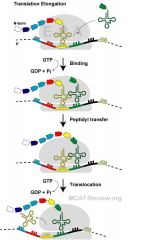
Elongation of the polypeptide chain involves addition of amino acids to the carboxyl end of the growing chain. The growing protein exits the ribosome through the polypeptide exit tunnel in the large subunit.
Elongation starts when the fmet-tRNA enters the P site, causing a conformational change which opens the A site for the new aminoacyl-tRNA to bind. This binding is facilitated by an elongation factor, a small GTPase. Now the P site contains the beginning of the peptide chain of the protein to be encoded and the A site has the next amino acid to be added to the peptide chain. The growing polypeptide connected to the tRNA in the P site is detached from the tRNA in the P site and a peptide bond is formed between the last amino acids of the polypeptide and the amino acid still attached to the tRNA in the A site. This process, known as peptide bond formation, is catalyzed by a ribozyme. Now, the A site has the newly formed peptide, while the P site has an uncharged tRNA (tRNA with no amino acids). In the final stage of elongation, translocation, the ribosome moves 3 nucleotides towards the 3'end of mRNA. Since tRNAs are linked to mRNA by codon-anticodon base-pairing, tRNAs move relative to the ribosome taking the nascent polypeptide from the A site to the P site and moving the uncharged tRNA to the E exit site. This process is catalyzed by another elongation factor. The ribosome continues to translate the remaining codons on the mRNA as more aminoacyl-tRNA bind to the A site, until the ribosome reaches a stop codon on mRNA(UAA, UGA, or UAG). |
|
|
|
Balance the following equation using the oxidation number method: HNO3(aq) + H3AsO3(aq) --> NO(g) + H3AsO4(aq) + H2O(l)
What are the first two steps? |
Step #1: Try to balance the atoms by inspection.
The H and O atoms are difficult to balance in this equation. You might arrive at the correct balanced equation using a trial and error technique, but if you do not discover the correct coefficients fairly quickly, proceed to Step #3. Step #3: Is the reaction redox? The N atoms change from +5 to +2, so they are reduced. This information is enough to tell us that the reaction is redox. (The As atoms, which change from +3 to +5, are oxidized.) Also, make sure the net charge is the same on both sides of the equation. If they are, the equation is balanced. If not, continue balancing equation. |
|
|
|
Balance the following equation using the oxidation number method: HNO3(aq) + H3AsO3(aq) --> NO(g) + H3AsO4(aq) + H2O(l)
What are the next two steps? |
Step #4: Determine the net increase in oxidation number for the element that is oxidized and the net decrease in oxidation number for the element that is reduced.
As +3 to +5 Net Change = +2 N +5 to +2 Net Change = -3 Step #5: Determine a ratio of oxidized to reduced atoms that would yield a net increase in oxidation number equal to the net decrease in oxidation number. As atoms would yield a net increase in oxidation number of +6. (Six electrons would be lost by three arsenic atoms.) 2 N atoms would yield a net decrease of -6. (Two nitrogen atoms would gain six electrons.) Thus the ratio of As atoms to N atoms is 3:2. |
|
|
|
Balance the following equation using the oxidation number method: HNO3(aq) + H3AsO3(aq) --> NO(g) + H3AsO4(aq) + H2O(l)
What are the last two steps? |
Step #6: To get the ratio identified in Step 5, add coefficients to the formulas which contain the elements whose oxidation number is changing.
2HNO3(aq) + 3H3AsO3(aq) --> NO(g) + H3AsO4(aq) + H2O(l) Step #7: Balance the rest of the equation by inspection. 2HNO3(aq) + 3H3AsO3(aq) --> 2NO(g) + 3H3AsO4(aq) + H2O(l) |
|
|
|
What is the cell structure of bacteria?
|
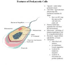
Lack of nuclear membrane, mitotic apparatus: Bacteria do not have a membrane-enclosed nucleus. Their genetic material is located in an irregular region called the nucleoid. Bacteria do not have spindles and asters that make up the eukaryotic mitotic apparatus. Instead, the prokaryotic cytoskeleton helps pull the replicated DNA apart.
Lack of typical eukaryotic organelles: Bacteria don't have Golgi, ER, mitochondria, chloroplasts. |
|
|
|
What are the Major classifications of bacteria?
|
Major classifications of bacteria by shape: bacilli (rod-shaped); spirilli (spiral shaped); cocci (spherical); vibrio (shaped like a comma).
eubacteria; archaea Eubacteria is the bacteria we encounter every day, while Archaea is the bacteria that inhabits extreme environments (high salt, temperature, or chemicals). Some bacteria might be named based on their shape - like Streptococcus pneumoniae - it's classified as a cocci (shaped like a sphere). |
|
|
|
What is the cell wall made of for bacteria?
|
Presence of cell wall in bacteria: bacterial cell wall is made of peptidoglycan, a polysaccharide-protein molecule. In contrast, plant cell wall is made of cellulose and fungi cell wall is made of chitin. As in other organisms, the bacterial cell wall provides structural integrity to the cell. The bacterial cell wall differs from that of all other organisms by the presence of peptidoglycan which is located immediately outside of the cytoplasmic membrane. Peptidoglycan is responsible for the rigidity of the bacterial cell wall and for the determination of cell shape. It is relatively porous and is not considered to be a permeability barrier for small substrates. In prokaryotes, the primary function of the cell wall is to protect the cell from internal turgor pressure caused by the much higher concentrations of proteins and other molecules inside the cell compared to its external environment. Turgor pressure or turgidity is the main pressure of the cell contents against the cell wall in plant cells and bacteria cells, determined by the water content of the vacuole, resulting from osmotic pressure. Since the cell wall is required for bacterial survival, but is absent in eukaryotes, several antibiotics stop bacterial infections by interfering with cell wall synthesis, while having no effects on human cells. While all bacterial cell walls contain peptidoglycan, not all cell walls have the same overall structures. There are two main types of bacterial cell walls, Gram positive and Gram negative, which are differentiated by their Gram staining characteristics. The Gram positive cell wall is characterized by the presence of a very thick peptidoglycan layer, which is responsible for the retention of the crystal violet dyes during the Gram staining procedure. Unlike the Gram positive cell wall, the Gram negative cell wall contains a thin peptidoglycan layer adjacent to the cytoplasmic membrane. This is responsible for the cell wall's inability to retain the crystal violet stain upon decolourisation with ethanol during Gram staining.
|
|
|
|
What is Flagellar propulsion?
|
Bacterial flagella is made of flagellin, a protein that arranges itself in a hollow cylinder to form the filament in bacterial flagellum. Flagella are complex structures that are composed of many different proteins, including flagellin. Flagellin makes up the whip-like tube and a protein complex that spans the cell wall and cell membrane to form a motor that causes the flagellum to rotate. This rotation is normally driven by proton motive force and are found in the body of the cell. In contrast, eukaryotic flagella is made of microtubules. The mechanism of the bacterial flagella is rotation. It is a motor at the base of the flagella that drives the rotation, powered by a proton or sodium gradient. (Compare this to eukaryotic flagella, which is powered directly by ATP). Flagella are whip-like structures protruding from the bacterial cell wall and are responsible for bacterial motility.
|
|
|
|
How do prokaryotic cells reproduce?
|
Reproduction by fission
DNA replicates Replicated DNAs separate by attaching to the cell membrane as the cell elongates (in contrast to mitosis, no spindle fibers needed). Cytokinesis divides the parent cell into two daughter cells. |
|
|
|
How do prokaryotic cells have a High degree of genetic adaptability?
|
Mutation
Transformation: bacteria take in plasmids and DNA fragments and integrates them into the genome. Transduction: bacteriophages undergoing lysogenic life cycle incorporate the viral DNA into the bacterial genome. Conjugation: Bacteria transfer DNA between one another through the sex pilus. |
|
|
|
How do prokaryotic cells become resistant to antibiotics through plasmids?
|
In addition to the nucleoid, many bacteria often contain small nonchromosomal DNA molecules called plasmids. Plasmids usually contain between 5 and 100 genes. Plasmids are not essential for normal bacterial growth and bacteria may lose or gain them without harm. They can, however, provide an advantage under certain environmental conditions. For example, under normal environmental growth conditions, bacteria are not usually exposed to antibiotics and having a plasmid coding for an enzyme capable of denaturing a particular antibiotic is of no value. However, if that bacterium finds itself in the body when the particular antibiotic that the plasmid-coded enzyme is able to degrade is being given to treat an infection, the bacterium containing the plasmid is able to survive and grow.
Plasmids are small molecules of double stranded, helical, nonchromosomal DNA. Like the nucleoid, the two ends of the double-stranded DNA molecule that make up plasmids covalently bond together forming a physical circle. Plasmids code for synthesis of a few proteins not coded for by the nucleoid. For example, R-plasmids, found in some gram-negative bacteria, often have genes coding for both production of a conjugation pilus (discussed later in this unit) and multiple antibiotic resistance. Through a process called conjugation, the conjugation pilus enables the bacterium to transfer a copy of the R-plasmids to other bacteria, making them also multiple antibiotic resistant and able to produce a conjugation pilus. |
|
|
|
How do prokaryotic cells become resistant to antibiotics through transposons?
|
Transposons (transposable elements or "jumping genes") are small pieces of DNA that encode enzymes that transpose the transposon, that is, move it from one DNA location to another. Transposons may be found as part of a bacterium's nucleoid (conjugative transposons) or in plasmids and are usually between one and twelve genes long. A transposon contains a number of genes, coding for antibiotic resistance or other traits, flanked at both ends by insertion sequences coding for an enzyme called transpoase. Transpoase is the enzyme that catalyzes the cutting and resealing of the DNA during transposition. Thus, such transposons are able to cut themselves out of a bacterial nucleoid or a plasmid and insert themselves into another nucleoid or plasmid and contribute in the transmission of antibiotic resistance among a population of bacteria.
Plasmids can also acquire a number of different antibiotic resistance genes by means of integrons. Integrons are transposons that can carry multiple gene clusters called gene cassettes that move as a unit from one piece of DNA to another. An enzyme called integrase enables these gene cassettes to integrate and accumulate within the integron. In this way, a number of different antibiotic resistance genes can be transferred as a unit from one bacterium to another. |
|
|
|
Explain how prokaryotic cells have exponential growth.
|
Exponential growth: Bacterial growth starts off being exponential because of the nature of binary fission. Typically the first organism splits into two daughter organisms, who then each split to form four, who split to form eight, and so on.
Later, when food becomes short, and it gets crowded, growth slows and eventually plateaus. Each generation of cells should be twice as numerous as the previous generation. However, the number of generations only gives a maximum figure as not all cells survive in each generation. |
|
|
|
Is mitosis same as binary fission?
|
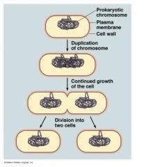
Binary fission, or prokaryotic fission, is the form of asexual reproduction and cell division used by all prokaryotes, some protazoa, and some organelles within eukaryotic organisms. This process results in the reproduction of a living prokaryotic cell by division into two parts which each have the potential to grow to the size of the original cell. Mitosis and cytokinesis are not the same as binary fission; specifically, binary fission cannot be divided into prophase, metaphase, anaphase, and telophase because prokaryotes have no nucleus and no centromeres. The ability of some multicellular animals, such as echinoderms and flatworms, to regenerate two whole organisms after having been cut in half, is also not the same as binary fission. Neither is vegetative reproduction of plants.
|
|
|
|
What are anaerobic and aerobic variants of prokaryotes?
|
Obligate aerobe = must have oxygen for growth. Obligate anaerobe = dies when oxygen is present. Facultative anaerobe = doesn't need oxygen for growth, but grows better with oxygen. Obligate aerobes require oxygen for aerobic cellular respiration. In a process known as cellular respiration, these organisms use oxygen to oxidize substrates (for example sugars and fats) in order to obtain energy. Facultative anaerobes can use oxygen, but also have anaerobic methods of energy production. Microaerophiles are organisms that may use oxygen, but only at low concentrations. Aerotolerant organisms can survive in the presence of oxygen, but they are anaerobic because they do not use it as a terminal electron acceptor. Almost all animals, most fungi, and several bacteria are obligate aerobes. Most anaerobic organisms are bacteria. Being an obligate aerobe, although advantageous from the energetical point of view, also means obligatory exposure to high levels of oxidative stress. Yeast is an example of a facultative anaerobe. Individual human cells are also facultative anaerobes: they switch to lactic acid fermentation if oxygen is not available. However, for the whole organism this cannot be sustained for long, and humans are therefore obligate aerobes. Prokaryotes also have inclusion structures in their bacterial cell that function as storage sites for energy or reservoirs for structural building blocks (i.e. amino acids). The inner membrane of a mitochondrion is analogous to the plasma membrane of a prokaryote. The enzymes for oxidative phosphorylation are embedded in the plasma membrane of prokaryotes like bacteria. Obligate anaerobes and Facultative anaerobes and obligate aerobes differ their ability to produce ATP. Since obligate anaerobes only produce ATP through anaerobic conditions, they produce less ATP than facultative and obligate aerobes. Even though they both produce ATP, they differ in their ability to produce it.
|
|
|
|
Why is conjugation so important for prokaryotes?
|
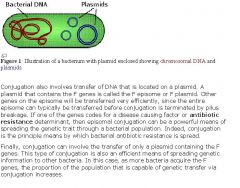
Conjugation is a mechanism whereby a bacterium can transfer genetic material to an adjacent bacterium. The genetic transfer requires contact between the two bacteria. This contact is mediated by the bacterial appendage called a pilus. Conjugation allows bacteria to increase their genetic diversity. Thus, an advantageous genetic trait present in a bacterium is capable of transfer to other bacteria. Without conjugation, the normal bacterial division process does not allow for the sharing of genetic information and, except for mutations that occur, does not allow for the development of genetic diversity. A pilus is a hollow tube constructed of a particular protein. One end is anchored to the surface of a bacterium. The other end is capable of binding to specific proteins on the surface of another bacterium. A pilus can then act as a portal from the cytoplasm of one bacterium to the cytoplasm of the other bacterium. once a channel has been formed, transfer of deoxyribonucleic acid (DNA) from one bacterium (the donor) to the other bacterium (the recipient) can occur. Conjugation requires a set of F (fertility) genes. Transfer of DNA from the genome of a bacterium can occur if the F set of genes is integrated in the bacterial chromosome. These F genes enter the pilus and literally drag the trailing genome along behind. Often the pilus will break before the transfer of the complete genome can occur. Thus, genes that are located in the vicinity of the F genes will tend to be successfully transferred in conjugation more often than genes located far away from the F genes.
|
|
|
|
Explain the significance of plasmids, extragenomic DNA, and transfer by conjugation for prokaryotes.
|
Plasmids are double stranded DNA. A plasmid can exist and replicate independently of the genomic DNA, or be integrated into it. Plasmids are inherited. Plasmids are not essential for growth and reproduction in the wild. Conjugation transfers genetic material between bacteria via a pillus or other bridge-like connection. A bacteria that is able to make a pillus (F+) has a plasmid that contains the genes to make a pillus. F+ bacteria can transfer the plasmid to an F- bacteria. Conjugation can also transfer some genomic DNA (because F+ plasmid can integrate into the chromosome through transformation). Bacterial conjugation is the transfer of genetic material between bacteria through direct cell to cell contact, or through a bridge-like connection between the two cells. The genetic information transferred is often beneficial to the recipient cell. Benefits may include antibiotic resistance or the ability to utilize a new metabolite. In order to perform conjugation, one of the bacteria, the donor, must use a conjugative or mobilizable genetic element, most often a conjugative or mobilizable plasmid. Similar to viruses, plasmids are not considered a form of "life" as it is currently defined. Unlike viruses, plasmids are "naked" DNA and do not encode genes necessary to encase the genetic material for transfer to a new host. Plasmid host-to-host transfer requires direct, mechanical transfer by "conjugation" or changes in host gene expression allowing the intentional uptake of the genetic element by "transformation".
|
|
|
|
What is transformation?
|
Transformation: incorporation into bacterial genome of DNA fragments from external medium
When a bacteria dies, it lyses and spills many DNA fragments into the environment. Another bacteria encounters these DNA fragments, takes them in, and integrates them into its own genome. If the DNA fragments contained an antibiotic resistant gene, then the transformation just made the bacteria antibiotic resistant. Transformation is the process by which bacteria pick up naked DNA from their environment. The DNA may come from a variety of sources, but most likely it is the remnants of DNA from dead bacterial cells. In order to become successfully transformed, bacteria must be competent. This means that the bacteria are expressing the appropriate enzymes (the 'transformation machinery') required to transport the exogenous DNA into the cell. Therefore, the correct genes must be expressed in order to carry out transformation. Expression of these genes depends on the growth conditions. |
|
|
|
How can prokaryotes regulate gene expression?
|
Regulation at the transcription level: some genes are actively transcribed, while others are not. Activaters and inhibitors modulate the transcription of a gene.
Regulation at the translation level: Some mRNA gets translated more. In prokaryotes, mRNAs with better Shine-Dalgarno sequence are translated more. In eukaryotes, translation regulation can involve adding more polyAs to mRNA (longer mRNA life time), modulating the translation machinary (phosphorylation of initiation factors), or storing mRNAs to be translated at a later time (mRNA masking). Prokaryotes regulate gene expression predominantly at the transcription level (eg. Operons, in which inducers increase transcription, and inhibitors decrease transcription). Eukaryotes have more regulation at other levels, and can also undergo RNA splicing, which can splice RNA in different ways to make different mRNAs. |
|
|
|
What is Transcription-translation coupling in prokaryotes?
|
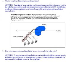
Transcription-translation coupling: in prokaryotes, translation occurs as the mRNA is being transcribed (the mRNA does not have to exit a nucleus before it needs to be translated as in eukaryotes). In prokaryotes the mRNAs undergo very little processing after being transcribed. There is a very short time interval between transcription and translation. In fact considerable translation may already take place before completion of transcription, and degradation of mRNA may begin. In eukaryotes the transcribed mRNA undergoes considerable processing before mature mRNA is formed. Transcriptional attenuation is a regulatory mechanism that causes premature termination of transcription by the operon under certain conditions, thereby preventing the expression of the mRNA required for expression of the corresponding gene products. The attenuator refers to a specific regulatory sequence that, when transcribed into RNA, forms hairpin structures to stop transcription when certain conditions are not met. An example is the trp gene in bacteria. Tryptophan levels are controlled by attenuator sequences through negative feedback control.
|
|
|
|
What are the Defining characteristics of the nucleus in the eukaryotic cell?
|
•Defining characteristics (membrane bound nucleus, presence of organelles, mitotic division)
◦Defining characteristics = what sets eukaryotes apart from prokaryotes. ◦Eukaryotes have a true nucleus (membrane-bound), while prokaryotes don't. ◦Eukaryotes have organelles (ER, Golgi, lysosomes, mitochondria), prokaryotes don't. ◦Eukaryotes divide by mitosis (all them chromosomes line up and stuff), prokaryotes undergo binary fission (no chromosomes, just a circular ring of DNA, no need for complex mitosis) Eukaryotes are fungi, animals, protists, and plants. Prokaryotes are bacteria. Then viruses are in their own groups - acellular parasites. The ribosome, found in both prokaryotes and eukaryotes, can be found in the cytoplasm or bound to the endoplasmic reticulum. It is involved in polypeptide synthesis. Membrane-bound organelles are found only in eukaryotes, and include the ER, Golgi apparatus, mitochondria, nuclei, and lysosomes. |
|
|
|
Explain the compartmentalization and storage of genetic information aspect of the nucleus.
|
◦compartmentalization: nuclear membrane / nuclear envelope surrounds the nucleus.
◦genetic information is stored inside the nucleus as DNA. |
|
|
|
What is the location and function of the nucleolus?
|
◦location is a region inside the nucleus.
◦function is to transcribe ribosomal RNA (rRNA) The nucleolus is a non-membrane bound structure composed of proteins and nucleic acids found within the nucleus. Ribosomal RNA (rRNA) is transcribed and assembled within the nucleolus. Through the microscope, the nucleolus appears like a large dark spot within the nucleus. Eukaryotic cells can have more than 1 nucleolus. Nucleoli are the organelles in the nucleus where ribosomes are made. Nucleoli are large when the cell is planning to make a lot of protein. Nucleoli are always round. |
|
|
|
What is the Nuclear envelope and nuclear pores?
|
◦nuclear envelope is a double membrane system made of an outer and an inner membrane. Also called nuclear membrane.
◦nuclear pores are holes in the nuclear envelope where things can pass into and out of the nucleus. Transcription occurs in the nucleus, and those transcribed RNA need to pass out of the nucleus. Things like transcription factors need to pass into the nucleus where they can access the DNA to be transcribed. |
|
|
|
Where is the site of ATP production?
|
•Mitochondria
◦site of ATP production: an apparatus called the ATP synthase makes ATP from ADP by utilizing the proton gradient as the driving force. The proton gradient is where the proton H+ concentration is higher in the inter-membrane space than the matrix of the mitochondria. |
|
|
|
Explain how the mitochondria does self-replication and has its own DNA and ribosomes.
|
■mitochondria replicate independently from the cell containing the mitochondria.
■mitochondria does not share the same genome with its host. ■mitochondria has their own ribosomes, which are different from the host's ribosomes in both sequence and structure. ■All these serve to support the endosymbiosis theory (symbiosis in which one organism lives inside the body of another and both function as a single organism). |
|
|
|
Explain the structure of the inner and outer membrane of the mitochondria.
|
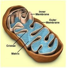
■Inner membrane surrounds the matrix.
■The folds of the inner membrane make up the cristae. ■Between the outer and inner membrane is the intermembrane space. ■The intermembrane space is high in protons H+. ■The outer membrane separates the mitochondria from the cytoplasm. |
|
|
|
What are Lysosomes?
|
•Lysosomes (spherical vesicle containing hydrolytic enzymes). Digests things like food and viral/bacterial particles. Things you want to digest gets into a vacuole by endocytosis (phagocytosis or pinocytosis), and then the vacuole fuses with the lysosome. Anything inside gets digested by the hydrolytic enzymes. Lysosomes digest excess or worn-out organelles, food particles, and engulfed viruses or bacteria. The membrane around a lysosome allows the digestive enzymes to work at the 4.5 pH they require. Lysosomes fuse with vacuoles and dispense their enzymes into the vacuoles, digesting their contents. The lysosomal membrane protects the cytosol, and therefore the rest of the cell, from the degradative enzymes within the lysosome. They are used for the digestion of macromolecules from phagocytosis (ingestion of other dying cells or larger extracellular material, like foreign invading microbes), and autophagy (wherein old or unneeded organelles or proteins, or microbes that have invaded the cytoplasm are delivered to the lysosome). Autophagy may also lead to autophagic cell death, a form of programmed self-destruction, or autolysis, of the cell, which means that the cell is digesting itself. Lysosomes carry hydrolases that degade nucleotides, proteins, lipids, phospholipids, and also remove carbohydrate, sulfate, or phosphate groups from molecules. The hydrolases are active at an acid pH which is fortunate because if they leak out of the lysosome, they are not likely to do damage (at pH 7.2) unless the cell has become acidic. Minerals (like magnesium and calcium) are obtained from diet and serve as cofactors for enzymes. They are not digested by lysosomes. Selective protein degradation occurs in the proteasome (using proteases like caspases), a large protein complex located in the nucleus and cytosol of eukaryotic cells. Proteasomal degradation of particular proteins is an essential mechanism by which cellular processes are regulated, such as cell division, apoptosis, differentiation and development. Proteins are usually tagged for selective destruction in proteolytic complexes called proteasomes by covalent attachment of ubiquitin, a small, compact protein that is highly conserved. However, some proteins may be degraded by proteasomes without ubiquitination.
|
|
|
|
What is the rough (RER) and smooth (SER)?
|
■rough ER has ribosomes studded over it, smooth ERs don't.
■RER deals with protein synthesis, folding, modification, and export. ■SER deals with biosynthesis of lipids and steroids, and metabolism of carbohydrates and drug detoxification. SER and RER are connected to the nuclear envelope. The Endoplasmic reticulum (SER and RER) consists of tubules vesicles, and sacs (cisternae) that branch forming a network. The network of endoplasmic reticulum allows increased surface area for the action or storage of key enzymes and the products of these enzymes. ■The Sarcoplasmic Reticulum is a special type of smooth endoplasmic reticulum found in smooth and striated muscle fibers whose function is to store and release calcium ions. The sarcoplasmic reticulum releases calcium ions when it is stimulated, causing muscle contraction, while it absorbs calcium ions during relaxation. Biosynthesis is an enzyme-catalyzed process in cells of living organisms by which substrates are converted to more complex products. The biosynthesis process often consists of several enzymatic steps in which the product of one step is used as substrate in the following step. Examples for such multi-step biosynthetic pathways are those for the production of amino acids, fatty acids, and natural products. The prerequisites for biosynthesis are precursor compounds, chemical energy (such as in the form ATP), and catalytic enzymes, which may require reduction equivalents (e.g., in the form of NADH, NADPH). Commonly known complex products of biosynthesis include proteins, vitamins, and antibiotics. Most organic compounds in living organisms are built in biosynthetic pathways. |
|
|
|
Where are the ribosomes in the endoplasmic reticulum?
|
◦RER (site of ribosomes): the ribosomes attach to the outside of rough ER and synthesizes proteins.
|
|
|
|
Explain the role in membrane biosynthesis for mitochondria.
|
SER (lipids), RER (transmembrane proteins). A transmembrane protein is a protein that spans the entire biological membrane.
■SER = makes lipids of the plasma membrane. ■RER = makes transmembrane proteins, carries them on its membrane, RER membrane forms vescicles and bud off, fuses with the plasma membrane, transmembrane proteins now on the plasma membrane. RER also makes secreted proteins. |
|
|
|
Explain the RER's role in biosynthesis of transmembrane and secreted proteins that cotranslationally targeted to RER by signal sequence.
|
■Transmembrane proteins and proteins that are to be secreted have a signal sequence right at the beginning. Both transmembrane proteins and secreted proteins travel within the cell inside of transport vesicles. ■When ribosome starts making those proteins, they make the signal sequence first.
■Signal sequence recruits a signal recognition particle that drags it to the RER. ■ribosome now on the RER continues making the protein, but snakes it into the lumen. ■Signal sequence is clipped off. The surface of the rough endoplasmic reticulum (RER) is studded with protein-manufacturing ribosomes giving it a "rough" appearance (hence its name).However, the ribosomes bound to the RER at any one time are not a stable part of this organelle's structure as ribosomes are constantly being bound and released from the membrane. A ribosome only binds to the ER once it begins to synthesize a protein destined for the secretory pathway. Here, a ribosome in the cytosol begins synthesizing a protein with a signal sequence until a signal recognition particle recognizes the sequence. This signal sequence allows the recognition particle to bind to the ribosome, causing the ribosome to bind to the RER and pass the new protein through the ER membrane. The signal sequence is then cleaved off within the lumen of the ER and the ribosome released back into the cytosol. Once the protein is in the lumen of the RER, it's put in a vesicle and sent to the golgi apparatus to be modified and ultimately sent to its proper destination. |
|
|
|
What is the structure of the endoplasmic reticulum?
|
The endoplasmic reticulum (ER) is a network of flattened sacs and branching tubules that extends throughout the cytoplasm in plant and animal cells. These sacs and tubules are all interconnected by a single continuous membrane so that the organelle has only one large, highly convoluted and complexly arranged lumen (internal space). Usually referred to as the endoplasmic reticulum cisternal space, the lumen of the organelle often takes up more than 10 percent of the total volume of a cell. The endoplasmic reticulum membrane allows molecules to be selectively transferred between the lumen and the cytoplasm, and since it is connected to the double-layered nuclear envelope, it further provides a pipeline between the nucleus and the cytoplasm.
|
|
|
|
What is the Golgi apparatus?
|
•Golgi apparatus (general structure; role in packaging, secretion, and modification of glycoprotein carbohydrates) . Looks like stacks of pancakes (known as cisternae). Each cisterna comprises a flattened membrane disk, and carries Golgi enzymes to help or to modify cargo proteins that travel through them. Modifies and/or secretes macromolecules (such as proteins and lipids) for the cell. ◦RER make protein → modified in the Golgi → leave the golgi in a vesicle and are secreted out of cell by exocytosis or used within the cell. Glycoprotein = protein with attached saccharides. Golgi can glycosylate proteins as well as modify existing glycosylations. Glycosylation affects protein's structure, function, and protect it from degradation. Glycosylation is the enzymatic process that links saccharides to produce glycans (polysaccharides), attached to proteins, lipids, or other organic molecules. This enzymatic process produces one of the fundamental biopolymers found in cells (along with DNA, RNA, and proteins). Glycosylation is a form of co-translational and post-translational modification. Glycans serve a variety of structural and functional roles in membrane and secreted proteins. The majority of proteins synthesized in the rough ER undergo glycosylation. It primarily modifies proteins delivered from the rough endoplasmic reticulum but is also involved in the transport of lipids around the cell, and the creation of lysosomes. In this respect it can be thought of as similar to a post office; it packages and labels items which it then sends to different parts of the cell. Enzymes within the cisternae are able to modify substances by the addition of carbohydrates (glycosylation) and phosphates (phosphorylation). In order to do so, the Golgi imports substances such as nucleotide sugars from the cytosol. These modifications may also form a signal sequence which determines their final destination.
|
|
|
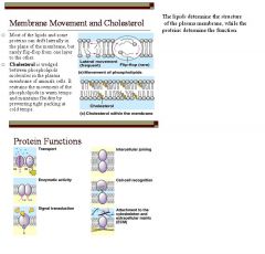
What is the General function of the plasma membrane?
|

•General function in cell containment. It is selectively permeable. The bilayer of molecules that surround cells, however, is mostly made up of phospholipids arranged in such a way that their hydrocarbon "tails" are all pointing into the center of the structure. Hydrocarbon molecules are strongly hydrophobic ("water fearing"), and it is this strongly hydrophobic layer of material that gives the cell membrane its "water proof" nature and allows it to act as a container for the cell and its contents. It would be useless to make a container of something that easily dissolved in water! The arrangement of hydrophilic heads and hydrophobic tails of the lipid bilayer prevent polar solutes (e.g. amino acids, nucleic acids, carbohydrates, proteins, and ions) from diffusing across the membrane, but generally allows for the passive diffusion of hydrophobic molecules. This affords the cell the ability to control the movement of these substances via transmembrane protein complexes such as pores and gates. Carbohydrates in the plasma membrane are used for cell-cell recognition - the ability for a cell to recognize one type of neighboring cell from another. This is important in cell sorting and organization as tissues and organs in development. Membrane thickness controls rate of diffusion - increasing the membrane thickness will decrease overall membrane diffusion. The plasma membrane is held together by hydrophobic interactions - they hydrophobic tails of the fatty acids interact with each other within the plasma membrane. Also, integral proteins that span the entire plasma membrane interactwith the plasma membrane through hydrophobic interactions.
|
|
|
What can diffuse across the plasma membrane and why?
|
Passage is controlled in part due to the hydrophobic core of the membrane. So hydrophobic molecules cross easily while polar and charged (ionic) molecules (hydrophilic molecules) have some difficulty. Hydrophobic molecules can get past the hydrophilic molecules because the bilayer is composed of hydrophobic lipids, it just has a hydrophilic head, but it's still a hydrophobic lipid. Proteins assist the control and transport of ions and polar molecules. The impermeability of the plasma membrane enables to Na-K pump to work (controls the pumping out of 3 Na and pumping in of 2 K ions). It also enables the control of Ca ions through calcium channels.
Hydrophobic and hydrophilic substances are also determined by dipole moments. Substances with a dipole moment (meaning it is polar and hydrophilic) can't readily cross a plasma embrane, while substances that do not have a dipole moment (meaning it is nonpolar and hydrophobic) can readily cross a plasma membrane. |
|
|
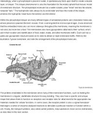
What are the components of the plasma membraine and the fluid mosaic model?
|
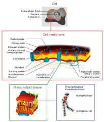
•Protein and lipid components, fluid mosaic model: the fluid mosaic model basically describes the membrane as protein boats floating in a sea of lipids.
The cell membrane (also called the plasma membrane or plasmalemma) is one biological membrane separating the interior of a cell from the outside environment. The cell membrane surrounds all cells and it is selectively-permeable, controlling the movement of substances in and out of cells. It contains a wide variety of biological molecules, primarily proteins and lipids, which are involved in a variety of cellular processes such as cell adhesion, ion channel conductance and cell signaling. The plasma membrane also serves as the attachment point for the intracellular cytoskeleton and, if present, the extracellular cell wall. According to the fluid mosaic model the biological membranes can be considered as a two-dimensional liquid where all lipid and protein molecules diffuse laterally (side by side in the plane of the plasma membrane) more or less easily. The components of a plasma membrane are integral proteins, peripheral proteins, glycoproteins, phospholipids, glycolipids, and in some cases cholesterol, lipoproteins. The lipids of the plasma membrane determine the structure (lipid bilayer - phospholipids with a hydrophilic and hydrophobic end arranged into a bilayer), while the proteins determine the function (some creates the channels, some are receptors, etc). |
|
|
|
What is osmosis of the plasma membrane?
|
•Osmosis: water diffuses freely across the membrane, but not ions. So osmosis occurs readily. Osmosis is the movement of water from a region of high water concentration to a region of lower water concentration through a semi permeable membrane. Facilitated diffusion of ions takes place through proteins, or assemblies of proteins, embedded in the plasma membrane. These transmembrane proteins form a water-filled channel through which the ion can pass down its concentration gradient. The transmembrane channels that permit facilitated diffusion can be opened or closed. They are said to be "gated". Some types of gated ion channels: ligand-gated, mechanically-gated, and voltage-gated. Many ion channels open or close in response to binding a small signaling molecule or "ligand". An example of a mechanically-gated ion channel is :Sound waves bending the cilia-like projections on the hair cells of the inner ear open up ion channels leading to the creation of nerve impulses that the brain interprets as sound. In so-called "excitable" cells like neurons and muscle cells, some channels open or close in response to changes in the charge (measured in volts) across the plasma membrane (voltage-gated channels). Some small, hydrophilic organic molecules, like sugars, can pass through cell membranes by facilitated diffusion. Once again, the process requires transmembrane proteins. In some cases, these — like ion channels — form water-filled pores that enable the molecule to pass in (or out) of the membrane following its concentration gradient. If you raise the level of extracellular substances in fluid, like glucose or NaCl, the water of the cells may leave the cells and cause cell dehydration because the cells are in a hypertonic solution. If you are given a question about whether a cell is hyper- or hypotonic, beyond what is being moved in and out of the cell, make sure that you also know the concentration of the cell inside and outside of the cell itself. Only then can you conclude the difference in concentration between the cell and its environment.
|
|
|
|
What is Passive and active transport of the plasma membrane?
|
•Passive and active transport: things that can't readily diffuse across the membrane are transported across the membrane either without energy (passive) or with energy (active). The diffusion of water is referred to as osmosis, not passive transport. Passive tranport is driven by the kinetic energy of the molecules being transported or by membrane transporters by facilitate crossing. Active transport depends upon the expenditure of cellular energy in the form of ATP hydrolysis. Another way of looking at the difference is to determine whether the transported solute is move "up" or "down" its concentration gradient - movement against an electrochemical gradient necessitates expenditure of energy and is therefore accomplished by active transport. A molecule or ion that crosses the membrane by moving down a concentration or electrochemical gradient and without expenditure of metabolic energy is said to be transported passively. Another name for this process is diffusion. All molecules and ions are in constant motion and it is the energy of motion - kinetic energy - that drives passive transport. Transport of uncharged species across a membrane is dictated by differences in concentration of that species across the membrane - that is, by the prevailing concentration gradient. For ions and charged molecules, the electrical potential across the membrane also becomes critically important. Together, gradients in concentration and electric potential across the cell membrane constitute the electrochemical gradient that governs passive transport mechanisms. Primary active transport consumes ATP directly (i.e. Na+/K+ pump). In secondary active transport, ATP consumption is used to create a concentration gradient, which is then used to co-transport compounds.
|
Adenine triphosphate (ATP) supplies energy to drive thermodynamically unfavored reactions (reactions that absorb energy, versus those spontaneous reactions that release energy). It works by losing a phosphate group, which is catalyzed by an enzyme. The reaction releases energy and its product is adenosine diphosphate (ADP) and a phosphate group. Even more energy can be extracted by removing the second phosphate group to produce adenosine monophosphate (AMP).
|
|
|
What are Membrane channels of the plasma membrane?
|
•Membrane channels: A family of biological membrane proteins which allow the passive movement of ions (ion channels), water (aquaporins) or other solutes to passively pass through the membrane down their electrochemical gradient. The lipid bilayer of biological membranes is impermeable to ions and polar molecules. Permeability is conferred by two classes of membrane proteins, pumps and channels. Pumps use a source of free energy such as ATP to drive the thermodynamically uphill transport of ions or molecules. Pump action is an example of active transport. Channels, in contrast, enable ions to flow rapidly through membranes in a downhill direction. Channel action illustrates passive transport, called facilitated diffusion. Although the Sodium-potassium pump spans the membrane like membrane channels, but they are not membrane channels, they are membrane pumps. Muscle contraction requires a great deal of calcium which is obtained from 1) the extracellular environment via calcium-gated channels and 2) from the sarcoplasmic reticulum (the muscle cell's smooth endoplasmic reticulum). Sodium-gated channels open during depolarization of NERVES, while most MUSCLE cells, including skeletal, cardiac, and smooth, rely on CALCIUM-gated channels.
|
|
|
|
What is the Sodium–potassium pump?
|
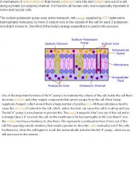
•Sodium-potassium pump: 3 sodium (NA+) out, 2 potassium (K+) in. Thus, the cell maintains a negative resting potential. Active transport is responsible for the well-established observation that cells contain relatively high concentrations of potassium ions but low concentrations of sodium ions. The mechanism responsible for this is the sodium-potassium pump, which moves these two ions in opposite directions across the plasma membrane. The carrier is an ATP-ase and that it pumps three sodium ions out of the cell for every two potassium ions pumped in. In order to maintain the cell membrane potential, cells must keep a low concentration of sodium ions and high levels of potassium ions within the cell. The three positive Na out while the two positive K in keeps a negative resting potential, because there is a net loss of ions out of the cell. A leaky potassium channel (where some K+ leaks out, making the cell more negative) is always open because it is ungated. It allows some K+ to flow down it's concentration gradient, so it is not voltage dependent.
|
Voltage-gated ion channels are a class of transmembrane ion channels that are activated by changes in electrical potential difference near the channel; these types of ion channels are especially critical in neurons, but are common in many types of cells. They have a crucial role in excitable neuronal and muscle tissues, allowing a rapid and co-ordinated depolarization in response to triggering voltage change. Found along the axon and at the synapse, voltage-gated ion channels directionally propagate electrical signals. They generally are composed of several subunits arranged in such a way that there is a central pore through which ions can travel down their electrochemical gradients. The channels tend to be ion-specific, although similarly sized and charged ions may sometimes travel through them. Examples include: 1) the sodium and potassium voltage-gated channels of nerve and muscle. 2) the voltage-gated calcium channels that play a role in neurotransmitter release in pre-synaptic nerve endings.
|
|
|
What are Membrane receptors and second messengers?
|
◦Many hormones can't cross the plasma membrane, so they bind to membrane recepters on the outside. Receptor binding triggers the production of second messengers. The term second messenger was coined upon the discovery of these substances in order to distinguish them from hormones and other molecules that function outside the cell as “first messengers” in the transmission of biological information. Second messengers cause a change inside the cell (through a protein kinase cascade). In cell physiology, a secondary messenger system (also known as a second messenger system) is a method of cellular signaling whereby a diffusible signaling molecule is rapidly produced/secreted, which can then go on to activate effector proteins within the cell to exert a cellular response. An effector is a molecule (originally referring to small molecules but now encompassing any regulatory molecule, including proteins) that binds to a protein and thereby alters the activity of that protein. Secondary messengers are a component of signal transduction cascades. Secondary messenger systems can be activated by diverse means, either by activation of enzymes that synthesize them, as is the case with the activation of cyclases that synthesize cyclic nucleotides, or by opening of ion channels to allow influx of metal ions, such as in Ca2+ signaling. These small molecules may then go on to exert their effect by binding to and activating effector molecules such as protein kinases, ion channels, and a variety of other proteins, thus continuing the signaling cascade. Receptors provide a high degree of specificity for a particular ligand and the receptor amplifies the stimulating signal, but the receptor never modifies the LIGAND, it just activates other enzymes after the ligand binds to it. Many second messenger molecules are small and therefore diffuse rapidly through the cytoplasm, enabling information to move quickly throughout the cell.
|
|
|
|
What are cell-signalling pathways?
|
■Contact signaling = physical contact triggers a change inside cell. Chemical signaling = chemical binding to receptor triggers a change inside cell. ■Nerves use neurotransmitters for chemical signalling. The endocrine system use hormones for chemical signalling. Electrical signaling = change in membrane potential triggers change in cell. Action potential along neurons (electrical signalling) propagates and cause release of neurotransmitters into synapse. Action potential along muscle (electrical signalling) cell membrane causes contraction. Neurotransmitters act on postsynaptic receptors to change the membrane potential. This change can be either 1) a direct depolarization, which causes an action potential 2) a direct hyperpolarization, which prevents an action potential, or 3) the activation of a second messenger that eventually leads to changes in the neuron firing rate. Why a second messenger? Remember, protein neurotransmitters are hydrophilic and cannot freely diffuse across the cell membrane. They require a second messenger (i.e. cAMP) to get their signal from the cell membrane to the nucleus.
|
|
|
|
What is membrane potential?
|
•Membrane potential: the resting potential of the cell membrane is negative because of the sodium-potassium pump.
Ion concentration gradient, number of functional channels, and the open or closed status of channels all affect the cell membrane potential. |
|
|
|
What is Exocytosis and endocytosis?
|
•Exocytosis and endocytosis: exo = getting stuff out, endo = taking stuff in. Endocytosis is a general term for a group of processes that bring macromolecules, large particles, small molecules, and even small cells into the eukaryotic cell. There are three types of endocytosis: phagocytosis, pinocytosis, and receptor-mediated endocytosis. In all three, the plasma membrane invaginates (folds inward) around materials from the environment, forming a small pocket. The pocket deepens, forming a vesicle. This vesicle separates from the plasma membrane and migrates with its contents to the cell’s interior. Exocytosis is the process by which materials packaged in vesicles are secreted from a cell when the vesicle membrane fuses with the plasma membrane. The initial event in this process is the binding of a membrane protein protruding from the cytoplasmic side of the vesicle with a membrane protein on the cytoplasmic side of the target site on the plasma membrane. The phospholipid regions of the two membranes merge, and an opening to the outside of the cell develops. The contents of the vesicle are released to the environment, and the vesicle membrane is smoothly incorporated into the plasma membrane. If the material taken in are particles such as bacterium or fragments of organic matter, it's phagocytosis. If the material taken in is liquid, it's pinocytosis. Receptor mediated endocytosis is an endocytotic mechanism in which specific molecules are ingested into the cell. The specificity results from a receptor-ligand interaction. Receptors on the plasma membrane of the target tissue will specifically bind to ligands on the outside of the cell. An endocytotic process occurs and the ligand is ingested.
|
|
|
What is Cell–cell communication (general concepts of cellular adhesion)?
|
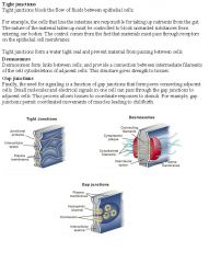
Gap junctions are protein channels that allow ions and small molecules to freely pass between cells without encountering the extracellular environment. Gap junctions directly connects the cytoplasm of two cells, which allows various molecules and ions to pass freely between cells. Tight junctions: stitches/glues two cells together, and does not allow stuff to flow through between the cells. A series of cells with tight junctions also effectively forms an impermeable barrier. Tight junctions hold cells together. They prevent the passage of molecules and ions through the space between cells. So materials must actually enter the cells (by diffusion or active transport) in order to pass through the tissue. This pathway provides control over what substances are allowed through. (Tight junctions play this role in maintaining the blood-brain barrier.) Desmosomes: connects two cells together by linking their cytoskeleton. Desmosomes are structures that join adjacent cells together. They also provide anchoring points for intermediate filaments between the two cells to help build a strong structural framework. Desmosomes are the most common type of intercellular junction in vertebrate epithelial cells and are used for strength.
|
Heart cells have gap junctions known as intercalcalated disks. Cardiac and smooth muscle cells contract as a group. As a result, both cell types depend on gap junctions (channels that link cells together) to spread an excitatory action potential between cells. Skeletal muscle cells can contract independently and DO NOT have gap junctions. Since cardiac cells have to contract in a coordinated manner and don't have the precise neural control that skeletal muscle cells do, they need to interact with each other: that's the function of the gap junction. Smooth muscle does have neural input, but it lacks the elaborate neuromuscular junction of skeletal fibers. Basically, skeletal muscle cells are more sophisticated (due to the voluntary nature of them), so they don't need gap junctions.
|
|
|
What is the general function of the cytoskeleton?
|
•General function in cell support and movement
|
|
|
|
What are Microfilaments?
|
•Microfilaments (composition; role in cleavage and contractility)
◦made of actin ◦responsible for cytokinesis. Supports cell shape by bearing tension. Common to all eukaryotic cells, these filaments are primarily structural in function and are an important component of the cytoskeleton, along with microtubules and often the intermediate filaments. Microfilaments range from 5 to 9 nanometers in diameter and are designed to bear large amounts of tension. In association with myosin, microfilaments help to generate the forces used in cellular contraction and basic cell movements. The filaments also enable a dividing cell to pinch off into two cells and are involved in amoeboid movements of certain types of cells. |
|
|
|
What are microtubules?
|
•Microtubules (composition; role in support and transport)
◦made of tubulin ◦responsible for mitotic spindle, they form the cilila/flagella, and they are used for intracellular transport of organelles and vesicles. Supports cell shape by resist compression, as opposed to microfilaments and intermediate filaments who bear tension. They also form the centrioles. They are organized by the centrosome. the centrosome is an organelle that serves as the main microtubule organizing center (MTOC) of the animal cell as well as a regulator of cell-cycle progression. Centrosomes are often associated with the nuclear membrane during interphase of the cell cycle. In mitosis the nuclear membrane breaks down and the centrosome nucleated microtubules can interact with the chromosomes to build the mitotic spindle. The spindle apparatus is the structure that separates the chromosomes into the daughter cells during cell division. It is part of the cytoskeleton in eukaryotic cells. Depending on the type of cell division, it is also referred to as the mitotic spindle during mitosis and the meiotic spindle during meiosis. |
|
|
|
What are Intermediate filaments ?
|
•Intermediate filaments (role in structural support). Their composition is varied. Supports cell shape by bearing tension. They are prominent in cells that withstand mechanical stress and are the most insoluble part of the cell. Like actin filaments (microfilaments), intermediate filaments function in the maintenance of cell-shape by bearing tension (microtubules, by contrast, resist compression. It may be useful to think of micro- and intermediate filaments as cables, and of microtubules as cellular support beams). Intermediate filaments organize the internal tridimensional structure of the cell, anchoring organelles and serving as structural components of the nuclear lamina and sarcomeres. They also participate in some cell-cell and cell-matrix junctions. They provide the anchoring points for desmosomes by connecting cells together.
|
|
|
|
What is the Composition and function of eukaryotic cilia and flagella?
|
◦made of microtubules (eukaryotic)
◦cilia can be for locomotion, sensory, or for sweeping mucus. ◦flagella is used for locomotion. There are two types of cilia: motile cilia and non-motile, or primary cilia, which typically serve as sensory organelles. Motile cilia are usually present on a cell's surface in large numbers and beat in coordinated waves. In humans, for example, motile cilia are found in the lining of the trachea (windpipe), where they sweep mucus and dirt out of the lungs. In female mammals, the beating of cilia in the Fallopian tubes moves the ovum from the ovary to the uterus. In comparison to motile cilia, non-motile (or primary) cilia usually occur one per cell; nearly all mammalian cells have a single non-motile primary cilium. In addition, examples of specialized primary cilia can be found in human sensory organs such as the eye and the nose. The outer segment of the rod photoreceptor cell in the human eye is connected to its cell body with a specialized non-motile cilium. The dendritic knob of the olfactory neuron, where the odorant receptors are located, also contains non-motile cilia. |
|
|
|
What are Centrioles?
|
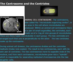
•Centrioles, microtubule organizing centers. They are basically barrel-shaped tubes of microtubules.
Centrioles are involved in the organization of the mitotic spindle. Centrioles are a very important part of centrosomes, which are involved in organizing microtubules in the cytoplasm. The position of the centriole determines the position of the nucleus and plays a crucial role in the spatial arrangement of the cell. In order for cells to do this, cells must mate. |
|
|
|
What is Interphase and mitosis (prophase, metaphase, anaphase, telophase)?
|
◦Interphase: G1 = Growth, S = Synthesis (replicate chromosomes), and ■G2 = Growth. Prophase = Prepare (condense chromatin into a highly ordered structures called a chromosomes, break down nuclear membrane, assemble mitotic spindle). Since the genetic material has already been duplicated earlier in S phase, the replicated chromosomes have two sister chromatids, bound together at the centromere. Although centrioles help organize microtubule assembly, they are not essential for the formation of the spindle, since they are absent from plants, and centrosomes are not always used in meiosis. Metaphase = Middle (Centromerers of chromosomes line up in the middle on metaphase plate, an imaginary line that is equidistant from the two centrosome poles). Proper chromosome separation requires that every kinetochore be attached to a bundle of microtubules (spindle fibres). Anaphase = Apart (Sister chromatids pulled apart to opposite sides of cell by microtubules pulling the centrosomes to the opposite sides of the cells). Telophase = Prophase in reverse = de-condense chromosomes, re-form nuclear membrane, break down mitotic spindle. Corresponding sister chromatids attach at opposite ends of the cell. A new nuclear envelope, using fragments of the parent cell's nuclear membrane, forms around each set of separated sister chromatids. Both now sets of chromosomes, now surrounded by new nuclei, unfold back into chromatin. Mitosis is complete, but cell division is not yet complete. Cytokinesis is often mistakenly thought to be the final part of telophase; however, cytokinesis is a separate process that begins at the same time as telophase. Cytokinesis is technically not even a phase of mitosis, but rather a separate process, necessary for completing cell division. In animal cells, a cleavage furrow (pinch) containing a contractile ring develops where the metaphase plate used to be, pinching off the separated nuclei. In both animal and plant cells, cell division is also driven by vesicles derived from the Golgi apparatus, which move along microtubules to the middle of the cell.
|
|
|
|
What are Mitotic structures and processes ?
|
◦Centrioles, asters, spindles: responsible for pulling apart the sister chromatids. Asters are radial microtubule arrays in animal cells which are located around each pair of centrioles. The cellular spindle apparatus includes the spindle microtubules, associated proteins, and any centrosomes or asters present at the spindle poles. The spindle apparatus is the structure that separates the chromosomes into the daughter cells during cell division. It is part of the cytoskeleton in eukaryotic cells. Two pairs of centrioles make up the centrosome, From each centrosome, microtubules grow into a "spindle" which is responsible for separating replicated chromosomes into the two daughter cells.
◦Chromatids, centromeres, kinetochores: sister chromatids are duplicated copies of the chromosome. Chromatids are joined at the centromere. There's a protein at the centromere called the kinetochore, where spindle fibers attach to pull the chromatids apart. ◦Nuclear membrane breakdown and reorganization: for most eukaryotes, the nuclear membrane breaks down at the beginning of mitosis, and reforms at the end of mitosis around each of the two newly formed nuclei. ◦Mechanisms of chromosome movement: chromatids move apart during anaphase by the spindle fibers. Microtubules cause the chromosome movement. |
|
|
|
What are the Phases of cell cycle?
|
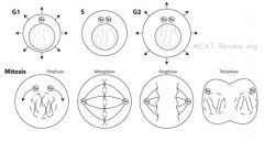
•Phases of cell cycle: G0, G1, S, G2, M. G0 = no more DNA replication or cell division. Examples include nerves and muscles. G1 = growth = make organelles, increase in cell size. G1 is marked by synthesis of various enzymes that are required in S phase, mainly those needed for chromosome replication. S = Chromosome replication and centrioles replicate while centrosomes divide. When it is complete, all of the chromosomes have been replicated, i.e., each chromosome has two (sister) chromatids. Thus, during this phase, the amount of chromatids in the cell has effectively doubled, though the ploidy of the cell remains the same. There are also two centrosomes, each with its own pair of centrioles. G2 = growth = make organelles, increase in cell size. Again, significant biosynthesis occurs during this phase, mainly involving the production of microtubules, which are required during the process of mitosis. M = mitosis.
|
|
|
|
What is Growth arrest?
|
•Growth arrest: a halt in the cell cycle. For instance, a cell's growth could be arrested between G1 and S or between G2 and M.
The cell cycle can be arrested for many reasons: ◦Too much genomic mutation/damage causes a cell to arrest in the cell cycle. ◦Contact inhibition: normal epithelial cells stop growing when it gets crowded and are touching adjacent cells. ◦Lack of food can also cause growth arrest. Growth arrest: a phenomenon occurring when a cell does not proceed through the cell cycle. |
|
|
|
What is apoptosis?
|
•Apoptosis (Programmed Cell Death) . ◦Apoptosis = death that is clean and healthy. ◦Apoptosis = activation of caspases that digest the cell from within. ◦No spilling of cell contents. ◦Afterwards, the apoptosed cell release chemicals that attracts macrophages, and gets engulfed. ◦Apoptosis can be brought upon by development (eg tadpole losing tail) or by immune response (infected/cancerous cells killed by cytotoxic T cells/natural killer cells). Caspases are a family of proteases (enzymes that cut peptide bonds in proteins) , which play essential roles in apoptosis (programmed cell death), necrosis and inflammation. Caspases are essential in cells for apoptosis, or programmed cell death, in development and most other stages of adult life, and have been termed "executioner" proteins for their roles in the cell. Apoptosis differs from necrosis, in which the cellular debris can damage the organism. In contrast to necrosis, which is a form of traumatic cell death that results from acute cellular injury, apoptosis, in general, confers advantages during an organism's life cycle. For example, the differentiation of fingers and toes in a developing human embryo occurs because cells between the fingers apoptose; the result is that the digits are separate. Excessive apoptosis causes atrophy, such as in ischemic damage, whereas an insufficient amount results in uncontrolled cell proliferation, such as cancer.
|
|
|
|
What is the cell body of the nerve cell (neural cell)
|
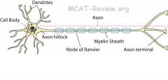
•Cell body (site of nucleus and organelles). Contains nucleus and organelles just like any other cell. Has well-developed RER and golgi (makes a lot of proteins). The neuron can be generally divided into three main functional parts, the cell body, the axon, and the dendrites. The cell body is the biosynthetic center of the cell. This is where cellular metabolism occurs, as well as the production of proteins and membrane. Some people believe that this production machinery , consisting of free ribosomes and rough endoplasmic reticulum (rER), is probably the most active and best developed of any cell in the body. The ribosomes and rER are the cellular organelles responsible for protein production and packaging. The cell body contains all of the basic cellular organelles of most other cells but is conspicuously lacking centrioles. Centrioles are organelles (the cellular equivalent of human organs) that are responsible for the formation of the mitiotic spindle, which is important for cellular replication. However, neurons do not divide (they are always in the G0 phase), so they don't need centrioles. However, some cells have been found to divide without centrioles (plant and animal cells). The rER is very well developed and often referred to as Nissl bodies and are the site of protein synthesis. The cell body contains intermediate filaments called neurofibrils, which are important in intracelluar transport and maintaing cell shape and integrity. Bundles of these neurofibrils are called neurofilaments. A collection of nerve cell bodies (neurons) in the CENTRAL NERVOUS SYSTEM is called a nucleus while a collection of nerve cell bodies (neurons) in the PERIPHERAL NERVOUS SYSTEM is called a ganglion.
|
|
|
What is the Axon (structure, function)?
|
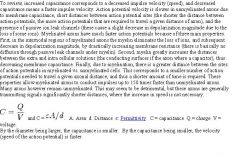
◦Axon = Conducting region of the nerve.
◦Axon terminals = secretory regions of nerve. ◦Other names for axon terminal = synaptic knob. An axon or nerve fiber is a long, slender projection of a nerve cell, or neuron, that conducts electrical impulses away from the neuron's cell body or soma. Neurons can have no more than a single axon (certain neurons in the CNS don't have any), responsible for the passing of messages from the neuron to other nerve cells or their effectors, targets of nerve impulses such as muscles. This is where the actual nerve impulse, or action potential, occurs, on the axon. Axons can be very long, or very short in length. Some axons are 3-4 feet in length, extending from the middle of the spine to the bottom of the feet. Long axons are sometimes called nerve fibers. Just as axonal length may vary so too does the diameter of the axon. The larger the diameter, the thicker the axon, the faster it delivers nerve impulses. The region of the axon that attaches to the cell body is called the axon hillock. It is at this junction that action potentials are generated. The axon hillock is where the numerous graded potentials are summed, and thus the site for determination of whether threshold has been met. From here the axon extends a distance until it terminates into what is known as the secretory component of the neuron. The secretory component is the actual part of the neuron that transmits the nerve impulse on to the next neuron. The conducting component of the neuron lies between the axon hillock and the secretory component, consisting entirely of a length of axon. It is responsable for the propagation of nerve impulses away from the cell body. Axons contain the same organelles as the cell body, except they lack Nissl bodies (rER), so are dependent on the cell body to supply them with proteins and membrane components. |
|
|
What are synaptic potentials?
|
In general, action potentials that reach the axon terminals cause a neurotransmitter to be released into the synaptic cleft. Neurotransmitters are small molecules that may open ion channels in the postsynaptic cell; most axons have the same neurotransmitter at all of their termini. The arrival of the action potential opens voltage-sensitive calcium channels in the presynaptic membrane; the influx of calcium causes vesicles filled with neurotransmitter to migrate to the cell's surface and release their contents into the synaptic cleft. Calcium channels are what release the neurotransmitters into the synaptic cleft, while sodium and potassium channels open in response to depolarization/repolarization. Depolarization caused is by the influx of sodium. When sodium gates open and sodium ions rush in, the inside temporarily becomes positively charged. Potassium gates then open and potassium ions rush out, restoring the negative charge.
The action potential cannot reverse its direction because membrane that has just been depolarized cannot be depolarized again until after a brief recovery (called refractory) period. During this period, the membrane is insensitive to stimulation. The refractory period is caused by the inactivation of the Na+ channels that originally opened to depolarize the membrane. These channels remain inactivated until the membrane repolarizes, after which they close, reactivate and regain their ability to open in response to stimulus. |
|
|
|
What are Dendrites (structure, function)?
|
•Dendrites (structure, function)
◦Receptive region of the nerve = gets input. ◦The branching helps to increase the surface area for reception. Branching of carbon chains lowers the freezing point/melting point. Dendrites are the branched projections of a neuron that act to conduct the electrochemical stimulation received from other neural cells to the cell body, or soma, of the neuron from which the dendrites project. Dendrites are the part of the cell that recieve signals and conduct those signals to the rest of the neuron. These electrical signals are not nerve impulses (action potentials) but are short distance signals called graded potentials, or receptor potentials (synaptic potentials). Motor neurons can have hundreds of dendrites. This is a large amount of surface area dedicated to signal and impulse reception, hinting that a substantial stimulation is required before an action potential can be generated. Dendrites contain most organelles present in the cell body. |
|
|
|
What are Myelin sheath, Schwann cells, oligodendrocytes, and insulation of axon?
|
◦Myelin sheath = Covers the axon intermittently, with gaps called nodes of Ranvier. ◦The purpose of myelin sheath is to speed up conduction by insulating the nerve in intervals. This intermittent insulation causes action potential to jump from one node of Ranvier to the next. ◦Schwann cells = makes myelin sheath in the peripheral nervous system by wrapping around the axon. Way to remember: P and S are close in alphabet. The peripheral nervous system is the part of the nervous system outside of the brain and spinal cord. Schwann cells are glia of the peripheral nervous system (PNS). Glial cells are cells that support nerve cells. Oligodendrocytes = the central nervous system analogue of Schwann cells, makes myelin sheath around CNS axons. Insulation of axon = achieved by the myelin sheath. Insulation occurs in intervals, which causes action potential to jump from one node of Ranvier to the next. ◦Myelin sheath is a good insulator because it is fatty and does not contain any channels. Oligodendrocytes are a variety of neuroglia and their main function is the insulation of axons (the long projection of nerve cells) in the central nervous system (the brain and spinal cord). Myelin made by different cell types varies in chemical composition and configuration, but performs the same insulating function. Myelinated axons are white in appearance, hence the "white matter" of the brain. Myelin is composed of about 80% lipid and about 20% protein. A single oligodendrocyte can wrap around multiple axons in the CNS while a schwann cell can only wrap around 1 axon at a time in the PNS. Since the myelin is made up of a lot of lipids, any irregularity in lipid production will affect the schwann cells and the oligodendrocytes.
|
|
|
|
What is the Nodes of Ranvier?
|
•Nodes of Ranvier (role in propagation of nerve impulse along axon)
◦Action potential jumps from one node of ranvier to the next. ◦This jumping of action potential speeds up conduction in the axon. The greatest concentration of sodium channels is found at the Nodes of Ranvier - each node has to open the sodium channels and close channels as the action potential is jumping from one to another. |
|
|
|
What is the Synapse?
|
•Synapse (site of impulse propagation between cells)
◦Synapse = conduction from one cell to the another. In the nervous system, a synapse is a structure that permits a neuron to pass an electrical or chemical signal to another cell. Synapses are essential to neuronal function: neurons are cells that are specialized to pass signals to individual target cells, and synapses are the means by which they do so. At a synapse, the plasma membrane of the signal-passing neuron (the presynaptic neuron) comes into close contact with the membrane of the target (postsynaptic) cell. Both the presynaptic and postsynaptic sites contain extensive arrays of molecular machinery that link the two membranes together and carry out the signaling process. In many synapses, the presynaptic part is located on an axon, but some presynaptic sites are located on a dendrite or soma (cell body). There are two fundamentally different types of synapses: In a chemical synapse, the presynaptic neuron releases a chemical called a neurotransmitter that binds to receptors located in the postsynaptic cell, usually embedded in the plasma membrane. Binding of the neurostransmitter to a receptor can affect the postsynaptic cell in a wide variety of ways. In an electrical synapse, the presynaptic and postsynaptic cell membranes are connected by channels that are capable of passing electrical current (called gap junctions), causing voltage changes in the presynaptic cell to induce voltage changes in the postsynaptic cell. |
|
|
|
What are transmitter molecules?
|
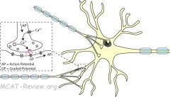
■Transmitter molecules = neurotransmitters
■Action potential → release of neurotransmitters by presynaptic axon terminal → picked up by receptor of postsynaptic neuron. ■Release of neurotransmitter = exocytosis of vesicles containing neurotransmitters. Triggered by calcium influx when action potential reaches axon terminal causes the calcium channels to open up. ■Neurotransmitter reception = diffusion of neurotransmitter across the synaptic cleft, binds to receptor, opens up ion channels that causes a change in membrane potential of the postsynaptic neuron (graded potential). If this graded potential is large enough, it will trigger a full-fledged, all-or-nothing action potential in the postsynaptic neuron. ■Neurotransmitters are quickly eliminated (destroyed by enzymes, reuptake by presynaptic terminal, or diffuse away) so that they don't persistently stimulate the postsynaptic neuron. |
|
|
|
What are examples of neurotransmitters?
|
■Acetylcholine (ACh)
■Norepinephrine (NE) ■Dopamine ■Serotonin ■Histamine ■ATP Acetylcholine neutrotransmitters are the main neurotransmitters of parasympathetic stimulation, while epinephrine and noephinephrine are the main neurotransmitters for sympathetic stimulation. Not only is acetylcholine the neurotransmitter of parasympathetic stimulation, it is the only only neurotransmitter used in the motor division of the somatic nervous system. In other words, acetylcholine must be released to move any voluntary muscle. |
|
|
|
What are synaptic knobs?
|
■Synaptic knob is another name for axon terminal.
■Contains vesicles of neurotransmitters waiting to be exocytosed. ■Action potential reaching the synaptic knob causes the calcium channels to open an influx of calcium in the neuron, which signals the vesicles to fuse with cell membrane to release the neurotransmitters into the synaptic cleft (exocytosis). The synapse is located at the end of each axonal end branch (axon terminal). Here the end branch forms a small button-like knob (axon terminal). This knob is adjacent to a tiny cleft or synapse. When a nerve impulse (action potential)reaches this knob, a molecule called a neurotransmitter is released from vesicles into the synapse. The neurotransmitter diffuses across the gap and binds to receptors on the membrane of the adjacent neuron or muscle cell (post-synaptic neuron). This will initiate an electrical change in the membrane of the adjacent neuron leading to a nerve impulse (action potential) in that cell. Neurons employ electrical signals (action potentials) to relay information from one part of the neuron to another. The neuron converts the electrical signal to a chemical signal in order to pass the information to another neuron. The target neuron then converts the message back to an electrical impulse to continue the process. |
|
|
|
What is fatigue in regards to synaptic activity?
|
■Continuous synaptic activity → depletion of neurotransmitters → fatigue.
Synaptic fatigue occurs when no neurotransmitter remains in presynaptic knobs. |
|
|
|
What is the propagation between cells without resistance loss in regards to synaptic activity?
|
■Action potential is all-or-nothing.
■As long as the neurotransmitters cause the postsynaptic cell to reach a certain threshold potential, the action potential induced is just as large as the presynaptic action potential. ■In summary, propagation between cells involves no resistance loss because the postsynaptic action potential is just as large as the presynaptic potential - all action potentials are all-or-nothing. |
|
|
|
What is the Resting potential (electrochemical gradient)?
|
◦Na+-K+ pump = 3 Na+ out, 2 K+ in = net positive on the outside.
◦Resting potential is -70 mV because the cell is more negative on the inside, and more positive on the outside. ◦Electrochemical gradient = combination of electrical and chemical gradient = both electrical potential and ion concentration gradient across membrane. An electrochemical gradient has two components. First, the electrical component is caused by a charge difference across the lipid membrane. Second, a chemical component is caused by a differential concentration of ions across the membrane. The combination of these two factors determines the thermodynamically favourable direction for an ion's movement across a membrane. |
|
|
|
What are the Stages of an action potential?
|
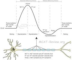
1.Resting: cell at rest, sodium-potassium pump maintaining negative resting potential (-70 mV). Lots of sodium outside, fewer of potassium inside (3 Na+ out, 2 K+ in). If the pump was to continue unchecked there would be no sodium or potassium ions left to pump, but there are also sodium and potassium ion channels in the membrane. These ion channels are normally closed, but even when closed, they “leak”, allowing sodium ions to leak in and potassium ions to leak out, down their respective concentration gradients. 2.Depolarization: sodium channels open, positive sodium rushes inside, membrane potential shoots up to +30 mV. Lots of sodium inside, lots of potassium inside, making the cell positive inside. 3.Repolarization: potassium channels open, sodium channels close, positive potassium rushes outside, membrane potential drops back down. Lots of sodium inside, lots of potassium outside (opposite of the resting state). 4.Hyperpolarization: potassium channels doesn't close fast enough, so the membrane potential actually drops below the resting potential for a bit. More potassium ions leave than they should and the sodium channels are still closed (preventing the cell from being more positive), so the inside of the cell is more negative than it needs to be. 5.Refractory period: the sodium-potassium pump works to re-establish the original resting state (more potassium inside, sodium outside). Until this is done, the neuron can't generate another action potential. Absolute refractory period = from depolarization to the cell having re-established the original resting state. Relative refractory period = After hyperpolarization till resting state re-established.
|
|
|
|
What are white muscle cells made of?
|
◦White muscle (fast fibers) = fast twitch, but fatigue easily.
■Anaerobic respiration (glycolysis) predominant. ■Few mitochondria because white muscles undergo mainly glycolysis. They are white because they do not need myoglobin for oxygen for krebs cycle - they use glucose for glycolysis. ■Equipped for short bursts of glycolysis: stores high amounts of glycogen. The fast twitch fibers rely on a well developed, short term, glycolytic system for energy transfer. Fast-twitch fibers allow one to generate a relatively high amount of force in a short period. It is worth noting here that fast-twitch fiber force production is no greater than slow-twitch fiber force production. The difference lies in the rate to which these fibers produce force. Fast fibers contain myosin with high ATPase activity and therefore have high shortening velocity. High shortening velocity means that the rate of the filaments sliding past one another is fast, so the muscle shortens quickly. Fatigue rapidly. Creatine (Cr) is one of the basic muscle energy stores, particularly in fast twitch glycolytic fibers. Creatine phosphate can anaerobically donate a phosphate group to ADP to form ATP during the first 2 to 7 seconds following an intense muscular or neuronal effort. |
|
|
|
What is the sodium–potassium pump for nerve cells?
|
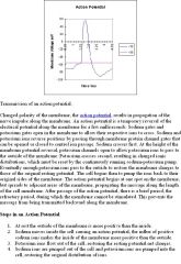
■3 sodium out.
■2 potassium in. ■net positive out. ■causes membrane to be more negative on the inside, hence negative membrane potential. Unequal pumping (3 Na+ out to 2K+ in) results in more positive charge on the outside compared to the inside. The membrane is polarized. The sodium-potassium pump mechanism is the same for all cells - 3 Na+ out and 2 K+ in. |
|
|
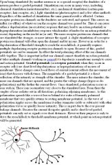
What is threshold, all-or-none in regards to action potentials?
|
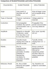
■When a stimulus (graded potential) depolarizes above a threshold value, an action potential will occur. Action potentials are all-or-none, meaning that if it occurs, all action potential have the same magnitude. One graded potential just barely makes the threshold value, another overshoots it a lot, but both will cause the same action potential. The all-or-none law is the principle that the strength by which a nerve or muscle fiber responds to a stimulus (the action potential) is not dependent on the strength of the stimulus (which correspond to the graded potentials). This is because the action potential will be the same strength as long as the stimulus reaches the threshold. If the strength of the stimulus is above the threshold, the nerve or muscle fiber will give a complete response or otherwise no response at all. The axon hillock is a specialized part of the cell body (or soma) of a neuron that connects to the axon. As a result, the axon hillock is the last site in the soma where membrane potentials propagated from synaptic inputs are summated before being transmitted to the axon. The axon hillock is located at the end of the soma and controls the firing of the neuron. If the total strength of the signal exceeds the threshold limit of the axon hillock, the structure will fire a signal down the axon. An action potential occurs in one direction because the recently depolarized area of the membrane is in absolute refractory period and cannot generate a potential. Propogation is the movement of the action potential in a wave and it increases with myelin sheaths.
|
|
|
|
What are Excitatory and inhibitory nerve fibers?
|
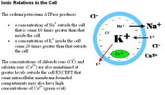
◦Excitatory = stimulates an action potential to occur. Excitatory synapse = receptor binding causes postsynaptic potential to be more positive (depolarization) = if it gets above threshold, action potential results. Inhibitory = inhibits an action potential from occuring. Inhibitory synapse = receptor binding causes postsynaptic potential to be more negative (hyperpolarization) = makes it more difficult to reach threshold. Certain external stimuli reduce the charge across the plasma membrane. Mechanical stimuli (e.g., stretching, sound waves) activate mechanically-gated sodium channels. Certain neurotransmitters (e.g., acetylcholine) open ligand-gated sodium channels. In each case, the facilitated diffusion of sodium into the cell reduces the resting potential at that spot on the cell creating an excitatory postsynaptic potential or EPSP. If the potential increased to the threshold voltage an action potential is generated in the cell. Despite their name, some neurotransmitters inhibit the transmission of nerve impulses. They do this by opening: 1)chloride channels and/or 2) potassium channels in the plasma membrane. In each case, opening of the channels increases the membrane potential by 1) letting negatively-charged chloride ions (Cl-) IN and 2) positively-charged potassium ions (K+) OUT. This hyperpolarization is called an inhibitory postsynaptic potential (IPSP). Although the threshold voltage of the cell is unchanged, it now requires a stronger excitatory stimulus to reach threshold.
|
|
|
|
What is summation and frequency of firing of nerve cells?
|
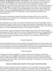
◦Summation = two or more nerves firing at the same time.
■Two subthreshold excitatory nerves firing at the same time can sum to reach the threshold. ■A threshold excitatory nerve and an inhibitory nerve firing at the same time, and the resultant signal won't reach the threshold. ◦Frequency = Firing, then quickly firing again. ■If the first fire is subthreshold, fire again before the previous depolarization dies, and the new depolarization will be even higher than the first time. |
|
|
|
Explain the Abundant mitochondria in red muscle cells.
|
•Abundant mitochondria in red muscle cells (ATP source)
◦Red muscle (slow fibers) = high endurance, but slow twitch. ■Aerobic respiration predominant. Red muscles are red because they are high in myoglobin and they use the oxygen of the myoglobin for krebs cycle. ■Many mitochondria because red muscles undergo aerobic respiration. ■Equipped to receive abundant oxygen supply: many capillaries, many myoglobin. ■High endurace, doesn't tire easily (fatigue-resistant). The slow twitch fibers generate energy for ATP re-synthesis by means of a long term system of aerobic energy transfer. They tend to have a low activity level of ATPase, a slower speed of contraction with a less well developed glycolytic capacity. Slow fibers contain myosin with low ATPase activity and therefore have low shortening velocity. Fatigue slowly. Low shortening velocity means that the filaments slide past one another slowly and and the muscle shortens slowly. They contain large and numerous mitochondria and with the high levels of myoglobin that gives them a red pigmentation they have been demonstrated to have high concentration of mitochondrial enzymes, thus they are fatigue resistant. These muscle cells use oxidative metabolism to generate ATP. |
|
|
|
What is the Organization of contractile elements (actin and myosin filaments, cross bridges)?
|
◦Actin filament = thin filament = has troponin and tropomyosin on it.
◦Myosin filament = thick filament = has myosin heads on it. ◦Cross bridge = myosin head binds to actin. A cross bridge is a distinctive structure consisting of two globular heads on each myosin molecule which interact in the presence of calcium ions with specific sites on a thin actin filament. The cross-bridges contain ATPase and generate the tension developed by a muscle fibre during a muscle action. |
|
|
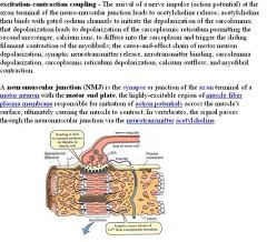
What are the sequence of biochemical events, starting with a nerve impulse, and ending with depolarization of the sarcolemma of a muscle fiber (i.e., excitation-contraction coupling). Name all the chemicals and cell parts involved.
|
Rigor mortis occurs because there is no ATP (because the person is dead) that binds to the myosin head to release it from the actin filament. Therefore, it stays in the contracted position.
|
|
|
What is the sliding filament model?
|
The sliding filament model describes a process used by muscles to contract. Each molecule of myosin in the thick filaments contains a globular subunit called the myosin head. The myosin heads have binding sites for 1)•the actin molecules in the thin filaments and 2) •ATP
Activation of the muscle fiber causes the myosin heads to bind to actin. An allosteric change occurs which draws the thin filament a short distance (~10 nm) past the thick filament. Then the linkage breaks (for which ATP is needed) and reform by the myosin binding to the actin farther along the actin filament to repeat the process. The linkage is the cross bridge between the myosin head and the actin filament. As a result, the filaments are pulled past each other in a ratchetlike action because multiple myosin heads bind to multiple actin filaments and pull the actin filaments toward the M line. The actin filaments are continually pulled in the presence of calcium until the muscle is maximally contracted. It doesn't relax until the calcium is gone. There is no shortening, thickening, or folding of the individual filaments, they just move past one another, specifically, the actin (thin filaments) moves past the myosin filaments and causes the sarcomere to shorten and the muscle cell to contract. The thick and thin filaments don't change in size, they just move closer and farther apart. They squeeze together and release. The myosin (thick) filament makes up the A zone. The A doesn't shorten decrease in width because it is the length of the myosin filament, but the A zones do move closer together to each other. As a muscle contracts, •the Z disks come closer together; •the width of the I bands decreases (because the thick filaments are getting closer); •the width of the H zones decreases (because the thin filaments are getting closer), but •there is no change in the width of the A band. Conversely, as a muscle is stretched, •the width of the I bands and H zones increases (because the thick and thin filaments are moving farther apart from each other), •but there is still no change in the width of the A band. The I band is different from the A band because the I band does not make up the length of the thin filament, if it did, then it would not shorten either. Instead, the I band area is the section of the thin filaments that are between the thick filaments. |
|
|
|
What is rigor mortis and what is troponin and tropomyosin used for?
|
◦Something counter-intuitive about the sliding filament model: ATP is not directly needed for the powerstroke. ATP binding is needed for detachment of myosin head to actin. ATP hydrolysis is needed for de-powerstroke (unbend myosin head).
◦Rigor mortis = no ATP after a person dies, myosin heads can't detach after power stroke, muscle remain in contracted position. ◦So what is troponin and tropomyosin there for? Ans: tropomyosin on actin blocks the myosin-binding sites on the actin filament, preventing the myosin heads on the thick filaments from forming cross bridges with the actin filaments. However, troponin moves tropomyosin out of the way at high Ca2+ levels (Ca2+ binds to troponin, and troponin moves tropomyosin). |
|
|
|
What are the the sequence of biochemical events in the skeletal muscle beginning with the depolarization of the T-tubules and ending with the start of the sliding filament mechanism, the "power stroke" of muscle contraction (i.e., sliding of actin past myosin). Name all the chemicals and cell parts involved.
|
The T tubules transmit the action potential to the sarcoplasmic reticulum (SR). A T-tubule (or transverse tubule) is a deep invagination of the sarcolemma, which is the plasma membrane, only found in skeletal and cardiac muscle cells. These invaginations allow depolarization of the membrane to quickly penetrate to the interior of the cell.
T-tubules are invaginations of the plasma membrane that are only found in striated muscle (skeletal and cardiac). Depolarization of the T-tubule membrane triggers the release of calcium from the sarcoplasmic reticulum and eventually muscle contraction. |
|
|
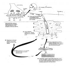
What is Calcium regulation of contraction and sarcoplasmic reticulum?
|
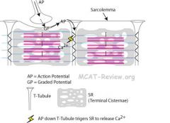
◦Sarcoplasmic reticulum (SR) = smooth ER in muscle = stores calcium, releases them when the muscle cell is stimulated. The SR is also called terminal cisternae where it meets T-tubules at the edges of the sarcomere. T-tubule = extension of the muscle cell membrane that runs deep into the cell, so that action potential can reach there. ◦Muscle contraction: 1.Nerve stimulates muscle. 2. Action potential runs along muscle cell membrane. 3.Goes deep into the muscle cell via T-tubules. 4.Stimulates the SR (terminal cisternae) to release calcium. 5.Calcium causes muscle to contract via the sliding filament mechanism.
Terminal cisternae are enlarged areas of the sarcoplasmic reticulum surrounding the transverse tubules. These discrete regions within the muscle cell store calcium (increasing the capacity of the sarcoplasmic reticulum to release calcium) and release it when an action potential courses down the transverse tubules, eliciting muscle contraction. The muscle cell obtains calcium from two sources: the extracellular space and the sarcoplasmic reticulum. |
|
|
What are the sequence of biochemical events in the "power stroke" of muscle contraction (i.e., sliding of actin past myosin). Name all the chemicals and cell parts involved.
|
The combination of the myosins binding to the actin and pulling it toward the M line results in the contraction, until it reaches a maximum contraction. A ratchet is a device that allows continuous motion in only one direction while preventing motion in the opposite direction.
|
|
|
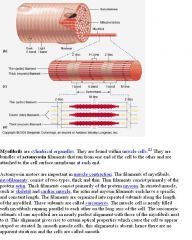
What are Sarcomeres?
|
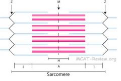
•Sarcomeres: "I" and "A" bands, "M" and "Z" lines, "H" zone. ◦I band = thinnest = thin filaments only = sides of the sarcomere. H zone = portion of the A band = center of the sarcomere, spans the M line. ◦A band = contains both thick and thin filaments, but it's the length of the thick filaments, center of the sarcomere and spans the H zone. ◦M line = line of myosin in the middle of the sarcomere, linked by M line proteins to the two Z lines. ◦Z line = line on the sides of the sarcomere, Z line proteins connect the filaments that span either side of the Z line. ◦Mnemonics :■I = thin like the letter I. ■M = middle line = myosin (linked by accessory proteins).
■Moving from middle to the side of sacromere = M HAIZ, the Muscle says HAIZ. Thin and thick filaments also contain accessory proteins. Proteins of the Z line serve as an embedding matrix or anchor for one end of the thin filaments, which extend toward the center of sarcomeres on either side of the Z line. Like Z line protein, the M line protein aggregate acts as an embedding matrix, in this case for the myosin thick filaments. Thick filaments extend from their point of attachment on both sides of the M line toward the two Z lines that define a sarcomere. The A band is the dark band and the I band is the light band. Mneumonic: Say "HI" to the muscle - both H bands and I bands contract during contraction (the A band does not change width in contraction or relaxation). The Z lines also move closer together during contraction. |
|
|
|
What are the sequence of biochemical events in the skeletal muscle beginning with the end of muscle cell/myofibril/sarcomere contraction and the restoration of the resting state of the skeletal muscle cell. Name all the chemicals and cell parts involved.
|
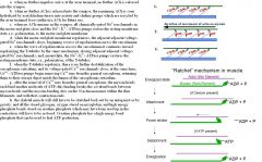
|
|
|
|
Explain the Presence of troponin and tropomyosin.
|
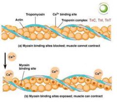
◦Tropomyosin = long protein that spirals along actin, blocks myosin- binding sites on the actin filament, preventing the myosin heads on the thick filaments from cross-linking with the actin filaments.
◦Troponin = binds to calcium when calcium is around, undergoes a conformational change and moves tropomyosin out of the way, enabling the myosin head of the thick filament to bind to actin. Actin, tropomyosin, and troponin are thin myofilaments. Myosin is a thick myofilament. |
|
|
What fuels muscle contraction?
|
|
|
|
|
What are Epithelial cells (cell types, simple epithelium, stratified epithelium)?
|
◦Squamous = flat.
◦Cuboidal = cube. ◦Columnar = column shaped. ◦Simple epithelium = single cell layer = good for absorption, secretion, filtration, diffusion. ■Simple squamous: endothelium, capillary wall, alveolar wall. ■Simple cuboidal: gland ducts, kidney tubules. ■Simple columnar: stomach and gut. ◦Stratified epithelium = two or more cell layers = good for protection against abrasion. ■Stratified squamous: skin. ■Stratified cuboidal/columnar: not common. Cells of the skin, the tympanic membrane, and the GI tract are routinely exposed to the external environment and are in constant need of division to replace the damaged cell population. These types of cells are called labile. Two other types exist: stable and permanent. Stable cells (like stem cells, pancreatic and liver cells) are normally arrested in G0 phase of the cell cycle but can re-enter cell division if stimulated or damaged. Permanent cells (i.e. nerve and cardiac cells) lose all mitotic activity during embryogenesis and are unable to divide even if damaged. |
|
|
|
What are Endothelial cells?
|
◦Endothelial cells = lines the inside of organs and blood vessels = they are simple squamous epithelium. The thin, single layer of endothelial cells facilitate diffusion.
|
|
|
What is the Connective tissue structure ?
|
◦Connective tissue structure = Cells + extracellular matrix.
■Cells: secrete the extracellular matrix (made of extracellular ground substance and extracellular fibers). ■Ground substance: glue that holds the matrix together. ■Fibers: mostly collagen, gives the matrix strength. Ground substance is a term for the non-cellular components of extracellular matrix containing the fibers. Most of the cells in multicellular organisms are surrounded by a complex mixture of nonliving material that makes up the extracellular matrix (ECM). Ground substance is the background material within which all other connective tissue elements are embedded. |
|
|
What are the types of connective tissue cells and tissues?
|
The major connective tissue types are: bone, cartilage, blood, and connective tissue proper. Connective tissue proper is divided into loose (areolar) tissue and dense tissue and includes fat, tendons, and ligaments. There are many more tissue Osteoblast cells make bone. Fibroblast cells secrete extracellular matrix. Chondroblast cells make cartilage. Hematopoietic stem cells make blood. Nomenclature: -blast = stem cell actively producing matrix. ■-cyte = mature cell, doing housekeeping. Fiber types: 1)Collagen = the most common fiber type. Very strong. Present in large amounts in dense connective tissue. 2)Elastic fibers = can stretch. 3)■Reticular fibers = can branch and form nets. Found in loose connective tissue. Tendons and ligaments are examples of dense connective tissue, while fat is a loose connective tissue. An easy way to remember tendons and ligaments is that they connect - they connect muscle to bone and bone to bone, respectively. Fibroblasts are the most common resident cells in ordinary connective tissue. Fibroblasts are responsible for secreting collagen and other elements of the extracellular matrix of connective tissue. Collagen is the most common protein in the body. As an essential structural element in the extracellular matrix of most connective tissues, including bone and cartilage, collagen confers toughness and tensile strength. Scars are made of collagen. Reticular fibers join connective tissues to other tissues and surround the liver and spleen. Elastic fibers are made of elastin and are stretchable. In ordinary connective tissue, elastic fibers help restore normal shape after distortion. Connective tissue proper may be distinguished as either loose or dense, depending on the proportion of the three types of fibers, but then there are also the blood, bone, and cartilage classes.
|
There are 2 types of bone: mature and immature. Woven bone is immature, mechanically weak, with a disorganized infrastructure of coarse collagen fibers. It is the first bone to form during development and in fracture repair. Lamellar bone is mature, strong, with layers of highly organized parallel collagen fibers. There are 2 types of lamellar bone: compact and spongy. Most fully developed bones have a rigid, outer, compact shell and an inner spongy zone. Compact bone is dense, closely-packed bony tissue. Spongy bone, also called cancellous or trabecular bone, is lighter than compact bone.
|
|
|
What is loose and dense connective tissue?
|
■Loose = loose fibers, lots of fluff (ground substance, cells) = anything that you don't associate with being fibrous = fat, paddings around organs.
■Dense = dense fibers predominantly collagen = genuinely fibrous, little fluff (ground substance, cells) = tendon, ligament. Dense connective tissue is so named because of high density of extracellular fibers, and relatively smaller proportions of ground substance and cells. Dense collagenous connective tissue is found wherever the tensile strength of collagen is of paramount importance. Examples include dermis (the layer of the skin which yields leather), tendons and ligaments, and organ sheaths (such as the sclera, or "white", of the eye). Dense elastic connective tissue is found wherever the elasticity of elastin is of paramount importance, as in the ligamentum flavum (flavum refers to the yellow color conferred by the elastin, connects adjacent verterbrae) and the aorta. Loose connective tissue has a relatively large proportion of ground substance, of cells, or of both cells and ground substance. In other words, loose connective tissue lacks the massive fibrous reinforcement that characterizes dense connective tissue. Nevertheless, the same types of fibers are still found, although fewer and more delicate. Loose connective tissue is easily distorted, permitting tissues on either side to move freely with respect to one another. However, when loose connective tissue is distorted sufficiently, it too becomes tough and resists further deformation. The intrinsic strength of collagen is the same in both loose and dense connective tissue. Adipose tissue converts excess glucose into fat. |
|
|
|
What is cartilage and extracellular matrix?
|
◦Cartilage = chondrocytes + matrix = elastic, flexible, used as padding in spinal discs, ends of bones, ear. Chondrocytes are mature cartilage cells. The ground substance of cartilage is characterized by a firm solid gel-like matrix (rather like very firm Jello®) reinforced by collagen and/or elastic fibers. Cartilage is relatively stiff and incompressible. However, since cartilage is not mineralized, it is more flexible and resilient than bone. ◦Extracellular matrix = secreted by cells = ground substance (glue) and fibers.
In biology, the extracellular matrix (ECM) is the extracellular part of animal tissue that usually provides structural support to the animal cells in addition to performing various other important functions. The extracellular matrix is the defining feature of connective tissue in animals. Extracellular matrix includes the interstitial matrix and the basement membrane. Interstitial matrix is present between various animal cells (i.e., in the intercellular spaces). Gels of polysaccharides and fibrous proteins fill the interstitial space and act as a compression buffer against the stress placed on the ECM. Basement membranes are sheet-like depositions of ECM on which various epithelial cells rest. Due to its diverse nature and composition, the ECM can serve many functions, such as providing support and anchorage for cells, segregating tissues from one another, and regulating intercellular communication. |
|
|
|
What is the Function of endocrine system?
|
The function of endocrine system is specific chemical control at cell, tissue, and organ level.
◦Endocrine system = make hormones = specific control of all target cells of that hormone. The anterior pituitary gland is responsible for the production of peptide hormones only (i.e. GH, FSH, LH, Prolactin, etc) and does not carry out steroid or fatty acid synthesis. The adrenal cortex (produces cortisol and aldosterone) and the liver (low density lipo-protein or LDL synthesis) are involved in cholesterol-based lipid synthesis. A lactating mammary gland is constantly active with milk production. Lipids make up a high percentage of milk. |
|
|
|
What are the Definitions of endocrine gland and hormone?
|
◦endo = within, crine = to secrete
◦endocrine glands secrete hormones directly into the bloodstream and the hormones are carried by blood and tissue fluids to the cells they act upon. Endocrine, autocrine, and paracrine are the 3 different kinds of chemical signalling. Exocrine is not a form of chemical signalling. ■endocrine: hormone, no duct, acts long distances ■exocrine: non-hormone secretions into ducts. Exocrine glands are glands that secrete their products (excluding hormones and other chemical messengers) into ducts (duct glands) which lead directly into the external environment. Products could be sweat, proteins, mucus, etc. ■autocrine: local chemicals, act short distances on themselves. Autocrine signaling is a form of signaling in which a cell secretes a hormone or chemical messenger that binds to autocrine receptors on the same cell, leading to changes in the cells. ■paracrine: local chemicals, act short distances on other cells. Paracrine signaling is a form of cell signaling in which the target cell is near ("para" = near) the signal-releasing cell. ◦hormone = chemicals that regulate metabolism and function of cells. The endocrine system is an information signal system like the nervous system. Hormones regulate many functions of an organism, including mood, growth and development, tissue function, and metabolism. |
|
|
|
What are the Major endocrine glands (names, locations, products)?
|
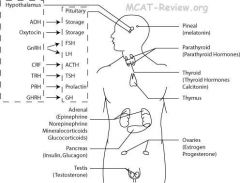
|
|
|
|
What does the Hypothalamus do?
|
◦Hypothalamus: Synthesizes and secretes releasing hormones that in turn stimulate or inhibit the secretion of the hormones in the anterior pituitary. ADH and oxytocin are two other hypothalamic hormones that travel to the posterior pituitary and are stored until they are released into circulation. The hypothalamus links the nervous system to the endocrine system via the pituitary gland ■The hypothalamus has releasing hormones/factors stimulates pituitary to release its hormone. ■GnRH = Gonadotropin Releaing Hormone = stimulates pituitary to release FSH and LH. FSH = Follicle Stimulating Hormone = Stimulate ovary follicles to mature, testis to produce sperm. LH = Luteinizing Hormone = LH surge triggers ovulation, stimulates testis to produce testosterone. ■CRF = Corticotropin Releasing Factor, also known as CRH - corticotropin releasing hormone (CRH). CRH stimulates the release of ACTH (adrenocorticotropic hormone, also known as corticotropin) which is a hormone involved in the stress response. ■TRH = Thyroid Releasing Hormone. TRH stimulates the release of thyroid-stimulating hormone. The main function of TRH is to stimulate TSH release, although TRH can also cause prolactin secretion. ■PRH = Prolactin Releasing Hormone. PRH stimulates the release of prolactin which stimulates breast to produce milk. ■GHRH = Growth Hormone Releasing Hormone. It's a releasing hormone for growth hormone to stimulate growth. ■ADH = Antidiuretic Hormone = Vasopressin = increase water reabsorption in kidney. This keeps water in the body and increases blood pressure. ■Oxytocin = stimulates uterine contractions during labor, also milk secretion during suckling. One of the major functions of the hypothalamus is it's function in homeostasis.
|
|
|
|
What does the anterior pituitary do?
|
◦Anterior Pituitary: produces and secretes FLAT PiG. Posterior pituitary stores and releases ADH and oxytocin.
■FSH = Follicle Stimulating Hormone = Stimulate ovary follicles to mature, testis to produce sperm. ■LH = Luteinizing Hormone = LH surge triggers ovulation, stimulates testis to produce testosterone. ■ACTH = AdrenoCorticoTropic Hormone = Stimulates adrenal cortex to stimulate the release cortisol from the adrenal cortex. ACTH is often produced in response to biological stress (along with corticotropin-releasing hormone from the hypothalamus). Cortisol is a glucocorticoid (it increases the blood glucose levels). ■TSH = Thyroid Stimulation Hormone = Stimulate thyroid to release thyroid hormones. Thyroid hormones are primarily responsible for the regulation of metabolism. There is no releasing or stimulating hormone from the hypothalamus for calcitonin secretion. Elevated blood calcium levels stimulate calcitonin secretion. ■PRL, also know as Prolactin = Stimulates breast to produce milk. Prolactin is unique among pituitary hormones in that its secretion is spontaneous in the absence of any stimulation from the hypothalamus. ■ignore ■GH = Growth Hormone = Stimulates growth of muscle and bone and increases fat consumption. Fat cells (adipocytes), for example, have growth hormone receptors, and growth hormone stimulates them to break down triglyceride and supresses their ability to take up and accumulate circulating lipids. |
|
|
|
What does the pineal gland do?
|
◦Pineal: makes melatonin, which makes you sleepy at night. Melatonin, a hormone that affects the modulation of wake/sleep patterns.
|
|
|
|
What does the thyroid and parathyroid gland do?
|
The thyroid produces thyroid hormones and calcitonin. ■Thyroid hormones: increase metabolism, requires iodine. The thyroid is composed of spherical follicles that selectively absorb iodine (as iodide ions, I-) from the blood for production of thyroid hormones. Twenty-five percent of all the body's iodide ions are in the thyroid gland. ■Calcitonin: turns blood Ca2+ into bone. It inhibits osteoclast activity in bones. Bone resorption is the process by which osteoclasts break down bone and release the minerals, resulting in a transfer of calcium from bone fluid to the blood. Lowers blood Ca2+. It acts to reduce blood calcium (Ca2+), opposing the effects of parathyroid hormone (PTH). Elevated blood calcium levels stimulate calcitonin secretion. Calcitonin also inhibits renal tubular reabsorption of calcium and phosphorus, causing them to be lost in urine. ◦Parathyroid: makes Parathyroid Hormone (PTH), which increases blood Ca2+ by bone resorption, dietary calcium absorption, and calcium reabsorption in kidneys. PTH acts to increase the concentration of calcium (Ca2+) in the blood, whereas calcitonin acts to decrease calcium concentration. It enhances the release of calcium from the large reservoir contained in the bones. Bone resorption is the normal destruction of bone by osteoclasts, which are indirectly stimulated by PTH. Thyroid-stimulating hormone is the chief stimulator of the thyroid gland and thyroid hormone synthesis. TSH is produced by the anterior pituitary and enhances all components needed for thyroid hormone synthesis, like the iodide transporter, thyroid peroxidase, and thyroglobulin. PTH is a peptide hormone, so works through the second messenger system (like other water-soluble hormones).
|
|
|
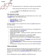
What does the adrenal gland do?
|
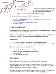
The adrenal gland releases epinephrine, noepinephine, mineralocorticoids, and glucocorticoids. ■Epinephrine and norepinephrine = fight or flight response. Norepinephrine and epinephrine are catecholamines and are stress hormones: they engage the fight-or-flight response, directly increasing heart rate, triggering the release of glucose from energy stores, and increasing blood flow to skeletal muscle. They are produced in the adrenal medulla. "AMEN" - the adrenal medulla produces epinephrine and norephinephrine.
■Mineralocorticoids = aldosterone = increase Na+ and water retention, raises blood pressure. Aldosterone acts on the kidneys to provide active reabsorption of sodium and an associated passive reabsorption of water. This in turn results in an increase of blood pressure and blood volume.The name mineralocorticoid derives from early observations that these hormones were involved in the retention of sodium, a mineral. ■Glucocorticoids = cortisol = stress hormone = increase blood sugar. The name glucocorticoid (glucose + cortex + steroid) derives from their role in the regulation of the metabolism of glucose, their synthesis in the adrenal cortex, and their steroidal structure. Cortisol (or hydrocortisone) is the most important human glucocorticoid. It is released in response to stress, or to a low level of blood glucocorticoids. Glucocorticoids also stimulate gluconeogenesis in the liver. Glucocorticoids are distinguished from mineralocorticoids by their specific receptors, target cells, and effects. Mineralocorticoids and glucocorticoids are produced in the adrenal cortex. Adrenal glands are located on top of the kidney. |
|
|
|
What does the thymus do?
|
◦Thymus: Thymus hormones (thymo-, thymic), stimulates T cells to develop.
|
|
|
|
What do the ovary and testis do?
|
◦Ovary: make estrogen.
◦testis: make testosterone. Luteinizing hormone (LH) is released by the anterior pituitary and CAUSES OVULATION. The release of LH is stimulated by an increase in estrogen. Progesterone is secreted by the ovary and the corpus luteum and maintains the pregnancy by inhibiting the loss of the endometrial lining. FSH is also produced by the anterior pituitary and stimulates the MATURATION of the OVA. FSH, estrogen, and progesterone do not cause ovulation. Estrogen and progresterone do not bind to LH receptors. LH binds to LH receptors. |
|
|
|
What does the pancreas do?
|
◦The pancreas releases glucagon and insulin.
■Glucagon = increases blood sugar (break down glycogen- called glycogenolysis, and it stimulates gluconeogenesis - synthesis of glucose from non-carbohydrates). Its effect is opposite that of insulin, which lowers blood glucose levels. Glucagon causes the liver to convert stored glycogen into glucose, which is released into the bloodstream. Gluconeogenesis results in the generation of glucose from non-carbohydrate carbon substrates and occurs mainly in liver. ■Insulin = lower blood sugar (stimulates glucose uptake by cells). Insulin is a hormone that is central to regulating the energy and glucose metabolism in the body. Insulin causes cells in the liver, muscle, fat, and several other tissues to take up glucose from the blood, storing it as glycogen in the liver and muscle. The only mechanism by which cells can take up glucose is by facilitated diffusion through a family of glucose transporters. Insulin stops the use of fat as an energy source. When insulin is absent, glucose is not taken up by body cells and the body begins to use fat as an energy source (fat metabolism). Insulin and glucagon, both produced by the pancreas, are peptide hormones. Insulin is synthesized by beta-cells, which are found in the pancreas. Somatostatin inhibits the release of both glucagon and insulin. |
|
|
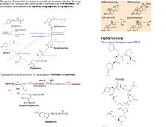
What are the Major types of hormones?
|
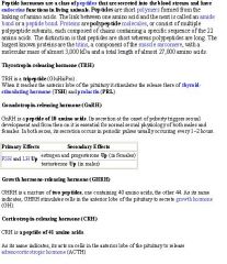
Most hormones are amino acid based (amino acid derivates and peptides). A few are steroids (made from cholesterol, only gonads and adrenal cortex produce these).
Hormones can be divided into three groups based on chemical structure: 1) amino acid derivatives (epinephrine, norepinephrine, thyroid hormones, and the pineal hormone called nelatonin) 2) peptide hormones (the largest class of hormones – all hormones secreted by the hypothalamus, anterior and posterior pituitary, heart, kidneys, thymus, digestive tract, and pancreas. Peptide hormones consist of amino acid chains ranging from the short chains of ADH and oxytocin to larger ones of GH and prolactin). 3) lipid derivatives covers two groups: a) steroid hormones (derived from cholesterol). Steroid hormones are released by the reproductive organs and the adrenal glands and are structurally similar to cholesterol. b) fatty acid-based hormones (includes prostaglandins). On average, hormone concentration ranges from 1 picogram to values no greater than a few micrograms/ml. These are tiny numbers. So tiny, in fact, that some hormones cannot even be detected in the blood. The rates of hormone production are correspondingly low, ranging from as little as a few micrograms per day to at most a few milligrams per day. So on average, endocrine hormone serum concentration is low and storage concentration is low. |
Remember that protein hormones DO NOT diffuse across the plasma membrane and thus if they are located on the outside of a plasma membrane, their initiate a cyclase inside the membrane (cAMP and IP3) and if they are located on the inside of the plasma membrane, they can't recognize hormones on the outside of the membrane and can't affect the outer surface.
|
|
|
What are lipids?
|
Along with proteins, nucleic acids, and carbohydrates, lipids are one of the major classes of biologically important molecules (or biomolecules). They are water-insoluble, organic compounds that are highly soluble in nonpolar organic solvents. Although the term lipid is often used informally as a synonym for fat, the latter refers to a subgroup of lipids called triglycerides. Unlike other groups of molecules, lipids comprise a broad and diverse range of structures, which also include phospholipids (components of cell membranes), sterols (most notably cholesterol, and the steroid hormones), and more complex lipid derivatives such as glycolipids (sugar-linked lipids). Cholesterol formed from squalene, a triterpene. Cholesterol is a sterol lipid (a combination steroid and alcohol) It is found in the cell membranes of all human body tissues, and transported in the blood plasma of all animals.Cholesterol is an important component of cell membranes, which enhances their fluidity. Cholesterol also aids in the manufacture of bile (which helps digest fats) and is also important for the metabolism of fat-soluble vitamins. Cholesterol is an important precursor of the steroid hormones. Steroid hormones produce their physiological effects by binding to steroid hormone receptor proteins, which causes changes in gene transcription and cell function. The five major classes of steroids are androgens, mineralocorticoids, glucocorticoids (all produced by adrenal cortex) and estrogen and progestagen (produces progesterone). The basic classes of lipids are as follows: fatty acids, glycerides, Nonglycerides (like cholesterol and the steroid hormones) and More complex lipid derivatives (like glycolipids).
|
|
|
|
What are lipid soluble hormones?
|
◦lipid-soluble hormones
■Able to cross the plasma membrane. ■Directly activate genes. Lipid-soluble hormones (steroid hormones and hormones of the thyroid gland) diffuse through the cell membranes of target cells. The lipid-soluble hormone then binds to a receptor protein that, in turn, activates a DNA segment that turns on specific genes. The proteins produced as result of the transcription of the genes and subsequent translation of mRNA act as enzymes that regulate specific physiological cell activity. Lipid-soluble hormones have a slow onset, but a long duration of action, while water-soluble hormones have a fast onset, but a short duration of action. |
|
|
|
What are water soluble hormones?
|
◦water soluble hormones can't cross the plasma membrane. They Bind to membrane receptors on the outside of cells. Secondary messengers then relay the signal inside the cell. Water-soluble hormones (the amino acid derivatives - polypeptide, protein, and most amino acid hormones) bind to a receptor protein on the plasma membrane of the cell. The receptor protein, in turn, stimulates the production of one of the following second messengers: 1) Cyclic AMP (cAMP) is produced when the receptor protein activates another membrane-bound protein called a G protein. The G protein activates adenylate cyclase, the enzyme that catalyzes the production of cAMP from ATP. Cyclic AMP then triggers an enzyme that generates specific cellular changes. 2) Inositol triphosphate (IP3) is produced from membrane phospholipids. IP3, in turn, triggers the release of Ca2+ from the endoplasmic reticulum, which then activates enzymes that generate cellular changes.
|
|
|
|
What is the cAMP pathway?
|
Water soluble hormones binds to a receptor and which can stimulate the production of one of the following second messengers : cAMP or Inositol triphosphate (IP3).
◦cAMP pathway: 1.Amino acid hormone binds membrane receptor. 2.G protein activated. 3. Adenylate cyclase activated. 4. Adenylate cyclase converts ATP into cAMP 5.cAMP made. 6.Protein kinase cascade. Adenylate cyclase is attached to the inner layer of the phospholipid bilayer; it is NOT located in the cytoplasm. cAMP is responsible for carrying the chemical stimulus into the cytoplasm and triggering a response, and so is called a second messenger. A protein kinase is a kinase enzyme that modifies other proteins by chemically adding phosphate groups to them (phosphorylation). The cAMP-dependent pathway, also known as the adenylate cyclase pathway, is a G protein-coupled receptor-triggered signaling cascade used in cell communication.G protein-coupled receptors (GPCRs) are a large family of integral membrane proteins that respond to a variety of extracellular stimuli. Each GPCR binds to and is activated by a specific ligand stimulus. When a GPCR is activated by its extracellular ligand, a conformational change is induced in the receptor that is transmitted to an attached intracellular G protein. The G protein binds to and activates an enzyme called adenylate cyclase, which, in turn, catalyzes the conversion of ATP into cyclic adenosine monophosphate (cAMP). The protein kinase enzyme only gets activated if cAMP is around. |
|
|
|
What are the functions of protein kinase and cAMP pathway?
|
Protein kinase A has several functions in the cell, including regulation of glycogen, sugar, and lipid metabolism. cAMP-dependent pathway is necessary for many living organisms and life processes. Many different cell responses are mediated by cAMP. These include increase in heart rate, cortisol secretion, and breakdown of glycogen and fat. This pathway can activate enzymes and regulate gene expression. The activation of preexisting enzymes is a much faster process, whereas regulation of gene expression is much longer and can take up to hours. The cAMP pathway is studied through loss of function (inhibition) and gain of function (increase) of cAMP. If cAMP-dependent pathway is not controlled, it can ultimately lead to hyper-proliferation, which may contribute to the development and/or progression of cancer.
|
|
|
|
What is the Phospholipid pathway?
|
Water soluble hormones bind to recepter proteins and stimulate the release of either cAMP or phospholipid second messengers. Inositol triphosphate (IP3) is produced from membrane phospholipids.
1.Amino acid hormone binds membrane receptor. 2.G protein activated and activates phospholipase C. 3.Phospholipase C activated and cleaves the membrane phospholipid. 4.Membrane phospholipid split into DAG and IP3. 5.DAG triggers protein kinase cascade. 6.IP3 releases Ca2+ from the ER. Phospolipase C performs a catalytic mechanism, generating inositol triphosphate (IP3) and diacylglycerol (DAG). These molecules then go on to modulate the activity of downstream proteins important for cellular signaling. IP3 is soluble, and diffuses through the cytoplasm and interacts with IP3 receptors on the endoplasmic reticulum, causing the release of calcium and raising the level of intracellular calcium. DAG remains tethered to the inner leaflet of the plasma membrane due to its hydrophobic character, where it recruits protein kinase C (PKC), which becomes activated in conjunction with binding calcium ions. This results in a host of cellular responses through stimulation of calcium-sensitive proteins. |
|
|
|
What is the mechanism of steroid hormone action?
|
This is the pathway of lipid soluble hormones. Lipid-soluble hormones are steroid hormones and hormones of the thyroid gland, even though thyroid hormones are amino acid based. The thyroid hormones are tyrosine-based hormones produced by the thyroid gland primarily responsible for regulation of metabolism. Tyrosine is one of the 20 amino acids that are used by cells to synthesize proteins. 1.Steroid hormones and thyroid hormones goes inside cell. 2.Goes into the nucleus. 3.Hormone binds receptor inside the nucleus. 4.Hormone-receptor complex turns certain genes on. 5. The proteins produced as result of the transcription of the genes and subsequent translation of mRNA act as enzymes that regulate specific physiological cell activity. Both the steroid and thyroid hormone-receptor complexes exert their action by binding to specific nucleotide sequences in the DNA of responsive genes. These DNA sequences are identified as hormone response elements, HREs. The interaction of steroid-receptor complexes with DNA leads to altered rates of transcription of the associated genes. Being lipids, steroid hormones enter the cell by simple diffusion across the plasma membrane. Thyroid hormones enter the cell by facilitated diffusion. Steroid and thyroid hormone receptors are found in the nucleus, cytoplasm, and surrounding plasma membrane. In the nucleus, the classic steroid and thyroid receptors mediate gene actions of steroid or thyroid hormones by acting as transcription factors that start gene transcription. Hormones binding to membrane receptors outside the cell send molecular cues through the membrane to activate messenger molecules - called intracellular second messengers - inside the cell. Receptors in the cell’s liquid cytoplasm can control both genomic or non-genomic hormone actions. Inactive (nonhormone-bound) steroid receptors in the cytoplasm may bind a ligand (any molecule that binds to a protein or other molecule), form a hormone-receptor complex, and move to the nucleus to trigger gene transcription. In this case, cytoplasmic receptors mediate genomic actions of the hormone. Alternatively, the hormone-receptor complex formed after hormones bind and activate inactive receptors in the cytoplasm can interact with other signaling molecules, such as cAMP, in the cell. In this case, cytoplasmic receptors are mediating non-genomic, rapid actions of steroid or thyroid hormones.
|
|
|
|
How are hormones transported?
|
•Transport of hormones (bloodstream): hormones travel to all parts of the body via blood and lymph. Although a few hormones circulate simply dissolved in the blood, most are carried in the blood bound to plasma proteins. For example, all the steroid hormones, being highly hydrophobic, are transported bound to plasma proteins. Albumin is a relatively non-selective transport protein found in all vertebrates. As the most abundant plasma protein, albumin's blood concentration often far exceeds even the flavonoids that flood the bloodstream after fruits and vegetables are eaten. Although albumin is not strongly attracted to particular molecules, there is so much of it in the blood that it simply mops up and shuttles stray fat soluble molecules. Albumin transports hormones and other compounds through blood, but it does not help those compounds to cross the plasma membrane - albumin is a big plasma protein and is too large to enter the intracellular space. That's why albumin prevents proteins bound to it from filtering out in the kidneys - it is too large to be filtered out in kidneys, so the proteins bound to it also don't get filtered out.
|
|
|
|
Explain the Specificity of hormones.
|
•Specificity of hormones: hormones target tissue
◦Specificity depends on the target cells having the receptors for the hormone, and non-target cells lacking receptors for the hormone. ◦Cells can either upregulate or downregulate the receptors they express. Downregulation is the process by which a cell decreases the quantity of a cellular component, such as RNA or protein, in response to an external variable. An increase of a cellular component is called upregulation. Hormone effect also depends on the concentration of the hormone in the cell, the number of receptors on the cell, and the affinity of the receptor for the hormone. Amines or derivatives of the amino acid tyrosine are a group of endocrine hormones that include: 1) thyroid hormone (synthesized by the thyroid gland) and 2 epinephrine and norepinephrine (secreted by the adrenal medulla). Epinephrine and noepinephrine are also called catecholamines. Oxytocin is a peptide hormone produced by the hypothalamus. Aldosterone is a cholesterol-derived steroid that is synthesized by the adrenal cortex. Acetylcholine is a neurotransmitter utilized by the parasympathetic nervous system and somatic nervous system. |
|
|
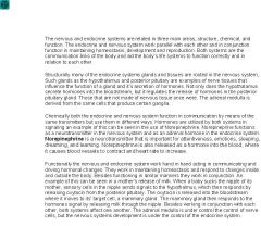
Explain the Integration of the endocrine system with nervous system.
|
The endocrine system functions in short-term and long-term control and works in conjunction with the nervous system, which mostly deals with short-term control, in regulating internal functions and maintaining homeostasis via feedback control. ◦The nervous system can modulate and overide normal control of hormones based on the status of the body. For example, the body's blood "normal" glucose level is set higher when you're under stress. ◦Hormones can modulate the nervous system. For example, low estrogen levels during menses give you a bad mood. ◦Normal control of hormones examples: 1) ■Humoral: glands directly respond to chemical levels in the blood (for example, when the parathyroid responds to low blood calcium). 2) ■Neural: glands release hormones when stimulated by nerves (for example, when under stress the sympathetic nervous system employs the fight or flight response). 3) ■Hormonal: glands release hormones when stimulated by other hormones (tropic hormones). Humoral: Pertaining to elements in the blood or other body fluids. Feedback control is the basic mechanism by which systems, whether mechanical, electrical, or biological, maintain their equilibrium or homeostasis. Tropic hormones are hormones produced and secreted by the anterior pituitary that target endocrine glands. Tropic hormones include TSH, ACTH, LH and FSH. The hypothalamus controls the release of tropic hormones by secreting a class of hypothalamic neurohormones called releasing hormones which are released and act on the anterior pituitary. The endocrine system is slower than the nervous system because hormones must travel through the circulatory system to reach their target.
|
|
|
|
What are the major functions of the nervous system?
|
1) high-level control and integration of body systems 2) response to external influences 3) sensory input 4) ◦integrative and cognitive abilities
■sensory = afferent ■nerve impulses conveyed to the CNS. 5) motor output ■motor = efferent ■nerve impulses from the CNS to effector organs. Effector organ is an organ that produces an effect in response to nerve stimulation. At a more integrative level, the primary function of the nervous system is to control the body. It does this by extracting information from the environment using sensory receptors, sending signals that encode this information into the central nervous system, processing the information to determine an appropriate response, and sending output signals to muscles or glands to activate the response. The evolution of a complex nervous system has made it possible for various animal species to have advanced perception abilities such as vision, complex social interactions, rapid coordination of organ systems, and integrated processing of concurrent signals. In humans, the sophistication of the nervous system makes it possible to have language, abstract representation of concepts, transmission of culture, and many other features of human society that would not exist without the human brain. |
|
|
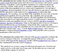
What is the Organization of vertebrate nervous system?
|
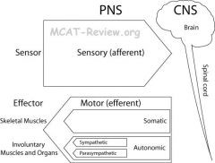
◦CNS = Central Nervous System = Brain and spinal cord
■Brain ■Spinal Cord ◦PNS = Peripheral Nervous System = Everything else ■Sensory = Afferent = Nerves carrying signal toward CNS. ■Motor = Efferent = Nerves carry signal toward effector organs. ■Somatic Nervous System = Voluntary = Controls skeletal muscles. ■Autonomic Nervous System = Involuntary = Effects visceral organs. Visceral organs are the internal organs. ■Sympathetic division = fight or flight response. ■Parasympathetic division = Rest. Both afferent and efferent come from French, evolved from Latin (the basis of many terms in medicine and biology) for the terms, respectively, ad ferens (Latin verb ferre: carry), meaning carrying into, and ex ferens, meaning carrying away. Ad and ex give an easy mnemonic device for remembering the relationship between afferent and efferent: afferent connection arrives and an efferent connection exits. |
|
|
|
What are Sensor and effector neurons?
|
◦Sensor = senses, carries sensory signals from the body to the CNS.
◦Effector = causes an effect = carries motor signals from the CNS to the body. |
|
|
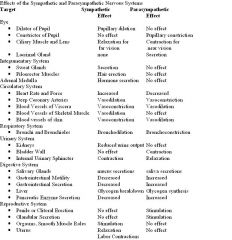
What are the Sympathetic and parasympathetic nervous systems?
|
◦Sympathetic = prepares body for activity = fight or flight response. It causes:
■Increase heart rate, blood pressure, thereby providing a mechanism for the enhanced blood flow to skeletal muscles. ■More blood flow to muscles, less to digestive system. The bronchi of the lungs dilates (relaxes) to let in more air and opening of the trachea. ■Pupil dilation - it's an effect of sympathetic nervous system to see farther distances. ■The liver to break down glycogen to release glucose into blood. ◦Parasympathetic = prepares body to rest. It causes: ■Decrease heart rate, blood pressure. ■Less blood to muscles, more to digestive system. ■Pupil constriction - an effect of parasympathetic nervous system to see closer distances. ■Promotes the synthesizes glycogen for storage from glucose. They are antagonistic to each other. It should be noted that the autonomic nervous system is always working. It is NOT only active during "fight or flight" or "rest and digest" situations. Rather, the autonomic nervous system acts to maintain normal internal functions and works with the somatic nervous system. Pupil dilation occurs when signals from sympathetic fibers cause the iris dilator muscles to contract. The vagus nerve is one of the main nerves of the parasympathetic nervous system. |
|
|
|
What is feedback loop?
|
■Feedback loop = positive feedback (reinforce initial event), negative feedback (counteracts initial event), or reflex arc (usually a type of negative feedback).
■positive feedback = uterine contraction lead to oxytocin release, which causes more uterine contraction. ■positive feedback = blood clotting platelets activated at wound site attract more platelet activation and clumping. ■negative feedback = drop in blood pressure causes ADH release, which increases the blood pressure. Conversely increase in blood pressure causes a drop in ADH. ■Reflex arc = withdrawal from a painful stimulus = negative feedback. ■Reflex arc = knee jerk = tapping the knee tenden causes sudden stretching of the muscle, which lead to contraction of that muscle that creates the knee jerk = negative feedback. A reflex arc is the neural pathway that mediates a reflex action. Pain is perceived in the central nervous system. Neurons in the central nervous system (sensory neurons), would send the pain signals to the central nervous system (brain and spinal cord) to perceive the pain. It is the afferent pain-perception pathway involved in feeling pain. Without the perception of pain, if a toe was stubbed, the toe would retract (from the sensory neuron synapsing in the CNS with the motor neurons that send signals for the muscle to retract), but the feeling of pain would be absent. Pain is a perceptive feeling from the brain, different from the muscle involvement in reflexes. |
|
|
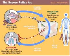
What is the reflex arc?
|
■Reflex arc = receptor → sensory neuron → integration center (the spinal cord where the association neuron is)→ motor neuron → effector
■receptor = site of stimulus ■sensory neuron = carries impulse from receptor to integration center ■integration center = connects sensory to motor neuron via synapse inside the CNS ■monosynaptic = no interneuron, direct synapse of sensory to motor. When a reflex arc consists of only two neurons in an animal (one sensory neuron, and one motor neuron), it is defined as monosynaptic. Monosynaptic refers to the presence of a single chemical synapse. ■polysynaptic = interneuron(s) present. By contrast, in polysynaptic reflex pathways, one or more interneurons connect afferent (sensory) and efferent (motor) signals. All but the most simple reflexes are polysynaptic, allowing processing or inhibition of polysynaptic reflexes within the spinal cord. ■motor neuron = carries impulse toward effector. ■effector = site of response to the stimulus A reflex arc is the neural pathway that mediates a reflex action. In higher animals, most sensory neurons do not pass directly into the brain, but synapse in the spinal cord. This characteristic allows reflex actions to occur relatively quickly by activating spinal motor neurons without the delay of routing signals through the brain, although the brain will receive sensory input while the reflex action occurs. There are two types of reflex arc - autonomic reflex arc (affecting inner organs) and somatic reflex arc (affecting muscles). |
|
|
|
What are examples of reflexes and its effects on flexor and extensor muscles?
|
■Examples of reflexes: knee-jerk, withdrawal from pain
■Effects on flexor and extensor muscles ■During the knee-jerk, in addition to contracting the extensor, the reflex relaxes the flexor. ■Golgi tendon reflex: sudden contraction of the quads (extensor), causes a negative feedback that relaxes the quads and contracts the hamstrings (flexor). Flexor muscle is a muscle which bends a joint. Extensor muscle is a muscle which straightens a joint. Hamstrings - flexor muscles on the back of the thigh. Quadriceps femoris-the main extensor muscle on the front of the femur (thigh bone). The flexor and extensor reflexes are two examples of the sequential ordering of muscular contraction and relaxation. Underlying this basic organization is the principle of reciprocal innervation—the contraction of one muscle or group of muscles with the relaxation of muscles that have the opposite function. |
|
|
|
What is the roles of spinal cord and brain on the reflexes?
|
■Spinal cord provides the synapse (or synapses if it's polysynaptic) for the reflex arc.
■Even though the reflex arc bypasses the brain, the brain is still aware of it happening. A reflex arc is the neural pathway that mediates a reflex action. In higher animals, most sensory neurons do not pass directly into the brain, but synapse in the spinal cord. This characteristic allows reflex actions to occur relatively quickly by activating spinal motor neurons without the delay of routing signals through the brain, although the brain will receive sensory input while the reflex action occurs. The spinal cord functions primarily in the transmission of neural signals between the brain and the rest of the body but also contains neural circuits that can independently control numerous reflexes and central pattern generators. The spinal cord has three major functions: A. Serve as a conduit for motor information, which travels down the spinal cord. B. Serve as a conduit for sensory information, which travels up the spinal cord. C. Serve as a center for coordinating certain reflexes. The spinal cord is the integration center for reflexes. |
|
|
|
What is efferent control in regards to reflexes?
|
■Brain can overide spinal reflexes (eg. you don't jerk away from getting a vaccine shot)
As a charactistic of all spinal reflexes, the brain can modify the reflex by sending impulses down pathways to the efferent motor neurons supplying the involved muscles to override the input from the receptors of the muscle, preventing the muscle from contracting in response to, for example, a painful stimulus. When our hand is pricked to obtain a blood sample, our pain receptors are stimulated, initiating the withdrawal reflex. However, our brain knows that we must be brave and not pull away, so the brain consciously overrides the reflex. The cerebellum is responsible for motor coordination and balance. The medulla oblongtata of the brain is needed to control respiration and heart rates. The hypothalamus synthesizes hormones (synthesizes ADH and oxytocin, which get stored in the posterior pituitary) and controls hormone release from the anterior pituitary (by synthesizing and secreting hormone-releasing factors). The cerebrum (cerebral cortex) is responsible for higher thought and memory. The hypothalamus holds such factors as blood pressure, body temperature, fluid and electrolyte balance, and body weight at a precise "set-point" for homeostasis. |
|
|
|
What are proprioceptive and somatic sensors in the skin?
|
Somatic (general) senses collect information about cutaneous sensations (tactile sensations on the surface of the skin). They involve touch, heat and pain receptors close to the surface (dermis-epidermis boundary) and pressure receptors deeper in the dermis. Proprioceptive sensors: sense the position of a body part, located in muscle and connective tissue. Proprioception means "sense of self". In the limbs, the proprioceptors are sensors that provide information about joint angle, muscle length, and tension, which is integrated to give information about the position of the limb in space.
Some somatic sensors are: ■mechanoreceptors - touch, pressure ■thermoreceptor - temperature change (a warm object will feel warm if your hand is cool, but won't feel warm if your hand is already warm) ■photoreceptor - light ■chemoreceptor - taste, smell ■nocireceptors - pain (caused by extreme heat, cold, pressure, chemicals) Electroreceptors: detect electrical currents in the surrounding environment. Sensory input begins with sensors that react to stimuli in the form of energy that is transmitted into an action potential and sent to the CNS. |
|
|
|
What is olfaction?
|
◦Olfaction:
1.Chemicals enter the nose via nostrils. 2.Gets into the nasal cavity. 3.Trapped in the mucus on top of the nasal cavity. 4.Picked up by the olfactory receptors on cilia (unlike most other cilia in the body, they are non-mobile, remaining stationary in the nose rather than wiggling around in the mucus like the cilia which line the trachea and intestines do, but they increase the surface area) of the olfactory sensory neuron. The olfactory sensory neuron has the cilia on it, and the cilia has the olfactory receptors on it. An olfactory sensory neuron is a transduction cell within the olfactory system. 5.Causes cell depolarization, and subsequent transduction of signal to the brain. Olfactory receptors expressed in the cell membranes of olfactory receptor neurons are responsible for the detection of odor molecules. Activated olfactory receptors are the initial player in a signal transduction cascade which ultimately produces a nerve impulse which is transmitted to the brain. The olfactory receptors are located in the cilia of the olfactory sensory neurons. |
|
|
|
How is the taste of food detected?
|
◦Taste:
1. Food dissolve in saliva. 2. Dissolved food comes in contact with taste bud. 3.Hair-like microvilli of taste receptors (which are located on top of the taste buds) picks up chemicals. 4.The taste buds (or taste receptor cells) send information detected by clusters of various receptors by releasing neurotransmitters and sending signals to brain. |
|
|
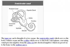
What is the structure of the ear?
|
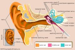
■Ear canal = auditory canal. ■Tympanic membrane = eardrum. ■Ear bones = malleus (hammer) → incus (anvil) → stapes (stirrup). These ear bones are known as the auditory ossicles which convert the sound waves striking the eardrum into mechanical vibrations. The smallest bones in the human body, the ossicles are named for their shape. The hammer (malleus) joins the inside of the eardrum. The anvil (incus), the middle bone, connects to the hammer and to the stirrup (stapes). The base of the stirrup, the footplate, fills the oval window which leads to the inner ear. ■Vestibule = contacts the oval window (where stirrup of the ossicles vibrates), is continuous with semicircular canals and cochlea. ■Cochlea = spiral = houses hair cells. The cochlea is the auditory portion of the inner ear. Its core component is the Organ of Corti, the sensory organ of hearing. Hair cells are sensory cells in the Organ of Corti, topped with hair-like structures called stereocilia. Stereocilia are mechanosensing organelles of hair cells, which respond to fluid motion or fluid pressure changes primarily for hearing and they are similar to microvilli. The cochlea is filled with a watery liquid, which moves in response to the vibrations coming from the middle ear via the oval window. As the fluid moves, thousands of "hair cells" are set in motion, and convert that motion to electrical signals that are communicated via neurotransmitters to many thousands of nerve cells. These primary auditory neurons transform the signals into electrical impulses known as action potential, which travel along the auditory nerve to structures in the brainstem for further processing. ■Semicircular canals = 3 of them perpendicular to one another = senses position and movement of the head, help you balance.
|
|
|
|
Why do photoreceptor cells undergo hyperpolarization instead of depolarization when stimulated by light?
|
Photoreceptor neurotransmitter glutamate is continously released in the dark in the retina. Thus the photoreceptor, whether it be rod or cone is in a depolarized state in the dark. On light stimulation the photoreceptor responds with a hyperpolarization, and transmitter release ceases and an action potential is triggered and conveys signals to the brain.
|
|
|
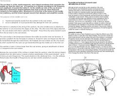
What is the mechanism of hearing?
|
1.Sound wave enters ear.
2.Hits ear drum (tympanic membrane) 3.Malleus (hammer) → Incus (anvil) → Stapes (stirrup) convert sound waves to mechanical waves. Because the oval window is much smaller than the ear drum, the force of the vibrations from the ear drum is amplified by the decrease in surface area of the oval window. This amplified force and the decrease in surface area increases the pressure on the inner ear (P=F/A). This middle ear also serves to transmit the energy (vibrations) transmitted through the air to fluid energy without lose of energy. Usually, when sound in air strikes a fluid boundary (a boundary between media with different acoustic impendances) there is a theoretical loss of 99.9% of the energy in a sound wave in air (due to reflection). The middle ear matches the acoustic impedance between the air and the fluid, thus maximizing the flow of energy from the air to the fluid of the inner ear. 4.Vibrates fluid in Cochlea. 5.Cochlear hair cells excited by vibrations, converts mechanical waves to electrical impulses and sends signal to brain. |
|
|
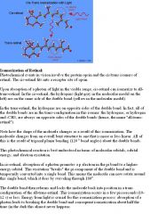
What are light receptors?
|
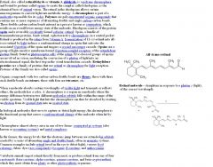
■Photoreceptor cells are located on the back of the retina. ■Rods = senses light and dark (used for night vision). They are more numerous than cones and more sensitive than cones to a single photon of light, yet they do not sense color. ■Cones = sense color (used for day vision), but are less sensitive than rods and require many more photons of light to be activated. ■Rhodopsin = GPCR responsible for light reception of rod cells, and photopsin in cone cells (the analog of rhodopsin). Rhodopsin is made of Retinal (chemical derived from Vitamin A) + Opsin (transmembrane protein). Retinal is attached to the light sensitive receptor of the GPCR. G protein-coupled receptors (GPCRs) comprise a large protein family of transmembrane receptors that sense molecules outside the cell and activate inside signal transduction pathways and, ultimately, cellular responses. Membrane or Transmembrane receptors are specialized integral membrane proteins that take part in communication between the cell and the outside world. Extracellular signaling molecules (usually hormones, neurotransmitters, cytokines, growth factors or cell recognition molecules) attach to the receptor, triggering changes in the function of the cell. This process is called signal transduction: The binding initiates a chemical change on the intracellular side of the membrane. In this way the receptors play a unique and important role in cellular communications and signal transduction. ■Light converts cis-retinal → trans-retinal. ■trans-retinal then causes hyperpolarization of photoreceptor cell because of the light stimulation, which prompts the chain of events that sends signal to the brain. ■Sends signal to brain via the optic nerve on the back of the retina. The back of the retina is also where the blind spot is - where there is a lack of photoreceptor cells.
|
|
|
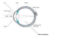
What is the structure of the eye?
|
1.Light first travels through the cornea 2.Through the pupil (hole in the iris muscle) 3.Lens = focuses light on retina. 4.Vitreous humor = fluid. 5.Retina = screen on the back of the eye = contains photoreceptors.
The vitreous humour is the clear gel that fills the space between the lens and the retina of the eyeball of humans and other vertebrates. It is often referred to as the vitreous body or simply "the vitreous". The retina is a light sensitive tissue lining the inner surface of the eye. The optics of the eye create an image of the visual world on the retina, which serves much the same function as the film in a camera. Light striking the retina initiates a cascade of chemical and electrical events that ultimately trigger nerve impulses. These are sent to various visual centers of the brain through the fibers of the optic nerve. The retina is a complex, layered structure with several layers of neurons interconnected by synapses. The only neurons that are directly sensitive to light are the photoreceptor cells. These are mainly of two types: the rods and cones. Rods function mainly in dim light and provide black-and-white vision, while cones support daytime vision and the perception of colour. The membranous photoreceptor protein opsin contains a pigment molecule called retinal. In rod cells, these together are called rhodopsin. In cone cells there are different types of opsins that combine with retinal to form pigments called photopsins. Three different classes of photopsins in the cones absorb different wavelengths of light, a differentation which eventually allows the visual system to distinguish color. The function of the photoreceptor cell is to convert the light energy of the photon into a form of energy communicable to the nervous system and readily usable to the organism: this conversion is called signal transduction. Photopsins are the photoreceptor proteins found in the cone cells of the retina that are the basis of color vision. |
|
|
|
What is visual image processing ?
|
■The lens of the eye, just like a convex lens in physics, forms a real image on the retina.
■Real images are inverted. ■The brain processes this inverted image to make it seem upright in your mind. ■The brain combines the two images from each eye to make a 3D image, from this 3D image, you can judge distance. ■Another reason for combining the two images from both eyes is that it gets rid of the blind spot in each eye. The visual cortex is the most massive system in the human brain and is responsible for processing the visual image. It lies at the rear of the brain above the cerebellum. Light entering the eye is refracted as it passes through the cornea because the light is passing through another medium. It then passes through the pupil (controlled by the iris) and is further refracted by the lens. The cornea and lens act together as a compound lens to project an inverted image onto the retina. |
|
|
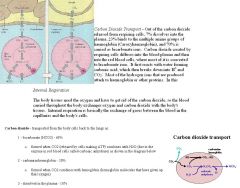
What are the functions of circulation and how does circulation deliver oxygen and carbon dioxide?
|
•Functions of the circulatory system are circulation of oxygen, nutrients, hormones, ions, and fluids; and removal of metabolic waste.
◦Oxygen delivery to tissues: 1. oxygen diffuses from alveolar walls into the blood of alveolar (lung) capillaries because the concentration (or pressure) of oxygen is greater in the alveoli than in the blood 2.oxygen binds to hemoglobin in red blood cells 3. oxygen gets transported to tissues 4. then oxygen used in cellular respiration ◦Carbon dioxide delivered out 1.cellular respiration makes CO2 2.CO2 gets transported by blood (either as dissolved CO2, dissolved bicarbonate ion, or bound to hemoglobin). Carbon dioxide reactions with water forming carbonic acid (H2CO3), which is then deprotonated to form bicarbonate (HCO3−). Bicarbonate serves a crucial biochemical role in the physiological pH buffering system. Bicarbonate is an alkaline, and a vital component of the pH buffering system of the body (maintaining acid-base homeostasis). 70 to 75 percent of CO2 in the body is converted into carbonic acid (H2CO3), which can quickly turn into bicarbonate (HCO3−). 3. Dissolved CO2 (or bicarbonate ion or CO2-Hb) travels to the lungs and and gets converted back to CO2 and diffuse from the blood of alveolar capillaries into the alveolar walls 4.exhaled out |
The tidal volume is the amount of air that moves into the lungs during each inspiration. Some of this air does not reach the alveoli so it's not available for gas exchange with the circulatory system. The volume of such gas in the air passageways (trachea, bronchi, and bronchioles) is called the respiratory dead space (nonalveolar respiratory volume). If you subtract the dead space per breath from the tidal volume per breath, you will get the quantity of air that enters the alveoli on each inspiration.
|
|
|
Explain how the circulatory system affects nutrients, hormones, ions, and fluids; and removal of metabolic waste.
|
■Nutrients are absorbed (either by diffusion or active transport) into blood stream in the small intestines.
■nutrients can also be released into the blood stream by cells. For example, glucagon causes glucose to be released into the blood stream. ■nutrients can be taken up by cells. For exampe, insulin causes cells to take in glucose from blood. ◦Hormones released by endocrine glands, circulate the blood in order to reach their target cells. ◦Fluids and ions circulate the blood and are regulated by how much reabsorption of water and salt occurs in the kidney. ◦Urea = metabolic waste, travels in the blood to the kidneys, where it is filtered out and passed in urine. Vasculature is anything that delivers blood to tissue, for example arteries, capillaries, and veins (vessels that carry deoxygenated blood). The lymphatic system does not deliver blood to tissue. Rather, it drains the extracellular environment, returning water, protein, nutrients, etc. back into circulation. |
Open circulatory systems are those in which the heart does not pump blood into capillaries, but into open cavities. Some insects have this kind of system.
|
|
|
What is the Role of the circulatory system in thermoregulation?
|
◦Vasoconstriction conserves heat. When it's cold, vasoconstriction occurs in the arterioles that feed the skin. Less blood flows near the surface of the skin, less heat lost.
◦Vasodilation cools you down. When it's hot, vasodilation occurs in the arterioles that feed the skin. More skin blood flow, more heat lost to the surroundings. |
|
|
|
What is the Four-chambered heart (structure, function)?
|
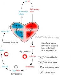
1.Deoxygenated blood returns to the heart: superior/inferior vena cava → right atrium
2.Deoxygenated blood gets pumped to the lungs: right atrium → right ventricle → pulmonary artery → lungs 3.Blood arrives at the lungs and gets oxygenated. 4.Oxygenated blood returns to the heart: lungs → pulmonary vein → left atrium 5.Oxygenated blood gets pumped to the body: left atrium → left ventricle → aorta The heart pumps oxygen, carbon dioxide, glucose, and other nutrients in the blood to the organs and takes the blood up from the organs after the exchange. So if the heart stops beating, glucose is not reaching the tissues. The cells of those tissues would have to use anaerobic metabolism in order to generate ATP. The cardiac conduction pathway begins with the SA node that is located in the right atrium. The signal then travels to the AV node (located between the atria and the ventricles), down the bundle of His, which splits into the right/left Purkinje fibers. The Purkinje fibers spread the conduction signal to the 2 ventricles, which contract simultaneously to expel blood. Summary: SA → AV → Bundle of His → Purkinje fibers. "SAAV BP" |
A normal heart has 2 major heart sounds, both correlating with the CLOSING of the valves. The first heart sound (lub) is the closing of the atrioventricular (mitral and tricuspid) valves. The second heart sound (dub) occurs during the closing of the aortic/pulmonic valves. (AV then AP) The closing of the mitral and tricuspid valves creates the 1st heart sound and happens at the beginning of systole. The closing of the aortic and pulmonary valves creates the 2nd heart sound and happens at the end of systole.
The SA node is the main pacemaker of the heart. It determines the heart rate and undergoes auto-depolarization stimulated by the influx of calcium. If the SA node is damaged, the AV node would take over as the main pacemaker of the heart. |
|
|
Explain how Blood goes through the heart including the valves.
|
1.Vena cava
2.Right atrium 3.Tricuspid valve (Mneumonic - tri - right) 4.Right ventricle 5.Pulmonary valve 6.Pulmonary artery 7.Lung 8.Pulmonary vein 9.Left atrium 10.Bicuspid (Mitral) valve 11.Left ventricle 12.Aorta |
The coronary arteries supply oxygen and nutrients to the cardiac muscle itself, not the rest of the body.
|
|
What is Systolic and diastolic pressure?
|
◦blood pressure = pressure blood exert on the walls of the blood vessel.
◦systolic pressure = The blood pressure when the heart is contracting. It is specifically the maximum arterial pressure during contraction of both of the ventricles of the heart (the right ventricle contracts blood into the pulmonary artery and the left ventricle contracts blood into the aorta). ◦diastolic pressure = Diastolic: Referring to the time when the heart is in a period of relaxation and dilatation (expansion). The diastolic pressure is specifically the minimum arterial pressure during relaxation and dilatation of both of the ventricles of the heart when both ventricles fill with blood from the veins. Blood pressure (BP) is a force exerted by circulating blood on the walls of blood vessels, and is one of the principal vital signs. During each heartbeat, BP varies between a maximum (systolic) and a minimum (diastolic) pressure. The mean BP, due to pumping by the heart and resistance in blood vessels, decreases as the circulating blood moves away from the heart through arteries. It has its greatest decrease in the small arteries and arterioles, and continues to decrease as the blood moves through the capillaries and back to the heart through veins. For each heartbeat, BP varies between systolic and diastolic pressures. Systolic pressure is peak pressure in the arteries, which occurs near the end of the cardiac cycle when the ventricles are contracting. Diastolic pressure is minimum pressure in the arteries, which occurs near the beginning of the cardiac cycle when the ventricles are filled with blood. An example of normal measured values for a resting, healthy adult human is 120 mmHg systolic and 80 mmHg diastolic (written as 120/80 mmHg, and spoken [in the US] as "one-twenty over eighty"). |
The heart has 2 atrial and 2 ventricular chambers. The ventricular musculature is much thicker in comparison to the atrium. The ventricle must generate a tremendous amount of pressure to force blood throughout the entire body. The left ventricle is the thickest overall chamber because it must force blood against the higher systemic resistance, while the right ventricle pumps against a low resistance of the pulmonary circulation.
|
|
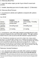
What is Pulmonary and systemic circulation?
|
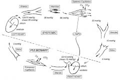
◦Pulmonary circulation = heart → lungs → back to heart = oxygenates blood. ◦Systemic circulation = heart → body → back to heart = delivers oxygenated blood to body ◦Pulmonary circulation = shorter than systemic circulation = less resistance = less blood pressure. ◦Systemic circulation: vasodilation when oxygen levels are low → vasodilation causes more blood flow → more blood flow to oxygen-starved tissue. ◦Pulmonary circulation: vasoconstriction when oxygen levels are low (opposite of systemic circulation) → less blood flow to low oxygen/blocked alveoli → more blood flow to good alveoli where gas exchange can occur. Pulmonary blood flow is directed away from poorly ventilated regions to better ventilated regions of the lung. Ventilation is increased to underventilated regions. Although pulmonary blood flow need not be as high as systemic blood flow (systemic blood flow overcomes the gravity of bringing blood from feet to heart), there are some influences of gravity: REGIONAL BLOOD FLOW DISTRIBUTION (Gravitational Effect): 1) Upright Posture • Pulmonary vascular hydrostatic pressure decreases at increasing heights above the heart causing vascular collapse as one moves towards the top (Apex) of the
lungs and further above the heart. • Blood flow is highest in the base of the lungs and decreases toward the apex because of the increase in passive vascular resistance. 2) Prone Posture: • All lung regions are near heart level, so pulmonary flow is more uniform. Pulmonary arteries carry the most deoxygenated blood, even more than the vena cava to the right atrium. |
|
|
|
What are the arteriol and venous systems?
|
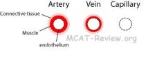
Arterial and venous systems (arteries, arterioles, venules, veins)
■Blood flows from artery → arteriole → capillary → venule → vein. |
|
|
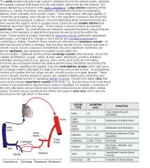
What is elastic and distributing arteries?
|
1) Elastic arteries. An elastic artery is an artery with a large number of collagen and elastin fibers which gives it the ability to stretch in response to each pulse. Elastic arteries include the largest arteries in the body, those closest to the heart. The pulmonary arteries, the aorta, and its branches together comprise the body's system of elastic arteries. ■Aorta and its major branches. ■Major function = provide elastic pipe for blood straight out of the heart. ■Lots of elastic tissue. ■Layers: endothelium, smooth muscle, connective tissue. ■Although capable of constricting and dilating, they are not active in vasoconstriction and do not regulate blood pressure or flow. The arteries needs to be thick from smooth muscle and collagen and elastic fibers to withstand the high pressure of blood flowing through it at all times. The endothelium is the thin layer of cells that line the interior surface of blood vessels, forming an interface between circulating blood in the lumen and the rest of the vessel wall. Endothelial cells line the entire circulatory system, from the heart to the smallest capillary. These cells reduce turbulence of the flow of blood allowing the fluid to be pumped farther. Endothelial tissue is a specialized type of epithelium tissue. More specifically, it is simple squamous epithelium.
2) Distributing arteries ■Major function = distribute blood to specific organs. Distributing arteries (or muscular arteries) are medium-sized arteries that draw blood from an elastic artery and branch into "resistance vessels" including small arteries and arterioles. ■Smooth muscle to constrict and dilate artery. ■Layers: endothelium, smooth muscle, connective tissue (elastic and collagen fibers). ■Major activity in vasoconstriction and vasodilation(they regulate the blood pressure and flow). |
|
|
|
What is the arteriole?
|
■Ranges from being like a smaller version of the artery, to being a larger version of the capillary with smooth muscles spiralling around it.
■Major function = controls blood flow to the capillaries. ■Active in vasoconstriction and vasodilation. The arterioles allow the body to control which tissues gets more blood. ■The arteriole is the most important site for vasoconstriction. Although other vessels are capable of vasoconstriction, you should always think of the arteriole when you see vasoconstriction. |
|
|

What is the capillary?
|
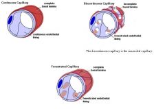
■Layer: Endothelium is made up of a single layer of cells.
■Major function: blood-tissue solute exchange. ■Not active in vasoconstriction. ■Diffusion is the major mechanism of gas and solute exchange, and the solutes or gas can be free molecules or bound to proteins. The principal function of the microcirculation is to permit the transfer of substances between the tissues and the circulation. This transfer occurs predominantly across the walls of the capillaries but some exchange occurs in the small venules also. Substances involved include water, electrolytes, gases (O2, CO2), nitrogenous wastes (like urea), glucose, lipids and drugs. Electrolytes and other small molecules like water cross the capillary endothelium through clefts of continuous capillaries. Lipid soluble substances (including oxygen and carbon dioxide) can also easily cross the thin capillary walls. Capillaries that are fenestrated pores that allow small molecules and some proteins to diffuse and capillaries that are sinusoidal allow red and white blood cells and larger proteins to diffuse. Epithelial cells in the capillaries also use pinocytosis to engulf dissolve materials of the liquid portion of the blood into a vesicle at the capillary surface. The resulting vesicles travel across the capillary cells and release their contents to surrounding tissues, while blood cells remain in the blood. |
|
|
|
What is the venule?
|
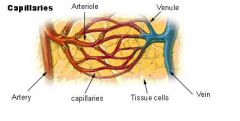
■Ranges from being like a large capillary to being like a small vein.
■Major function: merge of capillaries to be conducted to veins. ■No vasoconstriction of the post-capullary venules, but vasoconstriction starts to occur when the venules form into small veins. A venule is a small blood vessel in the microcirculation that allows deoxygenated blood to return from the capillary beds to the larger blood vessels called veins. Venules are blood vessels that drain blood directly from the capillary beds. Many venules unite to form a vein. Venule walls have three layers: An inner endothelium composed of squamous endothelial cells that act as a membrane, a middle layer of muscle and elastic tissue and an outer layer of fibrous connective tissue. The middle layer is poorly developed so that venules have thinner walls than arterioles. The post- capillary venules are extremely porous so that fluid and blood cells can still exchange easily from the bloodstream through their walls. Venules form when capillaries unite. As post-capillary venules join together and form larger venules, smooth muscle once appears and regulates capillary pressure. Capillaries do not function on their own. The "capillary bed" is an interweaving network of capillaries supplying an organ. The more metabolically active the cells, the more capillaries they will require to supply nutrients and carry away waste products. A capillary bed connects with the arteriole on one side of the bed and the venule on the other side of the bed. |
|
|
|
What is the vein?
|
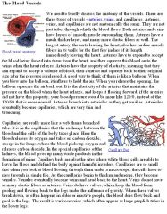
■Layers: endothelium, smooth muscle, connective tissue. The vein wall is much thinner than the wall of a comparable artery. As a result, the blood within the veins is carried under much lower pressure.
■Major function: returns blood back to the heart and regulates blood volume. ■Has valves to prevent the back flow of blood. ■Breathing, skeletal muscles, and valves help promote blood flow through the vein at low pressure back to the heart. ■Vasoconstriction and dilation can occur in the vein because of the smooth muscle. When blood leaves the heart, it is pumped around the body by the contractions of the heart, but the same is not true for blood returning to the heart. The blood flow within veins and venules is a lot slower than in arteries and arterioles. The blood is actually pumped by contractions in muscles in the extremities, the legs for example. Most blood is flowing against gravity when it is traveling back to the heart and lungs. Semi-lunar valves are present within each venule and vein to stop the blood from flowing back down the vein or venule. These valves are necessary because the blood is not being pumped as hard and to counteract the effects of gravity on the slower moving blood. |
|
|
|
What are the Differences between arteries and veins?
|

■Thickness: artery > vein > arteriole > venule > capillary
■Arteries are thicker, more muscular than veins. The smooth muscle of the large arteries are used to withstand the pressure of blood from the aorta, while the smooth muscle of the smaller arteries and arterioles are used to regulate blood pressure and blood flow to the organs. The smooth muscle of the venules are used to regulate the capillary pressure, and the smooth muscle of the veins are used to carry and regulate blood volume. Veins regulate blood volume by constricting the vein and decreasing and blood volume while increasing venous pressure or dilating the vein and increasing increasing blood volume while decreasing venous pressure before it reaches the right atrium the heart. Regulating blood volume alters the cardiac output. Blood volume is the amount of blood left after being distributed through the body and water and sodium has been ingested, by the kidneys into the urine, and lost through the gastrointestinal tract, lungs and skin. The amounts of water and sodium ingested and lost are highly variable. To maintain blood volume within a normal range, the kidneys regulate the amount of water and sodium lost into the urine. ■Veins have valves (called semi-lunar valves), arteries don't. ■Arteries carry oxygenated blood, veins carry deoxygenated blood. Exception: pulmonary arteries and veins are the other way round. Pulmonary arteries carry deoxygenated blood while the pulmonary veins carry oxygenated blood. That's the only thing to remember is opposite. Pulmonary arteries still carry blood away from the heart, just to the lungs. Pulmonary veins still carry blood towards the heart, just from the lungs. |
|
|
|
What are the Differences between artery and arteriole?
|
■arterioles are smaller.
■vasoconstriction and vasodilation occurs predominantly at the arterioles |
|
|
|
What are the pressure and flow characteristics of the arteriol and venous systems?
|
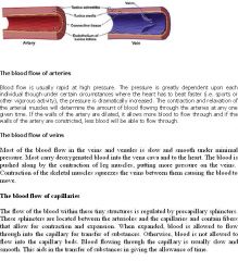
■Blood pressure of arteries > arterioles > capillaries > venules > veins
■Blood pressure is highest in the arteries (specifically the aorta) because it is right on the receiving end of the heart. ■Blood pressure is lowest in the veins (specifically the vena cava) because flow resistance brings the pressure down. ■Blood pressure can be regulated by vasoconstriction (increase bp), vasodilation (decrease bp), and hormones (ADH, aldosterone, renin, adrenaline all increases bp). ■Blood flows from artery → arteriole → capillary → venule → vein. ■Blood squirts from arteries, flows from veins, and oozes from capillaries. ■The elasticity of arteries causes blood to flow even when the heart is resting between pumps (this is why your diastolic blood pressure is not zero) |
Pulse pressure is the difference between the systolic pressure and the diastolic pressure. Blood pressure is measured from the brachial ARTERY (not from a vein or venous pressure). It is found from arterial pressure. The brachial artery is usually found in the arm.
|
|
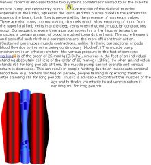
What are Adaptations that help blood flow through the vein at low pressure?
|
■Respiratory pump: product of inspiration and expiration
■Muscular pump: skeletal muscle squeezes on the veins when you exercise. ■When you're scared, smooth muscles around veins constrict and squeezes blood. Respiration produces cyclical variations in intra pleural and intra thoracic pressure. With each inspiration, the pressure is lowered with the thorax and hence also within the right atrium of the heart; this increases the pressure gradient and aids blood flow back to the heart. Simultaneously during inspiration, the diaphragm contracts and spreads out into the abdomen, increasing the vertical dimension of the chest cavity and raises the intra-abdominal pressure and increases the gradient to the thorax, again favouring venous return. With expiration, the pressure gradients are reversed and blood tends to flow in the opposite direction; fortunately this tendency is prevented by the valves in the medium sized veins. Thus venous return is maintained by changes in venomotor tone (degree of contraction of smooth muscle in vein), altering the capacity of the venous system, and by the skeletal muscle and respiratory pumps. Obviously it is also necessary to maintain an adequate circulating blood volume. If the blood volume is depleted for some reason, e.g. dehydration or haemorrhage, in the short term vasoconstriction can increase the effective circulating blood volume. However, the blood volume must be restored eventually by fluid replacement. The pressures in the central regions of the venous system directly reflect the blood volume; thus central venous pressure (CVP), or right atrial pressure, is a good indicator of blood volume, unlike arterial pressures which are reflexly regulated and controlled. |
|
|
What is a Continuous capillary?
|
Continuous capillaries have the thickest endothelial wall. They allow only water, and ions into their pathways. They are continuous in the sense that the endothelial cells provide an uninterrupted lining, and only allow small molecules, like water and ions to diffuse through tight junctions via vesicles which leave gaps of unjoined membrane which are called intercellular clefts. Tight junctions can be further divided into two subtypes: 1.Those with numerous transport vesicles that are primarily found in skeletal muscles, lungs, gonads, and skin. 2.Those with few vesicles that are primarily found in the central nervous system. These capillaries are a constituent of the blood-brain-barrier. Continuous capillaries have no pores on endothelial cells (unlike fenestrated capillaries). They may have clefts at cell boundaries. ■Exchange occurs through the clefts. They have low permeability and therefore allow only limited passage of substances across the capillary wall.■Continuous capillaries are the most abundant type in the human body and are found in skin, muscle, lungs, and the central nervous system. ■Blood-brain barrier = sealing of clefts by tight junctions.
|
|
|
|
What are capillary beds and what is a Fenestrated capillary?
|
The "capillary bed" is the network of capillaries supplying an organ. The more metabolically active the cells, the more capillaries they will require to supply nutrients and carry away waste products.
Fenestrated capillaries have “windows” that lets larger molecules in and out of the capillaries. ■Small pores, large enough for molecules, but not blood cells to leak through. ■Found in small intestines to facilitate nutrient absorption. ■Found in endocrine organs to allow passage of hormones. ■Found in kidneys to allow blood filtration. Fenestrated capillaries have pores in the endothelial cells and allow small molecules and limited amounts of protein to diffuse. Fenestrated blood vessels are primarily located in the endocrine glands, intestines, pancreas, and glomeruli of kidney. Fenestrated capillaries allow diffusion of proteins while continuous capillaries do not (because fenestrated capillaries have the pores that are big enough for proteins to go through) so they have greater permeability than continuous capillaries. |
|
|

What is a Sinusoidal capillary?
|
■Large pores, large enough for blood cells to leak through.
■Found in lymphoid tissues, liver, spleen, bone marrow. ■Large pores facilitate lymphocytes in their travel to tissues. They are very permeable capillaries. |
|
|
|
What is the mechanism of heat exchange for capillary beds?
|

■radiation - your body gives off infrared signal from the heat of our blood pumping. Radiation is the emission of electromagnetic energy (which your body does in the infrared wavelengths). When considering radiation absorbed by the skin from the sun, the body emits this radiation as well.
■conduction - you touch something cold, or take a hot bath and heat is transfered. ■evaporative cooling - you sweat, and your body cools as the sweat evaporates. Convection: Convection is the movement of heat by currents in the medium, ie., the wind of the air. So the heat of the blood gets moved by the currents of the wind or air. The skin temperature is affected by the surrounding air temperature. |
|
|
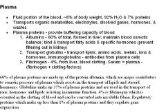
What is the Composition of blood?
|
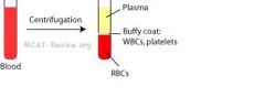
Blood is made up of plasma and blood cells and platelets. The average human being has 5 liters of blood in his body. 55% of the blood is made up of plasma constituting the fluid part of the blood. The cells and platelets that are present in our blood make up the other 45%.
■plasma = water and chemicals = mostly water, plasma proteins, electrolytes, gases, nutrients, wastes, and hormones. In general, water, accounting for roughly 75% of the total body weight, is the most abundant compound in the human body and in all cells (like cardiac cells, blood cells, skin cells, etc). All blood cells (i.e. red blood cells, white blood cells, platelets) are non-dividing. They are synthesized by the bone marrow, have a set lifespan, and when aged, are removed by the spleen. All living cells (skeletal and muscle cells, red blood cells, etc) have gated sodium-potassium ATPases and/or other pumps that are voltage dependent. They need this pump to be able to maintain the internal environment for depolarization. |
|
|
|
What are the different blood cells?
|
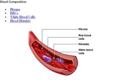
1) red blood cells (RBCs or erythrocytes)
■contain hemoglobin, transports O2 and CO2 ■no nucleus, which gives it a biconcave disk shape The lack of a nucleus in red blood cells (in humans and other mammals) is a specialization related to the ability of the erythrocyte to store additional hemoglobin. The function of the erythrocytes is to distribute oxygen to the tissues, and additional hemoglobin is advantageous in this function. ■most abundant cell in blood. 2) white blood cells (WBCs or leukocytes) ■larger than RBCs ■lobed or irregular shaped nuclei ■fights off pathogens 3) platelets - technically not cells, but they are cell fragments and are responsible for clotting blood. Platelets are produced from from big cells called megakaryocytes that break up into fragments. They are anuclear fragments. The megakaryocyte is a bone marrow cell responsible for the production of blood thrombocytes (platelets), which are necessary for normal blood clotting. |
|
|
|
What is erythrocyte production and destruction?
|
The major sites of erythrocyte production and destruction are the spleen and bone marrow.
■Bone marrow = makes RBCs from stem cells. ■Spleen = destroys aged and damaged RBCs. ■Other sites for RBC destruction include the LIVER and BONE MARROW. ■Components of hemoglobin from destroyed RBC gets recycled ■the iron gets recycled ■heme → bilirubin → bile → excreted in feces Heme is converted to bilirubin in the spleen and then the bilirubin is transported to the liver bound to the albumin protein. Then the bilirubin is secreted from the liver as bile and is excreted in feces and urine. ■protein (globin) = broken down to amino acids during degradation. In most humans, the hemoglobin molecule is an assembly of four globular protein subunits. Each subunit is composed of a protein chain tightly associated with a non-protein heme group. A heme group consists of an iron (Fe) ion (charged atom) held in a heterocyclic ring. The kidney releases the hormone erythropoietin (EPO) into the blood which activates the bone marrow to produce red blood cells when when the oxygen level is low in the kidney. EPO then stimulates the bone marrow to produce more red cells and thereby increase the oxygen-carrying capacity of the blood. EPO is the prime regulator of red blood cell production. Its major functions are to promote the differentiation and development of red blood cells and to initiate the production of hemoglobin, the molecule within red cells that transports oxygen. |
|
|
What is the regulation of plasma volume?
|
Plasma (blood) osmolarity is a measure of the concentration of substances such as sodium, chloride, potassium, urea, glucose, and other ions in blood. ■Higher plasma osmolarity → causes water to go into blood → leads to higher blood volume and lower plasma osmolarity as a result. ■Lower plasma osmolarity → causes water goes into tissues where there are more solutes → leads to lower blood volume and higher plasma osmolarity as a result. ■ADH (vasopressin): ↑ water reabsorption in kidney. Vasopressin is a peptide hormone that controls the reabsorption of water in the tubules of the kidneys by affecting the tissue's permeability. One of it's most important roles is to regulate the body's retention of water; it is released when the body is dehydrated and causes the kidneys to conserve water, thus concentrating the urine, and reducing urine volume. Antidiuretic hormone binds to receptors on cells in the collecting ducts of the kidney and promotes reabsorption of water back into the circulation by stimulating insertion of "water channels" or aquaporins into the membranes of kidney tubules. These channels transport solute-free water through tubular cells and back into blood, leading to a decrease in plasma osmolarity and an increase osmolarity of urine (making the urine more concentrated and the blood less concentrated). As noted above, ADH plays a role in lowering osmolarity by increasing water reabsorption in the kidneys, thus helping to dilute bodily fluids. To prevent osmolarity from decreasing below normal, the kidneys also have a regulated mechanism for reabsorbing sodium in the distal nephron. This mechanism is controlled by aldosterone, a mineralocorticoid steroid hormone produced by the adrenal cortex. The adrenal cortex directly senses plasma osmolarity. When the osmolarity increases above normal, aldosterone secretion is inhibited. The lack of aldosterone causes less sodium to be reabsorbed in the distal tubule. Remember that in this setting ADH secretion will increase to conserve water, thus complementing the effect of low aldosterone levels to decrease the osmolarity of bodily fluids. The net effect on urine excretion is a decrease in the amount of urine excreted, with an increase in the osmolarity of the urine. ADH directly lowers plasma osmolarity, while aldosterone indirectly lowers plasma osmolarity (because aldosterone stimulates sodium reabsorption, which causes water to reabsorption to follow.
|
|
|
What is coagulation, clotting mechanisms, role of liver in production of clotting factors ?
|
■Platelets contains enzymes and chemicals needed in the clotting process. Liver produces clotting factors (eg. fibrinogen), which circulates in blood plasma. Coagulation = liquid blood → gel. Clotting mechanism: 1) Platelet plug formation: wound + platelets → platelets clump at wound, release chemicals, activates clotting factors. 2) Coagulation: series of clotting factor/enzyme activation that ends in fibrinogen → fibrin. Fibrin being the fiber mesh that seals the clot. 3) Retraction and repair: After the wounded blood vessel repairs itself, the clot dissolves. Coagulation is a complex process by which blood forms clots. It is an important part of hemostasis (the cessation of blood loss from a damaged vessel), wherein a damaged blood vessel wall is covered by a platelet and fibrin-containing clot to stop bleeding and begin repair of the damaged vessel. Coagulation involves both a cellular (platelet) and a protein (coagulation factor) component. Coagulation begins almost instantly after an injury to the blood vessel has damaged the endothelium (lining of the vessel). Exposure of the blood to proteins stimulates a cascade that ends with fibrinogen converting to fibrin, which strengthens the platelet plug. Fibrin is made from fibrinogen, a soluble plasma glycoprotein that is synthesised by the liver. Glycoproteins are proteins that contain oligosaccharide chains (glycans) covalently attached to polypeptide side-chains. Clotting factors are substances in the blood that act in sequence to stop bleeding by forming a clot.
|
|
|
|
What is hemoglobin and hematocrit?
|
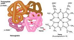
■hemoglobin = (heme + globin) x 4
■heme = group binding iron. A heme is a prosthetic group that consists of an iron atom contained in the center of a large heterocyclic organic ring ■globin = protein that surrounds heme. ■4 subunits of the heme-globin complex together and create the tetramer called hemoglobin. Each subunit is composed of a protein chain tightly associated with a non-protein heme group. ■hemoglobin can bind oxygen and carbon dioxide and carbon monoxide ■hematocrit = % volume of blood that is red blood cells, usually ~ 45% |
|
|
|
What is oxygen content for blood?
|

■each iron atom in hemoglobin can bind one oxygen.
■hemoglobin has 4 subunits containing 4 iron atoms. ■each RBC has hundreds of millions of hemoglobin molecules. |
|
|
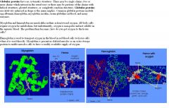
What is oxygen affinity for blood?
|
■hemoglobin has a sigmoidal oxygen binding curve. This is because oxygen binding to one subunit "relaxes" the conformation of the other subunits, and makes it easier for additional oxygen to bind.
■carbon monoxide binds hemoglobin tighter than oxygen. CO competes with oxygen at the heme binding site. Hemoglobin binding affinity for CO is 200 times greater than its affinity for oxygen, meaning that small amounts of CO dramatically reduce hemoglobin's ability to transport oxygen. ■fetal hemoglobin binds oxygen tighter than adult hemoglobin. ■myoglobin binds oxygen tighter than hemoglobin. Fetal hemoglobin has a slightly higher affinity for oxygen than does adult hemoglobin. This allows the fetus to extract oxygen more efficiently from the maternal circulation. Hemoglobin consists of four protein chains and four heme groups that carry oxygen from the lungs to the tissue cells. Myoglobin consists of a single protein chain and one heme group that stores oxygen in the muscle cells. Myoglobin has a stronger affinity for oxygen then hemoglobin. Because myoglobin only have 1 heme while hemoglobin has 4 heme, myoglobin binds reversibly to only one oxygen molecule, while hemoglobin binds four. These differences help to explain the different biological functions of hemoglobin and myoglobin. Hemoglobin is used to transport oxygen over large distances (from the lungs to all tissues), whereas myoglobin is present in muscle and behaves as a local "storage" reservoir of oxygen. At higher altitudes, there is less oxygen concetration, so there would be a decrease in affinity for hemoglobin and a right shift of the oxygen dissociation curve. |
|
|
|
What is the modification of oxygen affinity?
|
■Higher levels of carbon dioxide → lower oxygen affinity of hemoglobin.
■Lower pH → lower oxygen affinity. ■Higher temperature → lower oxygen affinity. ■Working muscle = hot, acidic, high CO2, needs oxygen. So, hemoglobin must unload its oxygen, and it does this by lowering its oxygen affinity. Carbon dioxide and protons occupies a different binding site on the hemoglobin. Carbon dioxide is more readily dissolved in deoxygenated blood, facilitating its removal from the body after the oxygen has been released to tissues undergoing metabolism. This increases the affinity for carbon dioxide by the venous blood because it carries this carbon dioxide to the heart, which will be carried to the lungs and exhaled out. Carbon dioxide reacts with water to give carbonic acid, which decomposes into bicarbonate and protons: CO2 + H2O → H2CO3 → HCO3- + H+ . Hence blood with high carbon dioxide levels is also lower in pH (more acidic). Carbon dioxide binds to hemoglobin and forms carbaminohemoglobin. This decreases hemoglobin's affinity for oxygen by the binding of carbon dioxide and protons. Conversely, when the carbon dioxide levels in the blood decrease (i.e., in the lung capillaries), carbon dioxide and protons are released from hemoglobin, increasing the oxygen affinity of the protein. Hemoglobin needs to release the oxygen that it binds to the tissues and if not, there is no point in binding it. The sigmoidal curve of hemoglobin makes it efficient in binding (taking up O2 in lungs), and efficient in unloading (unloading O2 in tissues).Hemoglobin can bind protons and carbon dioxide, which causes a conformational change in the protein and facilitates the release of oxygen. So in general, things like CO and CO2 can bind to hemoglobin and affect its affinity to oxygen. Increased temperature results in a release of oxygen by hemoglobin to the tissues (to produce water) via aerobic metabolism. Exercising muscle is acidic because of lactic acid and carbonic acid from increased carbon dioxide and hot because of increased blood flow and the metabolic rate. CO2 is increased because it is the product of aerobic respiration. |
|
|
Explain the equalization of fluid distribution in the lymphatic system.
|
■Interstitial fluid pressure > lymphatic pressure → lymph vessel flaps open → interstitial fluid enters lymphatic capillaries → lymphatic circulation merges with veins → returns the fluid to blood
■Interstitail fluid pressure < lymphatic pressure → lymph vessel flaps close → prevents lymph from leaking back out. The lymphatic circulation begins with blind ending (closed at one end) highly permeable superficial lymph capillaries, formed by endothelial cells with button-like junctions between them that allow fluid to pass through them when the interstitial pressure is sufficiently high. A valve system in place here prevents the absorbed lymph from leaking back into the ISF. Rhythmic contraction of the vessel walls through movements may also help draw fluid into the smallest lymphatic vessels, lymphatic capillaries. If tissue fluid builds up the tissue will swell; this is called edema. As the circular path through the body's system continues, the fluid is then transported to progressively larger lymphatic vessels culminating in the right lymphatic duct (for lymph from the right upper body) and the thoracic duct (for the rest of the body); both ducts drain into the circulatory system at the right and left subclavian veins. The system collaborates with white blood cells in lymph nodes to protect the body from being infected by cancer cells, fungi, viruses or bacteria. This is known as a secondary circulatory system. |
|
|
|
Explain the transport of proteins and large glycerides for the lymphatic system.
|

■fats get absorbed into the lacteals in the small intestine.
A lacteal is a lymphatic capillary that absorbs dietary fats in the villi of the small intestine. ■plasma protein that leaked into interstitial fluids get returned to the blood via the lymphatic system. Glycerides, more correctly known as acylglycerols, are esters formed from glycerol and fatty acids. Glycerol has three hydroxyl functional groups, which can be esterified with one, two, or three fatty acids to form monoglycerides, diglycerides, and triglycerides. |
|
|
|
Explain how the lymphatic system returns materials to the blood.
|
■cells and plasma proteins that leak out of the blood capillaries gets collected by the lymphatic capillaries and returned to the vein.
|
|
|
|
What is the Composition of lymph?
|
◦Composition of lymph (similarity to blood plasma; substances transported)
■Lymph = stuff that leaks out of the capillaries = mostly water, plasma protein, chemicals, and white blood cells. Lymph has a composition comparable to that of blood plasma, but it may differ slightly. Lymph contains white blood cells. In particular, the lymph that leaves a lymph node is richer in lymphocytes (a type of white blood cell, T cells, NK cells, and B cells). |
|
|
|
What is the source of lymph?
|
◦Source of lymph (diffusion from capillaries by differential pressure).
■blood plasma from capillaries → diffuses into interstitial fluid → then it becomes lymph → then gets returned to blood. Basically, this is fluid that has leaked out of the capillaries into the surrounding tissues. Much of it is pulled back into the blood by osmotic pressure, but whatever isn't is eventually returned to circulation. The driving force for lymph is the difference in pressure between the interstitial space and the lymph capillaries. An additional force that moves materials from the blood into the tissues is the pressure of the blood as it flows through the capillaries. Blood pressure is the force that filters, or “pushes” water and dissolved materials out of the capillary into the tissue fluid. Fluid is drawn back into the capillary by osmotic pressure, the “pulling force” of substances dissolved and suspended in the blood. Osmotic pressure is maintained by plasma proteins (mainly albumin), which are too large to go through the capillary wall. These processes result in the constant exchange of fluids across the capillary wall. The plasma solute osmotic pressure is great enough to draw most of the tissue fluid back into the blood except for about 10% which eventually is captured and returned to the blood by the lymphatic system. Osmotic pressure (also called oncotic pressure) : is the pressure created by the presence of large molecules that can’t diffuse and are prevented from moving through the capillary membrane. These large molecules are plasma proteins such as albumin. Albumin plays an essential role in maintaining this pressure. They encourage osmosis and they draw water toward them. Because capillary blood has a high content of plasma proteins, the capillary has a high oncotic pressure of 25mmHg. Osmotic pressure depends on water and a membrane that is permeable to water and not to other large molecules. |
|
|
|
What are Lymph nodes?
|
■Lymph nodes are concentrated with white blood cells and provides the growth and regulatory factors necessary for activation and maturation of immune cells. Pathogens, or germs, can set up infections anywhere in the body. However, lymphocytes, a type of white blood cell, will meet the antigens, or proteins, in the peripheral lymphoid organs, which includes lymph nodes. The antigens are displayed by specialized cells in the lymph nodes. Naive lymphocytes (meaning the cells have not encountered an antigen yet) enter the node from the bloodstream, through specialized capillary venules. After the lymphocytes specialize they will exit the lymph node through the efferent lymphatic vessel with the rest of the lymph. The lymphocytes continuously recirculate the peripheral lymphoid organs and the state of the lymph nodes depends on infection. During an infection, the lymph nodes can expand due to intense B-cell proliferation in the germinal centers, a condition commonly referred to as "swollen glands". An antigen is a molecule recognized by the immune system. Antigens are usually proteins or polysaccharides. This includes parts (coats, capsules, cell walls, flagella, fimbrae, and toxins) of bacteria, viruses, and other microorganisms. Lipids and nucleic acids are antigenic only when combined with proteins and polysaccharides. Vaccines are examples of immunogenic antigens intentionally administered to induce acquired immunity in the recipient. Exogenous antigens are antigens that have entered the body from the outside, for example by inhalation, ingestion, or injection. ■When pathogens or foreign antigens get inside a lymph node, lymphocytes that reside there get activated. ■Activation = lymphocytes start releasing chemicals that stimulate an immune response = proliferation, antibody production, release of cytokines. Antibodies are gamma globulin proteins that are found in blood or other bodily fluidsand are used by the immune system to identify and neutralize foreign objects, such as bacteria and viruses thru antigens. Cytokines are any of a number of small proteins that are secreted by specific cells of the immune system and that carry signals locally between cells, and thus have an effect on other cells thru cellular communication.
|
|
|
What are macrophages, neutrophils, mast cells, natural killer cells, and dendritic cells?
|
■macrophages = phagocytose pathogen and then act as antigen presenting cell. Antigen-presenting cell: A cell that can "present" antigen in a form that T cells can recognize it. The cells that can "present" antigen include B cells and cells of the monocyte lineage (dendritic cells and macrophages). Mononuclear leukocytes have a one-lobed nucleus and the 2 main types are monocytes and lymphocytes. Neutrophils, basophils, and eosinophils are called polymorphonuclear leukocytes because of the varying shapes of the nucleus, which is usually lobed into three segments. Neutrophils are recruited to the site of injury within minutes following trauma and are the hallmark of acute inflammation. Neutrophils are the most abundant white blood cells in humans. Myeloid precursors referr to the nonlymphocytic groups of white blood cells. ■neutrophils = Polymorphonuclear leukocytes = PMNs = phagocytose pathogen and destroys it. ■mast cells: release histamine during an allergic response and stimulates inflammation to the site. ■natural killer cells: kills infected/abnormal cells. They are a type of lymphocyte. The cells kill by releasing small cytoplasmic granules of proteins called perforin and granzyme that cause the target cell to die by apoptosis. The cytokines play a crucial role in NK cell activation. NK cells can react against and destroy another cell without prior sensitization to it but they must receive an activating signal. ■dendritic cells: the best antigen presenting cells. Dendritic cells are known as the most efficient antigen-presenting cell type with the ability to interact with T cells and initiate an immune response.
|
|
|
|
What are antigen-presenting cells?
|
An antigen-presenting cell (APC) or accessory cell is a cell that displays foreign antigen on its surface. These cells process antigens and present them to T cells.
|
|
|
|
How many types of leukocytes exist?
|
White blood cells (WBCs), or leukocytes are cells of the immune system defending the body against both infectious disease and foreign materials. Five different and diverse types of leukocytes exist, but they are all produced and derived from a multipotent cell in the bone marrow known as a hematopoietic stem cell. Leukocytes are found throughout the body, including the blood and lymphatic system. The types are polymorphonuclear leukocytes, monocytes, T cells, mast cells, and B cells.
|
|
|
|
What are T lymphocytes?
|
■Formed in the Bone Marrow, mature in the Thymus. ■cytotoxic T cells recognize antigen on infected cells, and signal for apoptosis. ■helper T cells recognize antigen on infected cells, and signal for activation of macrophages, T and B cells. T helper cells are a sub-group of lymphocytes (a type of white blood cell or leukocyte) that play an important role in establishing and maximizing the capabilities of the immune system. These cells are unusual in that they have no cytotoxic or phagocytic activity; they cannot kill infected host cells or pathogens, and without other immune cells they would usually be considered useless against an infection. T helper cells are involved in activating and directing other immune cells, and are particularly important in the immune system. They are essential in determining B cell antibody class switching, in the activation and growth of cytotoxic T cells, and in activating phagocytes such as macrophages. It is this diversity in function and their role in influencing other cells that gives T helper cells their name. Antibody class switching is a biological mechanism that changes a B cell's production of antibody from one class to another. T cells can also produce memory cells that will “remember” each specific pathogen encountered, and are able to mount a strong response if the pathogen is detected again. Cytotoxic T cells destroy cells infected by viruses and bacteria, they destroy tumor cells, and are also implicated in transplant rejection. Even though natural killer cells are lymphocytes, they are part of the innate immunity, as opposed to the T cells and B cells. B and T cells make memory cells (acquired immunity).
|
|
|
|
What are B lymphocytes and plasma cells?
|
■Formed in Bone marrow.
■B lymphocytes (B cells) form plasma cells and memory cells when exposed to antigen. Plasma cells are white blood cells that produce large volumes of antibodies. They are transported by the blood plasma and the lymphatic system. ■memory cells = stick around in case the same antigen attacks in the future. Plasma cells are mature B cells. Like all blood cells, plasma cells ultimately originate in the bone marrow; however, these cells leave the bone marrow as B cells, before terminal differentiation into plasma cells, which usually happens in lymph nodes. The Memory Cells are the second cell type produced by the division of B cells. These cells have a prolonged life span and can thereby "remember" specific intruders. T cells can also produce memory cells with an even longer life span than B memory cells. The second time an intruder tries to invade the body, B and T memory cells help the immune system to activate much faster. The invaders are wiped out before the infected human feels any symptoms. The body has achieved immunity against the invader. |
|
|
|
What is bone marrow?
|
■all blood cells arise from stem cells in the bone marrow.
■B lymphocytes (B-cells) leave the bone marrow as immature B cells, then differentiate into plasma cells outside of the bone marrow (usually in lymph nodes). |
|
|
|
What is the spleen?
|
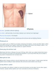
■Provides a site for WBCs to reside and proliferate.
■Removes pathogens from blood. ■Removes old RBCs and platelets. The spleen contains lymphocytes and another kind of white blood cell called macrophages, which engulf and destroy bacteria, dead tissue, and foreign matter and remove them from the blood passing through the spleen. |
|
|
|
What is the thymus?
|
◦thymus: T lymphocytes differentiate in the thymus.
The thymus is a specialized organ of the immune system. The only known function of the thymus is the production of T-lymphocytes (T cells), which are critical cells of the adaptive immune system. |
|
|
What do lymph nodes do?
|
■Provide a site for WBCs to reside and proliferate (like the spleen). ■Removes pathogens from lymph (like the spleen). ■Residing lymphocytes (B and T lymphocytes) monitor lymph for foreign antigens, and initiate an immune response (inflammatory response) when exposed to foreign antigens (like the spleen). Lymph nodes are garrisons of B, T, and other immune cells. Lymph nodes are found all through the body, and act as filters or traps for foreign particles. They contain white blood cells that use oxygen to process. Thus they are important in the proper functioning of the immune system. B cells are lymphocytes that play a large role in the humoral immune response (as opposed to the cell-mediated immune response, which is governed by T cells). The Humoral Immune Response (HIR) is the aspect of immunity that is mediated by secreted antibodies (as opposed to cell-mediated immunity, which involves T lymphocytes) produced in the cells of the B lymphocyte lineage (B cell). Cell-mediated immunity is an immune response that does not involve antibodies or complement but rather involves the activation of macrophages, natural killer cells (NK), antigen-specific cytotoxic T-lymphocytes, and the release of various cytokines in response to an antigen. T lymphocytes belong to a group of white blood cells known as lymphocytes, and play a central role in cell-mediated immunity. They can be distinguished from other lymphocyte types, such as B cells and natural killer cells by the presence of a special receptor on their cell surface called T cell receptors (TCR) for specific antigens. T cells do not use antibodies because the have receptors that target antigens. Antigens are also presented to them by monocytes. Production of antibodies is the main function of the humoral immune system whereas cell-mediated immunity uses cells to fight infection (T-cells, NK cells, macrophages)
|
|
|
What are the Basic aspects of innate immunity and inflammatory response?
|
Innate = first line of defense = kills anything that doesn't look right = not specific to a particular pathogen / antigen. ◦Skin: natural flora, has a layer of keratin as protection. In a healthy animal, the internal tissues, e.g. blood, brain, muscle, etc., are normally free of microorganisms. However, the surface tissues, i.e., skin and mucous membranes, are constantly in contact with environmental organisms and become readily colonized by various microbial species. The mixture of organisms regularly found at any anatomical site is referred to as the normal flora. ◦Mucus membranes: traps pathogen in mucus, and cilia moves it out. ◦Phagocytes (macrophages, neutrophils, dendritic cells): engulf pathogen. ◦Natural killer cells: destroy infected cells. ◦Antimicrobial proteins: eg, tears (lyse bacteria), interferons (interfere with virus replication), complement (enhance the immunity reaction). ◦Fever/inflammation: WBCs are more active at higher temperature, and inflammation recruits WBCs to site of infection by sending out chemical signals and making capillaries more permeable. Inflammation is the complex biological response of vascular tissues to harmful stimuli, such as pathogens, damaged cells, or irritants. It's not there for just trauma from injury or allergic responses, it's an endogenous antibacterial activity (Endogenous substances are those that originate from within an organism). Anti-inflammatory drugs that may prevent septic shock from a response to a bacteria also reduce the the endogenous antibacterial activities of other inflammatory cells like T cells, B cells, and other leukocytes that help fight infection. Inflammation is a protective attempt by the organism to remove the injurious stimuli as well as initiate the healing process for the tissue. The immune system has 2 major divisions: innate and acquired. Innate immunity is the one every individual is born with. It is NONSPECIFIC and includes barriers (i.e. skin), bodily secretions (i.e. stomach acid), and cells like macrophages and natural killers (NK). Acquired immunity is VERY SPECIFIC, designed to combat particular invaders, and develops as the individual matures. It has 2 subdivisions: CELL-MEDIATED and HUMORAL. Cell-mediated immunity is composed of T-cells that mature in the thymus. Humoral immunity is made of B-cells and antibodies.
|
|
|
|
What is the Concept of antigen and antibody?
|
◦Antibody = lock, Antigen = key. Each antibody is specific to the binding of an antigen.
◦Antibody is like a Y, the fork binds antigen ◦The tips of the fork are called hypervariable regions because they are unique to each antigen-specific antibody. Each of these variants can bind to a different target, known as an antigen. It is a small region at the tip of the protein is extremely variable, allowing millions of antibodies with slightly different tip structures, or antigen binding sites, to exist. ◦The antibody consists of 2 light chains and 2 heavy chains linked together by disulfide bonds. Recognition of an antigen by an antibody tags it for attack by phagocytes . Antibodies can also neutralize targets directly by binding to specific sites to prevent attachment. Lastly, they trigger destruction of pathogens by stimulating other immune responses such as using cytokines to initiate an amplifying cascade to kill the pathogen via apoptosis. |
|
|
|
What is the structure of the antibody molecule?
|
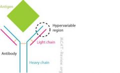
Antibodies contribute to immunity in three ways: they prevent pathogens from entering or damaging cells by binding to them; they stimulate removal of pathogens by macrophages and other cells by coating the pathogen; and they trigger destruction of pathogens by stimulating other immune responses such as the complement pathway.
The immune system can be subdivided into 2 major components: non-specific (innate) and acquired immunity. The non-specific immune system consists of barrier (i.e. skin, mucous membranes) and cells that non-specifically remove invaders (i.e. macrophages, dendritic cells, neutrophils, etc) The acquired immune system synthesizes cells directed against a specific invader. It has the capacity to remember past infections and responds rapidly in case of recurrence. The non-specific system does not have this ability and always responds in the same way to every infection. T and B cells make up the acquired immunity system, which has humoral and cell-mediated divisions. T cells (helper and cytotoxic) make up the cell-mediated immunity and B cells (which mature into plasma cells and make antibodies) make up the humoral immunity. |
|
|
|
What is the Mechanism of stimulation by antigen?
|
◦pathogen enters antigen-presenting-cell (APC) through phagocytosis or by infecting the cell
◦pieces of the pathogen gets displayed at the surface of APCs so that T cell receptors can recognize the antigen. The T cell receptors could be cytotoxic T cells or T helper cells. ◦T cell recepters recognize the presented antigen, and activates various immune responses. |
|
|
|
What happens with antigen presentation with an extracellular pathogen?
|
1.macrophage engulfs pathogen.
2.pieces of the pathogen becomes the antigen and gets presented at the macrophage's cell surface. 3.helper T cells recognize the presented antigen, and activates macrophages to kill pathogen that the macrophage has just engulfed. Helper T cells also activate B cells to produce antibodies against the pathogen. The host cell is preserved with extracellular pathogens because the host cell was not infected, it engulfed the pathogen. So it works with the Helper T cells. Macrophages are cells that search and ingest (phagocytize) foreign invaders as part of the non-specific immune response. After macrophages complete their meal, they inform local T lymphocytes (T-cells) of a possible infection. T-cells, mature in the thymus, serve to recognize surface markers on bacterial cells and they secrete molecules that destroy infected cells of the body. T-cells, however, do not physically engulf foreign invaders. Cytotoxic T cells destroy virally infected cells and tumor cells, and are also implicated in transplant rejection. T helper cell assist other white blood cells in immunologic processes, including maturation of B cells into plasma cells and activation of cytotoxic T cells and macrophages, among other functions. Memory T cells are a subset of antigen-specific T cells that persist long-term after an infection has resolved. They quickly expand to large numbers of effector T cells upon re-exposure to their cognate antigen, thus providing the immune system with "memory" against past infections. Natural killer cells are a type of lymphocyte (a white blood cell) and are part of the innate immunity because they do not respond to antigens, they respond to signals that tell it to kill the cell. |
|
|
|
What happens with antigen presentation with an intracellular pathogen?
|
1.pathogen invades host cell.
2.pieces of the pathogen gets presented on the host cell surface. 3.cytotoxic T cells recognize the presented antigen, and signals the infected cell to self-destruct. When the pathogen invades the host cell, the host cell is not preserved and the cytotoxic T cells signals for the host cell to self-destruct. |
|
|
|
What does saliva do and what are the parts of the digestive system?
|
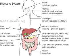
•saliva as lubrication and source of enzymes
◦saliva dissolves food. ◦saliva contains mucin, a protein that lubricates the bolus (chewed up food ball). ◦saliva contains amylase, which breaks down polysaccharides (starch and glycogen). ◦saliva also contains antibodies and lysozyme. Lysozymes kill pathogens by digesting the cell walls. Lysozyme is an enzyme found in tears, the mucus lining of stomach, and other secretions. It is responsible for breaking down the polysaccharide walls of many kinds of bacteria and thus it provides some protection against infection. Amylase is an enzyme that degrades carbohydrates and is released in only 2 locations, the MOUTH and the SMALL INTESTINE. Salivary amlyase is secreted by salivary glands to initiate carbohydrate digestion in the mouth. The pancreas supplies pancreatic amlyase, which is released into the duodenum of the small intestine. Carbohydrate digestion is completed in the small intestine by lactase and maltase, other pancreatic enzymes. The STOMACH only releases PEPSIN, an enzyme that digests only protein. |
|
|
|
What are the digestive activities of the mouth, stomach, small intestine, large intestine?
|
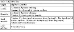
|
|
|
|
What are the major enzymes of the digestive tract?
|
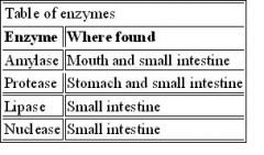
|
|
|
|
What is the epiglottal action?
|
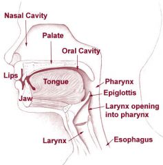
◦epiglottis = flap of cartilage that closes off airway when you're swallowing. The epiglottis is a flap of elastic cartilage tissue covered with a mucus membrane, attached to the root of the tongue. The epiglottis guards the entrance of the glottis, the opening between the vocal folds. It is normally pointed upward during breathing with its underside functioning as part of the pharynx, but during swallowing, elevation of the hyoid bone draws the larynx upward; as a result, the epiglottis folds down to a more horizontal position, with its upper side functioning as part of the pharynx. In this manner it prevents food from going into the trachea and instead directs it to the esophagus, which is posterior. The epiglottis is one of nine cartilaginous structures that make up the larynx (voice box). While breathing, it lies completely within the pharynx. When swallowing it serves as part of the anterior of the larynx. The pharynx is part of the digestive system and respiratory system of many organisms. Because both food and air pass through the pharynx, a flap of connective tissue called the epiglottis closes over the trachea when food is swallowed to prevent choking or aspiration. In humans the pharynx is important in vocalization.
The respiratory system begins with either nose or mouth. Then oxygen is propelled into PHARYNX and then LARYNX, which houses the voice box. The larynx continues into the TRACHEA, which divides into 2 main bronchi. |
|
|
|
What is the pharynx?
|
•pharynx (function in swallowing)
◦pharynx = throat = between mouth and esophagus. ◦muscular tube that squeezes and routes food to the esophagus when swallowing (closes off pathways to nasal cavity and airway). |
|
|
|
What is the esophagus?
|
•esophagus (transport function)
◦muscular tube that propels bolus (food) to the stomach by peristalsis. ◦peristalsis = squeezing stuff through a tube. Peristalsis is a series of organized muscle contractions that occur throughout the digestive tract with smooth muscle. |
|
|
|
How does the stomach store and churn food?
|
◦storage = the stomach is a muscular bag that is elastic and can stretch to store food.
◦churning = mechanical digestion = mixing food. The gastrointestinal tract is a large, muscular tube that extends from the mouth to the anus, where the movement of muscles and release of hormones and enzymes digest food. |
|
|
|
Explain how the stomach has low pH, gastric juice, and protection by mucus against self-destruction.
|
The stomach has nonspecific immune defense.◦Parietal cells secrete HCl that causes the pH to be very acidic. Parietal cells also secrete the intrinsic factor, a protein needed to absorb Vitamin B12 in the small intestines. ◦Gastric juice = HCl + pepsin + mucus + chymosin = secreted by the cells of the stomach. ◦Pepsin = protease that works best in acidic environment. ◦Goblet cells secrete mucus lining that protect the stomach from the acid and self-digestion. Chief cells of the stomach secrete the digestive enzymes (pepsin and chymosin) of the stomach. Gastric juice is a strong acidic liquid. Enteroendocrine cells are specialized endocrine cells of the gastrointestinal tract. They secrete the hormone gastrin into the bloodstream when peptides are detected in the stomach. This causes gastric glands in the lining of the stomach to secrete gastric juice. Its main components are digestive enzymes pepsin and chymosin, hydrochloric acid, and mucus. The mucus contains antiseptic enzymes (such as lysozyme), proteins such as lactoferrin, glycoproteins known as mucins that are produced by goblet cells in the mucous membranes and submucosal glands, immunoglobulins, and inorganic salts and it protects against infectious agents. Chymosin's role in digestion is to curdle or coagulate milk in the stomach, a process of considerable importance in the very young animal. If milk were not coagulated, it would rapidly flow through the stomach and miss the opportunity for initial digestion of its proteins. Pepsin is an enzyme whose precursor form (pepsinogen) is released by the chief cells in the stomach and that degrades food proteins into peptides. A protease breaks down proteins. A protease is any enzyme that conducts proteolysis, that is, begins protein catabolism by hydrolysis of the peptide bonds that link amino acids together in the polypeptide chain forming the protein. Most proteases work best in acidic conditions. Chymosin is secreted as an inactive proenzyme called prochymosin that, like pepsin, is activated on exposure to acid, which makes sense considering its mission. Because the parietal cells secrete HCl, this activates the pepsinogen and prochymosin. Goblet cells are glandular simple columnar epithelial cells whose sole function is to secrete mucin, which dissolves in water to form mucus. They are found scattered among the epithelial lining of organs, such as the intestinal and respiratory tracts.
|
|
|
|
Explain how the stomach does the production of digestive enzymes and how it's the site of digestion.
|
◦Chemical digestion: Stomach produces pepsin and chymosin, which digests proteins (secreted in an inactive form, gets activated in acidic environment)
◦Pepsin and chymosin are special in that they works best at very acid pH. ◦Mechanical digestion: Stomach churns food. |
|
|
|
What is the structure (gross) of the stomach?
|
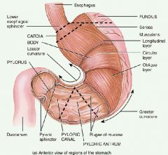
◦banana shaped bag that can stretch.
◦inner membrane densely folded (rugae), so can accomodate stretching. ◦sealed off on the top by the cardiac (gastroesophageal) sphinctor. ◦sealed off on the bottom by the pyloric sphinctor. Rugae is a term used in anatomy that refers to a series of ridges produced by folding of the wall of an organ. Most commonly the term is applied to the internal surface of the stomach. Bolus (masticated food) enters the stomach through the esophagus via the cardiac sphincter. The stomach releases proteases (protein-digesting enzymes such as pepsin) and hydrochloric acid, which kills or inhibits bacteria and provides the acidic pH for the proteases to work. Food is churned by the stomach through muscular contractions of the wall. The boluses are converted into chyme (partially-digested food). Chyme slowly passes through the pyloric sphincter and into the duodenum, where the extraction of nutrients begins. Depending on the quantity and contents of the meal, the stomach will digest the food into chyme anywhere between 40 minutes and a few hours. |
|
|
|
Explain how the liver produces of bile.
|
•production of bile: liver makes bile from cholesterol, stores it in gall bladder. Bile aids the process of digestion of lipids in the small intestine. Bile is stored in the gallbladder and upon eating is discharged into the duodenum. Bile is a complex fluid containing water, electrolytes, bicarbonate, and a battery of organic molecules including bile acids, cholesterol, phospholipids and bilirubin that flows through the biliary tract into the small intestine. The bicarbonate in bile is not secreted by the pancreas, the pancrease secretes bicarbonate in pancreatic juice to neutralize the acidic chyme from the stomach for the small intestine. There are two fundamentally important functions of bile in all species: 1)•Bile contains bile acids, which are critical for digestion and absorption of fats and fat-soluble vitamins in the small intestine. 2)•Many waste products, including bilirubin, are eliminated from the body by secretion into bile and elimination in feces. Because of the bicarbonates, bile is an alkaline substance.
|
|
|
|
Explain the liver's role in nutrient metabolism and vitamin storage.
|
◦Makes and stores glycogen from glucose.
◦Gluconeogenesis from glycerol and amino acids (amino acids undergo deamination to synthesize glucose). It is the synthesis of glucose from certain amino acids, lactate or glycerol.Glycogenolysis (the breakdown of glycogen into glucose). Glycogenesis (the formation of glycogen from glucose). The liver breaks down insulin and other hormones. The liver is responsible for protein synthesis. ◦Breaks down fat-soluble toxins, makes cholesterol, makes lipoproteins that are used to transport fats in the blood stream. The liver and the pancrease have lipase enzymes to break down fat, but the pancreatic lipase is used to digest lipids in the small intestine. The liver converts ammonia to urea. The liver converts bilirubin into bile hemoglobin. ◦Stores vitamins (A, D and B12) and iron. ◦Detox: metabolize alcohol, remove ammonia in blood. The liver also does lipogenesis, the production of triglycerides (fats). The liver produces and excretes bile (a greenish liquid) required for emulsifying fats. The liver produces albumin, the major osmolar component of blood serum. A lipoprotein is a biochemical assembly that contains both proteins and lipids whose function is to transport water-insoluble lipids in the water-based bloodstream. The liver plays the major role in producing proteins that are secreted into the blood, including major plasma proteins, factors in hemostasis (like fibrinogen) and fibrinolysis, carrier proteins, hormones, prohormones and apolipoproteins (for example, albumin). Apolipoproteins are proteins that bind to fats (lipids). They form lipoproteins, which transport dietary fats through the bloodstream. |
Glycogen is a storage polysaccharide sometimes known as animal starch. Plants do not produce glycogen, rather they produce their own "plant" starch. Glycogen is a highly branched molecule.
|
|
|
Explain the liver's role in blood glucose regulation and detoxification.
|
◦Blood glucose regulation by liver:
■Blood sugar too low: glucogenesis (breakdown glycogen into glucose). ■Blood sugar too high: glycogenesis (create glycogen from glucose). ◦Detoxification: metabolize alcohol (alcohol dehydrogenase), remove blood ammonia, inactivate various other drugs/toxins. |
|
|
|
What is the structure (gross) of the liver?
|
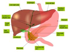
largest gland in body, spans both sides of the abdomen (though right side much larger). Ducts draining to duodenum and gall bladder. Bile can either drain directly into the duodenum via the common bile duct or be temporarily stored in the gallbladder via the cystic duct.
|
|
|
|
Where does bile get stored?
|
•storage in gall bladder
◦Gall bladder stores excess, unused bile, and concentrates it. Secretes it when needed. Bile is produced by the liver and flows into a duct into the gall bladder. |
|
|
|
What is the function of bile?
|
•function: bile is an emulsifying agent (not an enzyme). Bile breaks down large fat droplets into smaller microscopic droplets through emulsification. Bile breaks down large fat droplets into smaller microscopic droplets by breaking up the large droplets and incorporating them into bile micelles. By forming the micelles, the fat and water are mixed together (forming a colloid). Now that the fat and water are mixed, the enzymes can break the fat down (because the enzymes are water soluble). Emulsification (the process of mixing fat with water) happens because of these micelles. Bile form micelles to transport fat-soluble vitamins and complicated lipids for absorption in the small intestine. This increases the total surface area of the fat for lipase action. Bile acids are amphipathic. This means that they have both water-soluble and water-insoluble (or fat-soluble) parts. Emulsifying agents are amphipathic molecules that are able to mix fats with water. Micelle formation is essential for the absorption of fat-soluble vitamins and complicated lipids within the human body. This allows the absorption of complicated lipids (e.g., lecithin) and lipid soluble vitamins (A, D, E and K) within the micelle by the small intestine. A micelle is an aggregate of surfactant molecules dispersed in a liquid colloid. A typical micelle in aqueous solution forms an aggregate with the hydrophilic "head" regions in contact with surrounding solvent, sequestering the hydrophobic single tail regions in the micelle centre. To emulsify means to disperse a liquid into another liquid with which it is immiscible, making a colloidal suspension. Bile acts as an emulsifying agent in the digestive tract by dispersing ingested fats into small globules.
|
|
|
How is the pancreas involved in the production of enzymes and bicarbonate?
|
◦Pancreas is the major source for all the digestive enzymes.
■Amylase - digests starch - called pancreatic amylase. Amylase is also in saliva. ■Various proteases. A protease breaks down proteins. A protease is any enzyme that conducts proteolysis, that is, begins protein catabolism by hydrolysis of the peptide bonds that link amino acids together in the polypeptide chain forming the protein. Most proteases work best in acidic conditions. ■Lipase - digests fat - called pancreatic lipase. The liver also produces lipase to break down fat-soluble toxins (hepatic lipase). The pancreatic lipase combines with bile to digest lipids in small intestine. ■Nuclease - called pancreatic nuclease - digest nucleic acids (DNA and RNA) to nucleotides in the duodenum. ◦Pancreas makes HCO3- (bicarbonate) to neutralize the HCl from the stomach. This is an alkaline fluid known as pancreatic juice. Specific cells that line the pancreatic ducts, called centroacinar cells, secrete a bicarbonate- and salt-rich solution into the small intestine. |
|
|
|
How do enzymes transport to the small intestine?
|
◦Digestive enzymes of pancreas = flows into small intestine via duct.
Most of the digestive enzymes that act in the small intestine are secreted by the pancreas and enter the small intestine via the pancreatic duct. The four major classes of nutrients that undergo digestion are proteins, lipids (fats), carbohydrates, and nucleic acids. Proteins are degraded by proteases. Lipids are degraded by lipases. Carbs are degraded by amylase. Nucleic acids are degraded by nucleases. The small intestine is the site where most of the nutrients from ingested food are absorbed. Paracrine signaling is a form of cell signaling in which the target cell is near ("para" = near) the signal-releasing cell. The enzymes from the pancreas enter the small intestine in response to hormones. The small intestine is where most chemical digestion and nutrient absorption takes place. The stomach secretes protein-digesting enzymes and strong acids to aid in food digestion before sending partially-digested food (chyme) to the small intestines. The stomach releases proteases (protein-digesting enzymes such as pepsin) and hydrochloric acid, which kills or inhibits bacteria and provides the acidic pH for the proteases to work. Food is churned by the stomach through muscular contractions of the wall turning the boluses into chyme (partially digested food). Chyme slowly passes through the pyloric sphincter and into the duodenum, where further digestin occurs and the extraction of nutrients begins. |
|
|
|
What is the gross structure of the pancreas?
|
tadpole-shaped gland with duct leading to duodenum
|
|
|
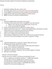
How does the small intestine absorb food molecules and water?
|
◦Small intestine is the major place for digestion and absorption.
◦Folds, villi, and microvilli increases the surface area for absorption. ◦Absorbs digested food into circulation (fats into lacteals, all other into capillaries). ◦Active transport occurs to absorb against concentration gradient. Water and lipids are absorbed throughout the small intestine by passive diffusion. Sodium is absorbed by active transport. Digested food is now able to pass into the blood vessels in the wall of the intestine through the process of diffusion. The small intestine is the site where most of the nutrients from ingested food are absorbed. The inner wall, or mucosa, of the small intestine is lined with simple columnar epithelial tissue. Structurally, the mucosa is covered in wrinkles or folds called plicae circulares, which are considered permanent features in the wall of the organ. They are distinct from rugae of the stomach which are considered non-permanent or temporary allowing for distention and contraction. From the plicae circulares project microscopic finger-like pieces of tissue called villi (Latin for "shaggy hair"). The individual epithelial cells also have finger-like projections known as microvilli. The function of the plicae circulares, the villi and the microvilli is to increase the amount of surface area available for the absorption of nutrients. Each villus has a network of capillaries and fine lymphatic vessels called lacteals close to its surface. The epithelial cells of the villi transport nutrients from the lumen of the intestine into these capillaries (amino acids and carbohydrates) and lacteals (lipids). The absorbed substances are transported via the blood vessels to different organs of the body where they are used to build complex substances such as the proteins required by our body. This is called diffusion. The food that remains undigested and unabsorbed passes into the large intestine. |
|
|
|
What is the function and structure of villi?
|
◦Villi = finger-like protrusions that line the wall of the small intestine.
◦Microvilli = same structure as villi but they line the surface of a single absorptive cell lining the villi. Microvilli are extensions of villi. Both the microvilli and villi increase the surface area of the small intestine to aid in absorption of nutrients. Digested nutrients (including sugars and amino acids) pass into the villi through diffusion. Circulating blood then carries these nutrients away via the villus capillaries (for nutrients like amino acids and sugars) and villus lacteals (for fat). It is important that the food is absorbed at a considerably fast rate so as to allow more food to be absorbed. (If the process is too slow, the concentration of the blood in the blood vessels and the food will be equal, thus, diffusion will not occur.) |
|
|
|
Explain how the small intestine does the production of enzymes and is the site of digestion.
|

◦The small intestine is the major place for digestion and absorption.
◦Pancreas is the major source for enzymes. However, the small intestine does make some of its own enzymes, including maltase and peptidase. The pancrease secretes chymotrypsin and trypsin to digest proteins in the small intestine. |
|
|
|
Explain how the small intestine does the neutralization of stomach acid.
|
◦The pancreas makes bicarbonate ion to neutralize the HCl from the stomach.
◦This neutralization facilitates enzymes as they digest in the small intestine, which would be denatured by stomach pH. |
|
|
|
What is the structure (anatomic subdivisions) of the small intestine?
|
1.Duodenum.
2.Jejunum. 3.Ileum. |
|
|
|
What does the large intestine absorb?
|
•absorption of water: The large intestine absorbs any remaining water that is not absorbed by small intestine.
The primary function of the colon is to absorb water and electrolytes (substances, such as salts, that in solution take on an electrical charge) from the ileal contents and to store fecal material until it can be evacuated by defecation. |
|
|
|
What is bacterial flora of the large intestine?
|
◦Ferment undigested carbohydrates. Without gut flora, the human body would be unable to utilize some of the undigested carbohydrates it consumes, because some types of gut flora have enzymes that human cells lack for breaking down certain polysaccharides. Bacteria turn carbohydrates they ferment into short chain fatty acids that can be used by host cells, providing a major source of useful energy and nutrients for humans.
Bacterial flora make gas. (flatus) ◦Produce vitamin K (important for clotting). The large intestine is populated by bacteria, many of which are essential for human nutrition. Some of these bacteria manufacture vitamin K (an important substance for blood clotting). The bacterial flora is also important for normal large intestine function by creating gas that aids peristalsis of fecal matter in the large intestine. |
|
|
|
What is the structure (gross) of the large intestine?
|
Lobes/pockets along its length due to muscle tone. Unlike small intestine, the large intestine has no villi or folds. The colon is the last part of the digestive system in most vertebrates; it extracts water and salt from solid wastes before they are eliminated from the body, and is the site in which flora-aided (largely bacteria) fermentation of unabsorbed material occurs. Unlike the small intestine, the colon does not play a major role in absorption of foods and nutrients. However, the colon does absorb water, potassium and some fat soluble vitamins. There are no villi in the large intestine and peristalsis is much less forceful than in the small intestine. As water is absorbed, the contents of the large intestine change from a watery liquid and are compressed into semisolid feces. Nerve endings in the large intestine signal the brain that it is time for a bowel movement. The fecal material moves through the colon down to several remaining inches known as the rectum and out through the anus. The large intestine is divided into the cecum, colon, then rectum. The main components of the large intestine include the cecum, appendix, colon, rectum, anus, and anal canal. E. Coli that inhabit the colon, inhabit all areas of the large intestine - like the appendix, the rectum, the cecum. So a ruptured appendix needs penicillin not from the E. Coli going from the colon to the appendix, but from E.coli travelling to the abdominal cavity, where it does not belong.
|
|
|
|
What does the rectum do?
|
Rectum (storage and elimination of waste, feces)
•Rectum stores feces. •The anal sphincter ties the end of the rectum. •During defecation, sphincter opens, feces are released through the anus. |
|
|
|
What is the sphincter muscle?
|
◦Cardiac sphincter (gastroesophageal sphincter): sphincter between esophagus and stomach. Prevents back flow of food.
◦Pyloric sphincter: between stomach and small intestine. Releases food into the small intestine Ileocecal sphincter: sphincter between the ileum and colon (between the small intestine and large intestine). ◦Anal sphincter: at the end of rectum. ties the end of the rectum. Sphincters: A band of muscle encircling one of the body's ‘tubes’, and so able to alter the tube by contracting/relaxing. In the urogenital tract, there are sphincters at the exit from the bladder. They occur also in the walls of small arterial blood vessels, regulating the flow into the capillaries bed, (pre-capillary sphincters). Those which assist retention of the excreta are partly under voluntary control (urine and feces); all the others, formed only of smooth muscle, are regulated by the autonomic nervous system; in general, sympathetic nerves constrict them; in the gut, parasympathetic nerves relax them. The thin sheet of circular muscle of the iris which constricts the pupil of the eye is also known as a sphincter — it is stimulated by parasympathetic nerves, in response to light and as part of accommodation for near vision. Dilation of the pupil is controlled by the sympathatic nervous system for far vision. |
|
|
|
What is peristalsis?
|
•peristalsis: involuntary movement of smooth muscles, squeezes food along the digestive tract.
Occurs in the esophagus, stomach, and small intestine. As opposed to the more continuous peristalsis of the small intestines, faecal contents are propelled into the large intestine by periodic mass movements. These mass movements occur one to three times per day in the large intestines and colon, and help propel the contents from the large intestine through the colon to the rectum. |
|
|
Explain how blood pressure is related to homeostasis in the excretory system.
|
◦If blood pressure or blood volume is too low:
■Renin-angiotensin pathway: Kidney (JGA cells) release renin, renin activates angiotension I, which in turn gets converted into angiotensin II. Angiotension II is a potential stimulator of the adrenal gland and stimulates aldosterone release, the end result is to raise blood pressure and volume. ■Aldosterone (aka mineralocorticoid): Adrenal glands release aldosterone, causes kidney (distal tubules) reabsorb more Na+, which in turn causes more water reabsorption. ■ADH (made in hypothalamus, stored in pituitary like oxycontin): causes more water reabsorption in the kidney tubules, raising blood pressure. High levels also cause vasoconstriction because vasoconstriction usually results in an increase in systemic blood pressure. ◦If blood pressure too high, all the above hormones stop releasing. Also, the heart can release ANP (Atrial natriuretic peptide), which antagonizes aldosterone and ADH and cause kidney to excrete both more Na+ and more water. ANP can also cause vasodilation. It is involved in the homeostatic control of body water, sodium, potassium and fat (adipose tissue). ANP acts to reduce the water, sodium and adipose loads on the circulatory system, thereby reducing blood pressure. When vessels dilate, the flow of blood is increased due to a decrease in vascular resistance. Therefore, dilation of arterial blood vessels (mainly arterioles) leads to a decrease in blood pressure. Atrial natriuretic peptide counteracts the effects of both ADH and aldosterone. The kidney also releases erythropoietin when oxygen levels are low in the blood and stimulate the bone marrow to produce more red blood cells. |
|
|
What is osmoregulation?
|
◦Ions of blood plasma are mainly Na+ and Cl-. Excess of water in body fluids leads to decreased osmotic pressure in blood because water moves from body fluids (interstitial fluids) to blood, lowering the pressure of the dissolved solutes (osmotic pressure) and increasing blood volume. ◦Blood osmolarity is determined predominantly by Na+ and Cl- ◦Blood osmolarity too low (not enough solute) → aldosterone, reabsorb Na+. Cl- follows. ◦Kidney tubules' secretion and reabsorption regulates osmolarity.
◦Other ions: 1) K+ is regulated by aldosterone. ■Aldosterone: reabsorb Na+, excrete K+ in urine. High levels of K+ in blood signals aldosterone release, low levels of K+ in blood inhibits aldosterone. Aldosterone is a hormone that increases the reabsorption of sodium and water and the release (excretion) of potassium in the kidneys. This increases blood volume and, therefore, increases blood pressure. ■Calcium regulated by PTH. ■PTH = parathyroid hormone = more Ca2+ reabsorption in kidney tubules (also, bone break down to release calcium and small intestine to absorb more calcium). Calcitonin works to decrease the amount of blood calcium levels. |
|
|
What is acid–base balance in regards to homeostasis?
|
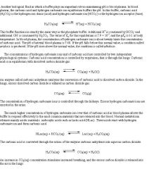
•acid-base balance: keep blood pH and cellular pH constant.
◦Buffer systems: Bicarbonate buffer system (blood and extracellular fluid), Phosphate Buffer System (inside cells). The inside of cells has K+ and hydrogen phosphate ions. ◦CO2 + H2O ↔ H2CO3 ↔ H+ + HCO3- ◦Breathing out CO2 decreases the acidity in blood. ◦Kidney tubules: ■Bicarbonate ion (HCO3-): excretion of it by kidneys makes blood more acidic. Reabsorption makes blood more basic. ■H+ excretion by kidneys gets rid of acidity. The kidneys are slower to compensate, but renal physiology has several powerful mechanisms to control pH by the excretion of excess acid or base. In responses to acidosis, tubular cells reabsorb more bicarbonate from the tubular fluid, collecting duct cells secrete more hydrogen and generate more bicarbonate. In responses to alkalosis, the kidney may excrete more bicarbonate by decreasing hydrogen ion secretion from the tubular epithelial cells. The respiratory system regulates the level of carbon dioxide (CO2) while the kidneys control the concentration of bicarbonate (HCO3-). The kidneys reabsorb bicarbonate from the blood when the blood is too acidic and excrete bicarbonate from the blood when it the blood is too basic. |
|
|
|
What is the removal of soluble nitrogenous waste?
|
◦Urine = concentrated urea in water, with some salt.
◦Urea = harmless form of toxic ammonia = nitrogenous waste. ◦Amino acids and Ammonia → Urea → peed out. Urea, also called carbamide, is an organic chemical compound which essentially is the waste produced when the body metabolizes protein. Naturally, urea is produced when the liver breaks down protein or amino acids, and ammonia. The kidneys then transfer the urea from the blood to the urine. Clearance is a direct measure of kidney function. The rate at which a particular chemical is removed from the blood indicates kidney efficiency. This rate of removal is called the renal clearance (creatinine clearance). |
|
|
|
What is the structure of the kidney?
|
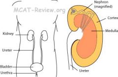
•cortex = outer shell of kidney = contains the convoluted tubules. •medulla = inner part of kidney = contains loop of Henle. The main function of the loop of henle is to to create a concentration gradient in the medulla of the kidney. it creates an area of high sodium concentration deep in the medulla, near the collecting duct. Water present in the filtrate in the collecting duct flows through aquaporin channels out of the collecting duct, moving passively down its concentration gradient. This process reabsorbs water into the blood and creates a concentrated urine for excretion. The collecting duct system of the kidney consists of a series of tubules and ducts that connect the nephrons to the ureter. The cortex is the outer part of the kidney. This is where blood is filtered. We call this process "ultra-filtration" or "high pressure filtration" because it only works if the blood entering the kidney in the renal artery is at high pressure. The medulla is the inside part of the kidney. This is where the amount of salt and water in your urine is controlled. It consists of billions of loops of Henlé. These work very hard pumping sodium ions. ADH makes the loops work harder to pump more sodium ions. The result of this is that very concentrated urine is produced. Aquaporins are proteins embedded in the cell membrane that regulate the flow of water. Aquaporins selectively conduct water molecules in and out of the cell, while preventing the passage of ions and other solutes. Here is the nephron anatomy: The glomerulus → Bowman's capsule → proximal convoluted tubue → Loop of Henle (descending and ascending limbs) → distal convoluted tubue → collecting ducts → minor calyx → major calyx → renal pelvis → out the kidney into the ureter.
|
|
|
|
What is the nephron, glomerulus, Bowman's capsule, and proximal tubule?
|
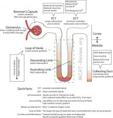
•Nephron = functional unit of kidney = glomerulus, Bowman's capsule, proximal tubule, loop of Henle, and distal tubule. The collecting duct is not part of the nephron and is shared by many nephron to connect to the ureter. •glomerulus = ball of fenestrated capillaries. •Bowman's capsule = Cup/Capsule that surrounds the glomerulus. •proximal tubule = convoluted tubule on the side of the Bowman's capsule = the major site for reabsorption (nutrient, salts and water) and secretion (except for K+, the secretion of which is the job of distal convoluted tubule in response to aldosterone). The glomerulus is the main filter of the nephron and is located within the Bowman's capsule. The glomerulus resembles a twisted mass of tiny tubes through which the blood passes. The glomerulus is semipermeable, allowing water and soluble wastes to pass through and be excreted out of the Bowman's capsule as urine. The filtered blood passes out of the glomerulus and into a vein to the heart. The Bowman's capsule contains the primary filtering device of the nephron, the glomerulus. Blood is transported into the Bowman's capsule. Within the capsule, the blood is filtered through the glomerulus and then passes out into the vein. Meanwhile, the filtered water and aqueous wastes are passed out of the Bowman's capsule into the proximal convoluted tubule. The proximal tubule is the portion of the duct system of the nephron leading from Bowman's capsule to the loop of Henle. The proximal convoluted tubule reabsorbs 2/3 of all water entering the nephron and the descending loop of Henle passively reabsorbs water. Sodium (Na+), potassium (K+) and chloride (Cl-) ions are reabsorbed from the urine by active transport in the ascending loop of Henle.
|
|
|
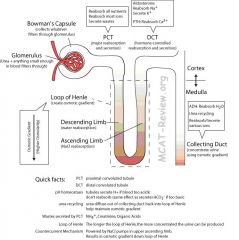
What is the loop of Henle, distal tubule, and collecting duct?
|
•loop of Henle = U shaped loop that dips into the renal medulla = countercurrent multiplier mechanism occurs here. ◦Descending limb = water reabsorption by osmosis because the sodium and chloride ions that exit the ascending limb create a hyperosmotic space in the medullary, to the water in the descending limb diffuses out in response to it. Descending limb is permeable to water, but not to solute. Here water leaves the loop and flows into the medullary space. The filtrate that is left behind from water becomes more and more concentrated as it descends down the loop. ◦Bottom of Loop of Henle = most concentrated with the solutes. ◦Ascending limb = salt reabsorption (permeable to salt, but not water). Here, salt leaves the Loop of Henle and is actively transported into the medullary space while the water is kept filtrate. As the filtrate ascends the loop of henle, it becomes less and less concentrated. Distal tubule = convoluted tubule on the side of the collecting duct = hormone-controlled reabsorption of salts and water. At this point, the body decides how much water and ions to reabsorb using hormones. The distal tubule is the primary site for the kidneys hormone based regulation of calcium (Ca). The distal tubule participates in calcium regulation by reabsorbing Ca2+ in response to parathyroid hormone. Sodium absorption and potassium excretion by the distal tubule is mediated by the hormone aldosterone. Collecting duct = the distal tubules of many nephrons drain here to the ureter= ADH-controlled reabsorption of water. The collecting duct system of the kidney consists of a series of tubules and ducts that connect the nephrons to the ureter. It participates in fluid balance through reabsorption of water and excretion of fluid entering collecting duct as urine by the antidiuretic hormone.
|
|
|
|
What is glomerular filtration?
|
◦Powered by hydrostatic pressure.
◦Both good stuff, bad stuff and ions are filtered out, as long as its small enough. ◦Good stuff: nutrients ◦Bad stuff: urea (creatinine and uric acid are also excreted as waste) ◦Good stuff reabsorbed by the proximal convoluted tubules, bad stuff are left in the tubules and are eventually peed out. Hydrostatic pressure is the pressure gradient of blood being delivered to the glomerulus, which tends to push the plasma filtrate from the capillaries of the glomerulus into the capsular space. A large amount of nutrients and water is filtered from the blood in the glomerulus. It is necessary to reabsorb most of the nutrients and water but leave wastes in the tubule. Selective reabsorption occurs in the proximal convoluted tubule. Glucose, vitamins, important ions and most amino acids are reabsorbed from the tubule back into the capillaries near the proximal convoluted tubule. The proximal convoluted tubule does not secrete waste into the medullary space, the waste stays in the tubules until it is eventually peed out. The glomerular membrane is thick compared to other basement membranes and is rich in negatively charged proteoglycans. The negatively-charged basement membrane repels negatively-charged proteins from the blood, helping to prevent their passage into Bowman's space, while positively charged proteins get filtered out of the blood. |
|
|
|
Explain the secretion and reabsorption of solutes in regards to the formation of urine.
|
◦Proximal convoluted tubules reabsorb all the good stuff (nutrients) and most of the ions. Bad stuff left in the filtrate (urea, ammonia, uric acid) to be peed out.
◦Loop of Henle reabsorbs water and salt using the countercurrent mechanism. ◦Distal convoluted tubules selectively reabsorb or secrete stuff based on hormonal control. ◦Collecting duct reabsorb water to concentrate urine if ADH present, so it's ability to secrete and absorb is affected by hormones like the distal convoluted tubule. ◦Regulation of blood pH: kidneys excrete H+ when blood too acidic (or absorb more HCO3-), excrete HCO3- when blood too basic (or absorb more H+). The tubular cells of the distal convoluted tubules of the kidneys secrete the H+ and HCO3- and other ions and wastes into the tubular fluid, which eventually gets excreted out as urine. The body relies on homeostatic mechanisms to excrete excess and unnecessary substances. For instance, if someone were to ingest excess NaCl, aldosterone would not increase in the blood in order to reabsorb the excess NaCl. Aldosterone is only added if there are low levels of NaCl, and the kidney tubules need to reabsorb more NaCl instead of it leaving as urine. Instead, the body's homeostatic mechanisms will excrete the excess NaCl. |
|
|
|
What is the concentration of urine determined by?
|
◦The distal convoluted tubule contains dilute solution of filtrate (which includes waste like urea).
◦The collecting duct concentrates it by water reabsorption (facilitated diffusion through aquaporins) when ADH is present. ◦Water reabsorption in the collecting duct is possible because the loop of Henle created very high osmolarity (very concentrated) of fluid outside the ascending loop at the bottom. So ADH at the collecting duct decides whether the aquaporins should open and let water diffuse down it's concentration gradient (similar to the way the water diffuses down its concentration gradient out of the descending limb) or keep the aquaporins closed and let the water stay in the urine and get excreted out. |
|
|
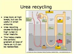
What is the countercurrent multiplier mechanism (basic function) ?
|
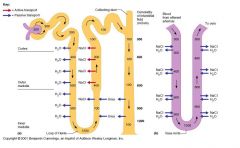
◦What does the Countercurrent multiplier do? It creates an osmotic gradient down the loop of Henle, which is used by the collecting duct to concentrate urine.
◦What drives the creation of this gradient? NaCl pump on ascending limb. ◦What's countercurrent? Descending limb: water flow out of filtrate, impermeable to salt. Ascending limb: salt flow out of filtrate, impermeable to water. ◦What's multiplier? The gradient-producing power of each individual NaCl pump multiplies down the length of the loop of Henle. Longer the loop of Henle, greater the osmotic gradient, more concentrated urine can be produced. ◦What is urea recycling? Urea at the bottom of collecting duct leaks out into the interstitial fluid and back into the filtrate. Contributes to the high osmolarity at the bottom of the loop of Henle so that H20 can be reabsorbed. |
|
|
|
Explain the Storage and elimination of the ureter, bladder, and urethra.
|
•Collecting ducts drain into the ureter.
•Ureters drain into the bladder. •Bladder stores urine: its special epithelium (transitional epithelium) can squish to accomodate storage of large amounts of urine. •Urine gets peed out of the bladder through the urethra. |
|
|
|
What are the functions of the muscular system?
|
◦support, mobility
■Support = muscles maintain your posture when you sit/stand, muscles also stabilize joints, help prevent dislocations. ■Mobility = you move because of skeletal muscles. Your guts move because of smooth muscles. Your blood flow because of pumping action of the heart. Your body has two types of muscle (really more than this, but the others are in your internal organs), movers and stabilizers. Movers are big muscles that move your body parts, hence the name. Stabilizers are muscles that hold your parts in place and prevent you from being damaged while the movers are moving you. |
|
|
|
Explain how the muscular system is involved in peripheral circulatory assistance.
|
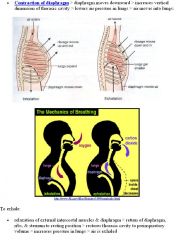
■Heart is a muscle that pumps blood.
■Contraction of skeletal muscles around the deep veins help squeeze the blood through those veins. ■Diaphragm contraction (breathing) sucks blood into the chest cavity, and also squeezes on abdominal veins (from the diaphragm pushing on it). Abdominal breathing is also known as diaphragmatic breathing. The diaphragm is a large muscle located between the chest and the abdomen. When it contracts it is forced downward causing the chest to expand. This causes a negative pressure within the chest forcing air into the lungs. The negative pressure also pulls blood into the chest improving the venous return to the heart. Diaphragmatic contraction is used for inspiration. Diaphragmatic relaxation is used for expiration and the pressure in the pleural cavity increases. |
Gauge pressure is a measure of the pressure compared to local atmospheric pressure. In other words, local atmospheric pressure is arbitrarily given a value of zero. So when a biologist says there is negative pressure inside your chest, there is still pressure in your chest; it is just less pressure than atmospheric pressure. The higher pressure of the atmosphere pushes air into your lungs.
|
|
|
Explain how the muscular system is involved in thermoregulation.
|
◦thermoregulation (shivering reflex)
■Muscles generate heat when you shiver in response to cold. |
|
|
|
What are the Structural characteristics of skeletal, smooth, and cardiac muscle; striated vs nonstriated?
|
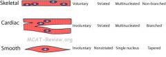
◦Skeletal muscle = striated, voluntary, shaped like long fibers, multinucleated.
◦Smooth muscle = nonstriated, involuntary, shaped like almonds (tapered ends), one nucleus per cell (uninucleated). Smooth muscle do NOT have T-tubules or TROPONIN! ◦Cardiac muscle = striated, involuntary, branched, shaped like fibers cross-linked to one another, multinucleated. ◦Striated = due to sacromere structure (A bands dark, I bands light). Skeletal and cardiac muscles have sarcomeres. ◦Nonstriated = smooth muscles don't have sarcomeres so they're not striated. They still have myosin, actin, and use the sliding filament mechanism. They just are not organized into sarcomeres. Myofilaments, actin, and myosin are necessary for the contractil apparatus and are present in all muscle cells. |
|
|
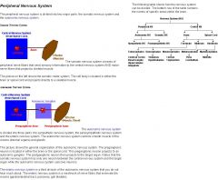
What are motor neurons?
|
◦motor neurons = efferent neurons = signals muscles/organs to do stuff = the opposite of sensory neurons. ■Somatic motor neurons = controls skeletal muscles. ■Autonomic motor neurons = sympathetic and parasympathetic divisions = controls involuntary (smooth, cardiac) muscles. The autonomic nervous system is composed of sympathetic and parasympathetic components. Both components rely on a 2-neuron motor pathway while the somatic nervous system only relies on one. The 2 motor neurons are the preganglionic neuron and postganglionic neuron. The sympathetic nervous system has a short preganglionic neuron and a long post-ganglionic neuron. The parasympathetic nervous system has a long preganglionic neuron and a short postganglionic neuron. Fibers from the ganglion to the effector organ are called postganglionic fibers. Fibers from the CNS to the ganglion are known as preganglionic fibers. Sympathetic preganglionic fibers tend to be shorter than parasympathetic preganglionic fibers because sympathetic ganglia are often closer to the spinal cord than are the parasympathetic ganglia. A ganglion is a biological tissue mass, most commonly a mass of nerve cell bodies in the PERIPHERAL NERVOUS SYSTEM. A nucleus is a collection of nerve cell bodies in the CENTRAL NERVOUS SYSTEM. Ganglia are composed mainly of somata and dendritic structures which are bundled or connected together. Ganglia provide relay points and intermediary connections between different neurological structures in the body, such as the peripheral and central nervous systems. The somatic nervous system works by the sensory neuron relying a message to the CNS, which then sends the message to the effector organs via the motor neuron. The ganglion synapses for the autonomic nervous system, but not the somatic nervous system. Both the autonomic nervous system and somatic system compose of the peripheral nervous system.
|
|
|
|
What are neuromuscular junctions and motor end plates?
|
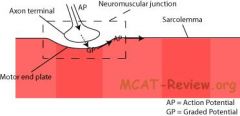
■neuromuscular junction = nerve (axon terminal) meets muscle (motor end plate).
■motor end plate = part of muscle cell membrane (called the sarcolemma) that synapse with the motor neuron, has receptors for the neurotransmitters. The motor end plate is the highly-excitable region of muscle fiber plasma membrane responsible for initiation of action potentials across the muscle's surface, ultimately causing the muscle to contract. ■what happens at the neuromuscular junction? Action potential of nerve reach axon terminal → release neurotransmitters into synapse → receptors on motor end plate (sarcolemma) picks this signal → graded potential created → if reaches threshold, then action potential created → action potential travels down the sarcolemma and cause muscle to contract. |
|
|
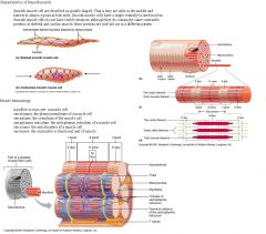
What are voluntary and involuntary muscles?
|
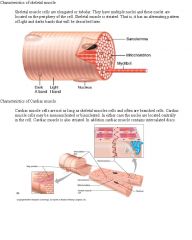
■voluntary = you can control = skeletal muscles, eg. Biceps.
■involuntary = you can't control = smooth (eg. gut) and cardiac (heart) muscles. |
|
|
|
What is sympathetic and parasympathetic innervation?
|
■sympathetic = fight or flight = heart beat faster, pupil dilation, raise blood pressure, blood to muscles, less blood to digestive system.
■parasympathetic = rest and digest = opposite of sympathetic = heart slower, pupil constriction, lower blood pressure, blood to digestive system. ■Both sympathetic and parasympathetic are motor neurons that innervate involuntary muscles. |
|
|
|
What are the functions of the skeletal system?
|
◦structural rigidity and support: bone forms the body's framework. ◦calcium storage: bone stores minerals, especially calcium. When blood calcium is low, parathyroid hormones signal bone tissue to break down and release calcium. ◦physical protection: rib cage protects internal organs. Skull protects brain. Spine protects spinal cord. Many large bones also shelter bone marrow that contains stem cells that make blood cells and platelets. Fat is stored in yellow bone marrow as an energy reserve.
|
|
|
|
What is the specialization of bone types and structures?
|
■Long bones: shaped like a rod. eg. arm, leg, finger bones.
■Short bones: shaped like a cube. eg. wrist, ankle bones. ■Flat bones: bones that are flat. eg. sternum, shoulder blades, ribs, skull. ■Irregular bones: complicated shapes. eg. vertebrae, hip. |
|
|
|
What are joint structures?
|
■Joint = where bone meets bone.
■Joints can be mobile or non-mobile. ■Mobile joints (filled with synovial fluid) have a fluid-containing cavity to lubricate movements of the bones. ■Non-mobile joints connect bone to bone with cartilage or fiber. ■Ball and socket joint: shoulder, hip. ■Hinge joint: elbow. ■Gliding joint: wrist. This type of joint allows bones to glide past each other. There are gliding joints in your ankles, wrists and spine. ■Immobile joint: plates of the skull, rib-to-sternum. ■The joint type that allows most freedom of movement = ball-and-socket. |
|
|
|
Explain the difference between endoskeleton versus exoskeleton.
|
■Endoskeleton = what we have, skeleton on the inside.
■Exoskeleton = what insects have, skeleton (chitin) on the outside. Chitin is a tough, protective, semitransparent substance, primarily a nitrogen-containing polysaccharide, forming the principal component of arthropod exoskeletons |
|
|
|
What is Cartilage (structure, function)?
|
◦Cartilage = cells + extracellular matrix. Cartilage is a connective tissue.
◦Cartilage cells = chondrocytes. ◦Extracellular matrix = secreted by the cells, contains fiber meshworks (collagen and elastic) that give the cartilage its characteristic properties (flexibility and resilience) ◦Functions ■Flexibility: ear, nose, epiglottis, end of ribs ■Resilience, compressibility: Ends of bones in joints, meniscus of knee, between vertebrae (vertebral discs). |
|
|
|
What are Ligaments and tendons?
|
◦Ligament = connect bone to bone, stabilize joints like muscles.
◦Tendon = connect muscle to bone, anchors muscle and are capable of withstanding tension. The bones support the knee and provide the rigid structure of the joint, the muscles move the joint, and the ligaments stabilize the joint. Muscles begin and end with segments of strong, inelastic tissue called tendons. Tendons anchor the muscle to its associated bones and aid in transmitting the force of the contracting muscle across joints to produce motion. Tendons and ligaments are dense collagenous connective tissue. |
|
|
|
What is the bone structure?
|

◦Macroscopically: bone = solid structure with canals inside where blood vessel runs, and holes where cells can reside, the whole thing surrounded by membrane (periosteum) that contains stem cells (osteoblasts). The osteoclasts lie in the in extracellular matrix. ◦Microscopically: bone = cell + extracellular matrix = arranged in cylinders called osteons, with blood vessel and nerve running through the middle of the cylinder. ◦Cell = osteocytes (mature bone cells). The osteons contain the osteocytes, while the periosteum contains osteoblasts and osteoclasts. Osteoblasts secrete the extracellular matrix and also build bone and take up calcium from the bloodstream. Osteoclasts break down bone and release calcium into the bloodstream (called bone resorption -they reabsorb bone). Bone resorption is the process by which osteoclasts break down bone and release the minerals, resulting in a transfer of calcium from bone fluid to the blood.
The osteoclasts reabsorb collagenous tissue into the cells. Osteoblasts build bone by creating bone tissue. Bone tissue is a mineralized connective tissue. It is formed by cells, called osteoblasts, that deposit a matrix of Type-I collagen and also secrete calcium, magnesium, and phosphate ions that ultimately combine chemically within the collagenous matrix into a crystalline mineral, known as bone mineral, in the form of hydroxyapatite. The combination of hard mineral and flexible collagen makes bone harder and stronger than cartilage without being brittle. Remodeling or bone turnover is the process of resorption followed by replacement of bone with little change in shape and occurs throughout a person's life. Osteoblasts and osteoclasts, coupled together via paracrine cell signalling, are referred to as bone remodeling units. Bone is a dynamic tissue that is constantly being reshaped by osteoblasts, which build bone, and osteoclasts, which resorb bone - the stimulation of osteoclasts, then stimulates osteoblasts. |
Osteocytes are mature osteoblasts that have been trapped in bone that they recently synthesized. So osteocytes could be found in woven (immature) bone. Osteoblasts deposit bone while osteoclasts resorb bone.
|
|
|
What is the calcium–protein matrix?
|
◦calcium-protein matrix: the extracellular matrix of bone consists of calcium salts, collagen fibers (protein), and ground substance (glue the fits it all together). Collagen is the main protein of connective tissue. Ground substance of bone is a major component of bone consisting of proteoglycans that contain chondroitin sulfate and hydroxyapatite. A chondroitin chain can have over 100 individual sugars, each of which can be sulfated (SO4 2-) in variable positions and quantities. Chondroitin sulfate is an important structural component of cartilage and provides much of its resistance to compression. Hydroxyapatitie is a major component and an essential ingredient of normal bone and teeth. Hydroxyapatite makes up bone mineral and the matrix of teeth. It is hydroxyapatite that gives bones and teeth their rigidity.
|
|
|
|
How do bones grow in diameter and in length?
|
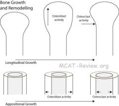
■Growth in length:
■Lengthwise bone growth occurs at the ends of long bones at the knobs. ■Osteoblasts' role in lengthwise bone growth is to add bone tissue at the bone ends. ■By itself, osteoblasts will lengthen the knobs at the ends of the bone. ■Osteoclasts' role in bone growth is to remodel bone tissue by chipping away the knobs until it's the right size and shape. ■Growth in diameter: ■Osteoblasts' role in diameter growth of bones is to add bone tissue to the outside of the bone. ■Osteoclasts' role in diameter growth of bones is to remove some bone tissue from the inside of the bone (bones are hollow). ■Without osteoclasts, diameter growth will result in bones that are too thick and too heavy. Even with osteoclasts, bones still grow thicker, just not unwieldly thick. |
Osteoclasts are similar to macrophages because they break down bone (phagocytize bone).
|
|
Explain gas exchange and thermoregulation of the respiratory system.
|
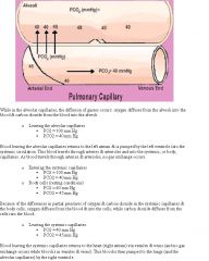
◦In lungs: oxygen diffuses into blood. Carbon dioxide diffuses out of blood. ◦The mechanism of this gas exchange follows Henry's law because O2 and CO2 are gases that are dissolved in the blood. When a gas is in contact with the surface of a liquid, the amount of the gas which will go into solution is proportional to the partial pressure of that gas. A simple rationale for Henry's law is that if the partial pressure of a gas is twice as high, then on the average twice as many molecules will hit the liquid surface in a given time interval, and on the average twice as many will be captured and go into solution. ◦When blood reaches the lungs, the partial pressure of oxygen in the blood is less than the partial pressure of oxygen in the lungs because the body used them up. Therefore, oxygen diffuses into blood. ◦The partial pressure of CO2 in blood that reaches the lungs is higher than the partial pressure of CO2 in the lungs because of the body releases them. Therefore, CO2 diffuses out of blood and is exhaled out of the lungs. ◦Thermoregulation: Panting increases evaporation of water across the moist surfaces of the lungs and the tongue and mouth and aids in cooling the body down. When water evaporates its change in state from liquid to a gas takes up a great deal of energy (because the bonds need energy to break and become a gas) and lowers the temperature of the surface on which it occurs. This is the process of evaporation. Additional evaporative heat loss occurs through breathing. As you breath in and out you are exchanging heat via evaporation to the outside world.
|
|
|
|
How does the respiratory system protection against disease and particulate matter?
|
◦Nostril hair filters out particles.
◦Mucus lining of respiratory tract traps pathogens and particles. ◦Cilia on mucus lining of respiratory tract sweeps pathogen and particles out, where you either spit it out or swallow it into stomach acid. ◦Macrophages reside in alveoli. An alveolar macrophage (or dust cell) is a type of macrophage found in the pulmonary alveolus, near the pneumocytes, but separated from the wall. Activity of the alveolar macrophage is relatively high, because they are located at one of the major boundaries between the body and the outside world. These macrophages reside on respiratory surfaces and clean off particles such as dust or microorganisms. |
|
|
What are the resiliency and surface tension effects of breathing?
|
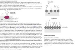
◦Lung is elastic, it recoils as soon as you relax after breath intake. If not for the rib cage, the lung would collapse even further. ◦Surface tension causes the lung to collapse. Surfactants produced in the alveoli decreases surface tension, and helps alveoli to stay open. Surfactants increase the ability of the lungs to stretch and prevent atelectasis (collapse of the lung) at the end of expiration. The walls of alveoli are coated with a thin film of water & this creates a potential problem. Water molecules, including those on the alveolar walls, are more attracted to each other than to air, and this attraction creates a force called surface tension. This surface tension increases as water molecules come closer together, which is what happens when we exhale & our alveoli become smaller (like air leaving a balloon). Potentially, surface tension could cause alveoli to collapse and, in addition, would make it more difficult to 're-expand' the alveoli (when you inhaled). Both of these would represent serious problems: if alveoli collapsed they'd contain no air & no oxygen to diffuse into the blood &, if 're-expansion' was more difficult, inhalation would be very, very difficult if not impossible. Fortunately, our alveoli do not collapse & inhalation is relatively easy because the lungs produce a substance called surfactant that reduces surface tension.
|
|
|
What is differential pressure for the respiratory system?
|
◦Differential pressure = difference between intrapulmonary (inside lung) pressure and intrapleural (outside lung) pressure.
■Intrapulmonary pressure = atmospheric pressure (lung is open to the outside, so has same pressure as outside). ■Intrapleural pressure = less than atmospheric pressure = pressure within the thoracic cage against the lungs, prevent lung from collapsing. During breath intake, intrapleural pressure decreases even further, causing the lung to expand. ◦Negative pressure mechanism in breathing is just a fancy term for sucking. You breathe in by establishing negative pressure in the lung (sucking). However, when someone gives you mouth-to-mouth, that's positive pressure. Intrapulmonary pressure: Pressure within the lungs. It is usually greater than intrathoracic pressure, causing the lungs to remain slightly inflated after expiration. Gasses are able to move in and out of the lungs through muscular energy exerted on the thorax and changes between intrathoracic and atmospheric pressures. The pressure within the lungs and thorax must be less than atmospheric pressure for inspiration to occur. Air then flows from an area of higher pressure to one of lower pressure. As the diaphragm and intercostal muscles work to increase the size of the thorax, intrathoracic pressure decreases below atmospheric pressure and air moves into the lungs. During exhalation, the inspiratory muscles relax, and the elastic recoil of the lung tissues, combined with a rise in intrathoracic pressure, causes air to move out of the lungs |
|
|
|
How does the skin Function in homeostasis and osmoregulation?
|
•Heat homeostasis:
◦Too cold: hair stands up (goose bumps), vasoconstriction decreases blood supply at skin (less heat loss). ◦Too hot: sweat (evaporative cooling), vasodilation increases blood supply at skin (more heat loss). •Water homeostasis: Insulates body against water loss. •Osmoregulation: sweat excretes salts and nitrogenous wastes (urea, uric acid, ammonia) Heat and cold receptors are located in the skin. When the body temperature rises, the hypothalamus sends a nerve signal to the sweat-producing skin glands, causing them to release about 1-2 liters of water per hour, cooling the body. The hypothalamus also causes dilation of the blood vessels of the skin, allowing more blood to flow to the skin, causing heat to be convected away from the skin surface. Within the body, blood convection is used to move the heat from the inside of your body to your skin. Convection is the movement of heat by currents in the medium. When body temperature falls, the sweat glands constrict and sweat production decreases. The vasoconstriction causes the blood flow to the inner organs, and keep heat inside. If the body temperature continues to fall, the body will engage in thermiogenesis, or heat generation, by raising the body's metabolic rate and by shivering. Water loss occurs in the skin by two routes: 1) evaporation 2)sweating The skin pH is acidic (around 5.6) and it's part of the innate immunity and the acidic pH makes it difficult for pathogens to enter the body. |
|
|
|
What are other functions of the skin besides homeostasis and osmoregulation?
|
◦protect against UV radiation by making melanin (absorbs UV). Melanin is a pigment that absorbs harmful UV-radiation and transforms the energy into harmless amounts of heat
◦make vitamin D upon exposure to sunlight. Vitamin D is important for calcium absorption in the small intestine. When blood calcium levels are low, PTH levels increase and as a result, Vitamin D levels increase. PTH enhances the absorption of calcium in the small intestine to elevate blood levels of calcium indirectly by stimulating the active form of vitamin D in the kidney. Vitamin D induces synthesis of a calcium-binding protein in intestinal epithelial cells that facilitates efficient absorption of calcium into blood. ◦Act as blood reservoir. Vasoconstriction in skin shunts blood to other organs. ◦Sense touch, pressure, pain, heat, cold. ◦Protection. |
|
|
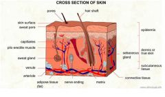
How does the skin Function in thermoregulation?
|
◦Hairs help insulate the body by trapping air in them.
◦Normally hair lies at an angle to the skin, with erectile muscle attaching to it. ◦When it's cold, erectile muscles contract, and the hair stands up. This erect position helps hair to trap more air, helping to retain heat. It's known as goose bumps. •Fat layer for insulation: fat in hypodermis act as insulation. •sweat glands, location in dermis: produce sweat, cools the body by evaporative cooling. •Vasoconstriction and vasodilation in surface capillaries: ◦When it's cold: vasoconstriction of arterioles happens when arterioles carrying blood to the capillaries shrink (constrict), reduce blood supply to skin capillaries. It leads to less heat loss at skin surface by rerouting the blood away from the skin and toward the warmer core of the body. This prevents blood from losing heat to the surroundings and also prevents the core temperature dropping further. It is impossible to prevent all heat loss from the blood, only to reduce it. In extremely cold conditions excessive vasoconstriction leads to numbness and pale skin. Frostbite only occurs when water within the cells begins to freeze, this destroys the cell causing damage. ◦When it's hot: vasodilation of arterioles increase blood supply to the skin capillaries. Leads to more heat loss at skin surface. Vasodilation is the process of relaxation of smooth muscle in arteriole walls allowing increased blood flow through the artery. This redirects blood into the superficial capillaries in the skin increasing heat loss (by convection or conduction). Convection is broadly the movement of any molecules through any fluid state, including liquids and gasses. Blood is an example of convection movement. Conduction is transfer of heat through direct contact. Body temperature is around 37 degrees Celsius. |
|
|
|
How does the skin function in Physical protection?
|
•nails, calluses, hair
◦nail = hard keratin = tougher than the soft keratin on skin. ◦calluses = extra thick layer of dead keratin-packed cells on the surface of skin. ◦hair = hard keratin. •protection against abrasion, disease organisms ◦Keratin protect skin against abrasion. ◦The tight seal made from keratin-packed cells and glycolipids form a barrier against pathogens. Glycolipids are produced and fill in the spaces between the dying keratinocytes. Glycolipids are molecules containing carbohydrates and fats. Glycolipids provide waterproofing for the body; keeping excess fluids and water-soluble compounds from escaping or entering the body. Keratinocytes are specialized cells in the epidermis that produce keratin. •Chemical protection: Sweat is acidic, contains antimicrobial agents. Sebum (oil that lubricates skin and hair) inhibits the growth of some microorganisms like some bacteria. •Natural flora: good bacteria on the surface of skin don't cause harm to you, and they fight off bad bacteria that can harm you. |
|
|
|
What is the structure of the skin and what is the epidermis?
|
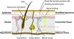
◦Epidermis = stratified squamous epithelial tissue = protection
■Keratinocytes = cells that produce keratin = dominates the epidermis. ■Keratinocytes start off like normal cells at the bottom of the epidermis, but gets flatter as you go up, and becomes dead, they are like keratin plates at the surface of the skin. ■Melanocytes = cells that make melanin, the skin pigment and protects agains UV radiation. ■Dendritic cells (Langerhans cells) = phagocytes that eat pathogen and present foreign antigens to activate immune response. Dendritic cells are a type of monocyte. |
|
|
|
What is the dermis of the skin?
|
◦Dermis = connective tissue = blood, nerve supply
■Fibroblasts = make fiber and ground substance (non-cellular substance that acts as the glue) for the extracellular matrix that makes up connective tissue. ■Contains the hair follicles, sweat glands, and oil (sebum) glands. ■Contains the blood vessels and nerves. |
|
|
|
What is the hypodermis?
|
◦Hypodermis = adipose tissue = absorbs shock, protecting from injury, and provides insulation.
|
|
|
|
Explain how the skin has relative impermeability to water.
|
•relative impermeability to water: due to layer of dead, keratin-packed cells sealed with glycolipids.
◦Keratin is water insoluble, and layers of dead, keratin-packed cells reside on the skin surface. ◦Glycolipids seal the space between the dead keratin-packed cells. ◦Sebum (skin oil) contribute some. But oil glands are not present everywhere (absent in palms and soles). Sebum acts to protect and waterproof hair and skin, and keeps them from becoming dry, brittle and cracked by dehydration. |
|
|
|
What are the male gonads ?
|
■male: testes
■makes sperm in the seminiferous tubules. ■makes testosterone. ■external. Seminiferous tubules are located in the testes, and are the specific location of meiosis, and the subsequent creation of gametes, the sperm cells. Luteinizing hormone in males stimulates testosterone release. Testosterone is necessary for the proper development of testes, as well as the penis and seminal vesicles. It is also important for secondary male sex characteristics. Follicle-stimulating hormone in males stimulates sperm maturation, as it stimulates ova maturation in females. Once estrogen levels reaches a certain threshold, it stimulates a surge in LH, the surge in LH marks ovulation. |
|
|
|
What are the female gonads?
|
■female: ovaries
■houses immature egg, which matures monthly after puberty. ■makes estrogen. ■internal. Prolactin is produced by the anterior pituitary and stimulates the production of milk. Oxytocin is produced by the hypothalamus and causes the ejection of milk (in addition to causing contractions of the uterus). Estrogen stimulates the development of mammary glands. While estrogens are present in both men and women, they are usually present at significantly higher levels in women of reproductive age. They promote the development of female secondary sexual characteristics, such as breasts, and are also involved in the thickening of the endometrium and other aspects of regulating the menstrual cycle. In males, estrogen regulates certain functions of the reproductive system important to the maturation of sperm and may be necessary for a healthy libido. |
|
|
|
What are genitalia for men?
|
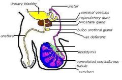
■male: testes, penis, and various ducts and glands.
■sperm made in the seminiferous tubules. ■sperm are stored and matured in the epididymis. ■sperm travels through vas deferens → ejaculatory duct → urethra → penis ■mnemonic: seven up = Seminiferous tubules, Epididymis, Vas deferens, Ejaculatory duct, nothing, Urethra, Penis. |
|
|
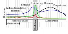
What are genitalia for women?
|
■Monthly cycle: primary oocyte matures into secondary oocyte every month. To prepare for fertilization, the endometrium thickens. If fertilization doesn't occur, menses occur, and the cycle begins anew. ■GnRH (Gonadotropin-releasing hormone)= stimulates release of FSH and LH. GnRH is released from the hypothalamus. ■FSH = folicle stimulating hormone = stimulates growth and maturation of follicle and the primary oocyte within it. It also stimulates the production of estrogen. FSH is released from the anterior pituitary. ■Follicle = houses primary oocyte and produces estrogen. ■Estrogen = normally inhibits LH and FSH from negative feedback, but estrogen causes LH surge when it reaches a certain threshold. Estrogen levels vary during a menstrual cycle, but reach the highest level before ovulation. Ovulation is the process in a female's menstrual cycle by which a mature ovarian follicle ruptures and discharges an ovum (secondary oocyte). In a classical negative feedback loop, sex steroids inhibit secretion of GnRH, so they therefore inhibit LH and FSH. ■Estrogen reaches this threshold right before ovulation→ surge of LH occurs. ■LH = luteinizing hormone = the surge of LH triggers ovulation by stimulates the outer cells of the follicle and releasing the mature egg. The surge of the LH also turns the follicle into corpus luteum. Corpus luteum = makes estrogen and progesterone, which are needed to maintain the endometrium. LH is necessary for the maintenance of the newly formed corpus luteum. LH is released from the anterior pituitary.■LH surge triggers rupture of follicle. The luteal phase is the latter phase of the menstrual cycle and begins with the formation of the corpus luteum and ends in either pregnancy or luteolysis.
|
|
|
|
Explain what happens when there is no fertilization and when there is fertilization for women.
|
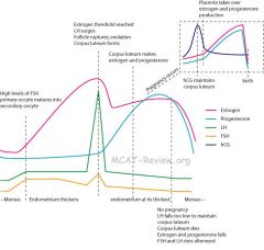
■No fertilization → LH falls → corpus luteum dies → estrogen and progesterone fall → endometrium sloughs off and is expelled from the vagina (menses) → cycle begins anew with FSH and LH re-rising.
■Fertilization occurs → implanted embryo (implantation means embryo adheres the wall of the uterus) releases hCG → hCG mimicks LH to maintain corpus luteum → estrogen and progesterone maintained by corpus luteum → placenta takes over the responsibility of making estrogen and progesterone later on and the corpus luteum degrades without embryo/fetal loss. Human chorionic gonadotropin (hCG) signals the corpus luteum to continue progesterone secretion, thereby maintaining the thick lining (endometrium) of the uterus and providing an area rich in blood vessels in which the zygote(s) can develop. |
|
|
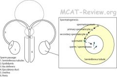
What are the differences between male and female structures?
|
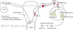
■male: mostly external. Shared passage with urinary tract.
■female: mostly internal. Separate passage from urinary tract. |
|
|
|
What is spermatogenesis?
|
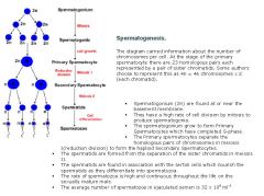
•Gametogenesis by meiosis
◦Male = spermatogenesis = occurs in the seminiferous tubules. 1.Spermatogonium (2n) = stem cell. Spermatogonium are stem cells because they are incapable of fertilizing an egg cell but can give rise to cells that develop into sperm. During spermatocytogenesis primitive cells called spermatogonia proliferate by mitosis. Mitosis of spermatogonium can either create more spermatogonium or create primary spermatocyte. Mitosis ends when a spermatogonium yields two primary spermatocytes. 2.Spermatogonium (2n) → mitosis → primary spermatocyte (2n). Occurs after puberty. 3.Primary spermatocyte (2n) → meiosis I → Secondary spermatocyte (n). 4.Secondary spermatocyte (n) → meiosos II → spermatid (n). 5.Spermatid (n) → mature → sperm (n). The fancy name for sperm is spermatozoa. The diploid number of primary spermatocytes is halved during meiosis. A primary spermatocyte is transformed into two secondary spermatocytes during meiosis I - these cells then in turn are converted into (1N) spermatids during meiosis II. Males have an almost unlimited capacity to produce germ cells (spermatogonia). Sperm cells (the male gametes) are matured spermatids that have already undergone differentiation. This differentiation process is called spermiogenesis. |
|
|
|
What are the differences in formation of the ovum and sperm?
|
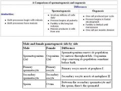
Basically, the secondary oocyte divides in meiosis II straight into the ovum, whereas the secondary spermatocyte divides straight into the spermatid in meiosis, then the spermatid matures into the sperm. The ovum is already matured after meiosis II.
|
|
|
|
What is oogenesis?
|
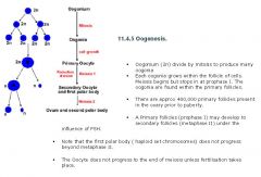
•Gametogenesis by meiosis
◦Female = oogenesis = occurs in the ovaries, then fallopian tubes. 1.Oogonium (2n) = stem cell. Oogonium are stem cells, immature ovum, that are incapable of being fertilized, but give rise to a mature ovum that can be fertilized. 2.Oogonium (2n) → mitosis → primary oocyte. 3.Primary oocyte (2n) arrests at prophase I (occurs before birth). The meiotic resting phase lasts until puberty. Then one primary oocyte comes out of arrest every month and completes the first meiosis creating a secondary oocyte (this happens every month between puberty and menopause). 4.Primary oocyte (2n) → meiosis I → secondary oocyte (n). Once the secondary oocyte is created, it ruptures from ovary follicle into the fallopian tube. This is the ovulation period. 5.Secondary oocyte (n) arrests at metaphase II of meiosis II. Comes out of arrest if fertilization occurs. Fertilization completes the second meiosis. |
In oogenesis, the cytoplasm is divided unequally and only one ovum, with the bulk of the cytoplasm, is produced plus two or three inert polar bodies. Oogenesis differs from spermatogenesis in that with spermatogenesis, the cytoplasm is equally divided during meiosis and four viable sperm are produced from one diploid cell.
|
|
|
What are the differences in morphology for the ovum and sperm?
|
■Sperm = motile = flagella.
■Egg = non-motile = round. |
|
|
|
Explain the relative contribution to next generation for the ovum and sperm.
|
■Sperm contributes DNA only (the egg actively destroys sperm mitochondria).
■Egg contributes DNA + everything else (mitochondria, organelles, epigenetics). The term ‘epigenetics’ refers to the mechanism by which non -genetic factors influence gene expression and thereby, inheritance. Epigenetics plays a key role in cellular differentiation of a newly formed embryo and is the reason why all the cells, which has the same DNA content, eventually function differently. The single-celled fertilized egg undergoes multiple rounds of cell division to become the multicelled embryo with further undergoes cell differentation to form different cell types with different functions. |
|
|
|
What is the Reproductive sequence and what is fertilization and implantation?
|
•Reproductive sequence (fertilization, implantation, development, birth)
1.fertilization: sperm + egg → zygote 2.implantation: 1.zygote 2.morula (an embryo consisting of cells in a solid ball) 3.blastocyst (structure formed after formation of the morula, but before implantation) 4.the blastocyst is the one that implants in the endometrium |
|
|
|
What is the process for development and birth?
|
Development: 1.zygote 2.blastocyst 3.implantation 4.gastrulation 5.organogenesis
Birth: ■Babies switch from getting oxygen from mom's blood → breath on their own. ■Babies switch from getting nutrients from mom's blood → suckling on breasts for nutrients. ■Babies undergo fetal circulation (which bypasses lungs and liver in the womb) and get oxygen from their mom's blood using fetal hemoglobin, which has a higher affinity for oxygen than regular hemoglobin→ normal circulation. In fetal circulation, blood coming thru the umbilical vein is shunted around the liver (not through it) through the ductus venosus. Upon birth, the ductus venosus closes off, and the adult remnant is called the ligamentum venosum. Also, the foramen ovale and ductus arteriosus closes. In an embryo, blood is shunted around the lungs 2 ways: 1) the foramen ovale allows blood to flow from the right atrium to the left atrium, bypassing the ventricles and the lungs. 2) the ductus arteriosus connects blood from the pulmonary artery to the the aorta, bypassing the lungs. Gastrulation is a phase early in the development of most animal embryos, during which the morphology of the embryo is reorganized to form the three germ layers: ectoderm, mesoderm, and endoderm. The embryo froms from blastula to gastula. Organogenesis is when individual organs develop within the newly formed germ layers. If the foramen ovale and/or ductus arteriousus failed to close upon birth, then there would be a decreased partial pressure of oxygen in the systemic arteries and increased partial pressure of carbon dioxide in the systemic arteries because the ducts shunts deoxygenated blood away from the lungs and straight into the aorta to go into circulation. So there would be a mixing of oxygen and carbon dioxide, decreasing the partial pressure of oxygen. REMEMBER: ductus ARTERIOSUS - moves around the ARTERY - which artery? Pulmonary artery. Where does it go? AORTA (another large artery). Foramen ovale - well, a big oval that goes from Right atrium to Left atrium (like a circle). |
The umbilical vein carries oxygenated blood from the mother to the fetus. The umbilical artery (a for away from fetus) carries deoxygenation blood from the fetus to the mom.
|
|
|
What are the stages of early development?
|
a. fertilization
b. cleavage c. blastula formation d. gastrulation e. neurulation |
|
|
|
What are the general features of fertilization?
|
1. Sperm meets egg
2. Acrosomal reaction causes sperm to penetrate egg 3. Cortical reaction causes egg to prevent additional sperm from penetrating 4. Egg completes meiosis II 5. Sperm and egg nuclei fuse During fertilization, a sperm must first fuse with and then penetrate the female egg in order to fertilize it. Fusing to the egg usually causes little problem, whereas penetrating through the egg's hard shell can present more of a problem to the sperm. Therefore sperm cells go through a process known as the acrosome reaction which is the reaction that occurs in the acrosome of the sperm as it approaches the egg. The acrosome is a cap-like structure over the anterior half of the sperm's head. As the sperm approaches the egg, the membrane surrounding the acrosome fuses with the plasma membrane of the egg (specifically the zona pellucida), exposing the contents of the acrosome. The contents include surface antigens and numerous enzymes which are responsible for breaking through the egg's tough coating and allowing fertilization to occur. |
|
|
|
What is the process of cleavage?
|
Normal mitotic cell division is when a cell grows then divides, grows again, then divides.
Cleavage is when there are mitotic divisions without cell growth. Cell division with no significant growth, producing a cluster of cells that is the same size as the original zygote, is called cleavage. |
|
|
|
What are the cell movements of gastrulation?
|
Cells from the surface migrate inwards. Gastrulation occurs slightly different for different animals. Some by invagination, some by migration (called ingression), some by splitting. Cell movements by splitting is called delamination. It is the splitting of one cellular sheet into 2 or more parallel sheets. In mammals, the cells start migrating inward at the primitive streak. During the early stages of development, the primitive streak is the structure that will establish bilateral symmetry, determine the site of gastrulation and initiate germ layer formation. In bilateral symmetry (also called plane symmetry), only one plane, called the sagittal plane, will divide an organism into roughly mirror image halves. A sagittal plane is an imaginary plane that travels vertically from the top to the bottom of the body, dividing it into left and right portions.
During gastrulation, cell movements result in a massive reorganization of the embryo from a simple spherical ball of cells, the blastula, into a multi-layered organism. During gastrulation, many of the cells at or near the surface of the embryo move to a new, more interior location. The primitive streak, extends through this midline, becoming the first symmetry-breaking event in the embryo, and marks the beginning of gastrulation. This process involves the ingression of mesoderm and endoderm stem cells and their migration to their ultimate position, where they will differentiate into the three germ layers (endoderm, mesoderm, ectoderm) that will give rise to all the tissues of the adult organism. |
|
|
|
What are the steps of blastula formation?
|
1. fertilization produces zygote
2. cleavage produces a solid ball of cells called the morula 3. morula hollows out into the blastocyst (mammals form into blastocyte). 4. Blastocyte implants into the endometrium |
|
|
|
What are the primary germ layers of gastrulation?
|
Gastrulation is the formation of primary germ layers. (endoderm, mesoderm, ectoderm)
The cells that migrate inwards form the endoderm. The cells that remain outside is the ectoderm. The cells in the middle are the mesoderm. |
|
|
|
What is neurulation?
|
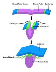
ectoderm → brain and spinal cord and skin.
The ectoderm forms the brain and spinal cord by folding into a tube. Neurulation is a part of organogenesis. It eventually forms the central nervous system. The process begins when the notochord induces the formation of the central nervous system (CNS) by signaling the ectoderm germ layer to form the thick and flat neural plate. The neural plate folds in upon itself to form the neural tube, which will later differentiate into the spinal cord and the brain, eventually forming the central nervous system. The notochord is a flexible, rod-shaped body composed of cells derived from the mesoderm and defines the primitive axis of the embryo and in most vertebrates it is replaced by the vertebral column. In human embryology, formation of neural plate is the first step of neurulation. It is created by a flat thickening opposite to the primitive streak of the ectoderm. During the stage of neural plate formation the embryo consists of three cell layers: the previously mentioned ectoderm that eventually forms the skin and neural tissues, the mesoderm that forms muscle and bone, and the endoderm that will form the cells lining the digestive and respiratory tract. As the neural plate develops, it becomes surrounded by neural folds, which eventually create the cylindrical neural tube. This process is termed primary neurulation. |
|
|
|
What are the Major structures arising out of primary germ layers?
|
endoderm = innermost layer = guts, lungs, and digestive internal organs (liver, pancreas).
mesoderm = middle layer = muscle, blood and bone tissues, and internal organs (kidney and gonads). ectoderm = outermost layer = skin and nerves (including the brain). The endoderm forms: the stomach, the colon, the liver, the pancreas, the urinary bladder, the lining of the urethra, the epithelial parts of trachea, the lungs, the pharynx, the thyroid, the parathyroid, and the intestines. The mesoderm forms: skeletal muscle, the skeleton, the dermis of skin, connective tissue, the urogenital system, the heart, blood (lymph cells), and the spleen. The ectoderm forms: the central nervous system, the lens of the eye, cranial and sensory, the ganglia and nerves, pigment cells, head connective tissues, the epidermis, hair, and mammary glands. |
Most of the other organs and systems of the body (besides the digestive system and the nervous system) are mesodermal, including the excretory system, the reproductive system, the muscular and skeletal systems, and the circulatory system. If you cannot identify a particular tissue as being specifically endodermal or ectodermal, there is a very good chance that it is one of the many mesodermal-derived tissues.
|
|
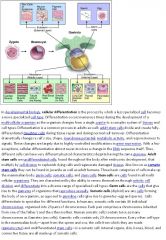
What is cell specialization, determination, and differentiation?
|
Determination = irreversible commitment to become a certain cell type along a developmental path. A cell at the determination stage may not display any specific characteristics that would yet identify it as a specific type of cell. After determination, the cell then differentiates.
Differentiation = becoming a cell type and adopting its specialized functions. A fully differentiated cell is called specialized. A cell can be determined but not yet differentiated. The design and shape of a cell is very much dictated by its function and the conditions under which it works. Cell specialization is the cell performing a specific function for a larger organ or tissue. It is an adaptation to do a particular job in a cell. Unicellular organisms tend to have fairly complex cells, which makes sense considering that these organisms must depend on only one cell to do everything. Multicellular organisms exhibit much greater specialization. For example, epidermal cells produce keratin to protect skin against abrasion, myocyte produce actin and myosin to make muscles contract, and neurons make neurotransmitters to transmit electrochemical impulses. As cells become more specialized, they may contain organelles that are not common to all cells. CELL SPECIALIZATION occurs because many forms of life have many levels of organization. In most plants, animals, and fungi, cells are organized into different types of tissues. TISSUES are groups of cells that carry out a common function. All cells do not perform the same function because of cell specialization. |
|
|
|
What are the tissue types of cell specialization?
|
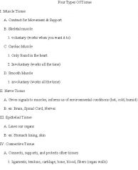
Epithelial: skin, lining of organs
Connective: blood, bone, tendons, ligaments, cartilage Nervous: brain, spinal cord, nerves Muscle: skeletal, smooth, and cardiac muscle |
|
|
|
What is Cell communication in development?
|
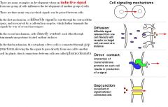
Induction: one group of cells changing the behavior of an adjacent group of cells.
Inducer = the one that sends the signal for the other to change. Responder = the one that gets the signal and changes. For example, the optic vesicle is able to induce the ectoderm to develop into lens. Induction mechanisms: physical touching of cells (juxtracine) or by releasing chemicals to nearby cells (paracrine). |
|
|

What is Gene regulation in development?
|
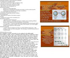
Most differential gene expression is regulation at the level of transcription initiation.
Differential gene transcription: Modification of DNA (methylations, addition of a methyl group) can shut off genes. DNA that is not methylated turn on genes, so methylated DNA suppresses the expression of viral genes and other deleterious elements which have been incorporated into the genome of the host over time. Methylation of DNA is a common method of gene silencing. This modification can be inherited through cell division. DNA methylation is typically removed during zygote formation and reestablished through successive cell divisions during development. DNA methylation is a crucial part of normal organismal development and cellular differentiation in higher organisms. DNA methylation stably alters the gene expression pattern in cells such that cells can "remember where they have been"; in other words, cells programmed to be pancreatic islets during embryonic development remain pancreatic islets throughout the life of the organism without continuing signals telling them that they need to remain islets. Modification on histones (acetylations, adding an acetyl group to histones) that wrap the DNA can shut on genes. Histone acetylation is linked to transcriptional activation. Often, DNA methylation and histone deacetylation work together in gene silencing. The combination of the two seems to be a signal for DNA to be packed more densely, lowering gene expression. To make or not to make transcription factors can regulate what genes get transcribed. Differential RNA processing: 1) Selecting what RNA make it outside the nucleus to be translated. 2) Alternative splicing of RNA (exons can be spliced differently and create different proteins). |
|
|
|
What is Gene regulation in development in regards to Translation regulation and Post-translational regulation?
|
Translation regulation: 1) Some mRNA are made to last longer than others (more proteins translated off of it), and some are made to be rapidly degraded (less proteins translated off of it). 2) Selective inhibition of translation of stored RNA in the oocyte. Get translated only when needed after fertilization. Oocyte controls early development by storing mRNAs and translating them only after fertilization.
Post-translational regulation: 1) Some proteins are inactive until modified by certain enzymes. 2) Active proteins can be selectively marked for degradation by ubiquitin. Posttranslational modification (PTM) is the chemical modification of a protein after its translation. Proteins are in a continual state of flux, being synthesized and degraded. In addition, when proteins become damaged they must be degraded to prevent aberrant activities of the defective proteins and/or other proteins associated with those that have been damaged. One of the major mechanisms for the destruction of cellular proteins involves a complex structure referred to as the proteosome. Proteins that are to be degraded by the proteosome are first tagged by the polypeptide ubiquitin. Proteasomes are very large protein complexes that degrade unneeded or damaged proteins by proteolysis, a chemical reaction that breaks peptide bonds. Enzymes that carry out such reactions are called proteases. Proteasomes are part of a major mechanism by which cells regulate the concentration of particular proteins and degrade misfolded proteins. |
|
|
|
Explain Programmed cell death as a form of a developmental mechanism.
|
apoptosis = programmed cell death.
During apoptosis, strong proteases are activated and they digest the cell from within. In mammals, the proteases are called caspases. The spaces between our fingers are created by apoptosis. The tail of a tadpole undergoes apoptosis when it morphs into a frog. Apoptosis is carried out by caspases, which are proteases (they degrade peptide bonds in proteins). These caspases are able to carry out cell death because of 1) digestion of structural proteins in the cytoplasm and 2)degradation of chromosomal DNA (which are complexed with histone proteins). The cytoskeleton in the cell is also proteinaceous and it gets broken down and causes cell shrinkage. The nuclear envelope and cell membrane have proteins embedded in them, so they get broken down too. The cell breaks apart into several vesicles called apoptotic bodies, which are then phagocytosed by other cells in the body. |
|
|
|
What is a Phenotype and genotype?
|
Phenotype: what is observed. For example, height, color, whether the organism exhibits a trait.
Genotype: the genetic make up. For example, homozygous dominant (TT), heterozygous (Tt), homozygous recessive (tt). Be careful if a question asks for a percentage of CARRIERS of recessive genes versus the percentage of those affected by the recessive gene. The term carrier describes an organism that carries two different forms (alleles) of a recessive gene (alleles of a gene linked to a recessive trait) and is thus heterozygous for that the recessive gene. Although carriers may act to convey and maintain recessive genes within a population by passing them on to offspring, the carriers themselves are not affected by the recessive trait associated with the recessive gene. |
|
|
|
How do you solve probability calculations for genetics with the rule of addition?
|
Rule of addition (the rule of or) is that the probability of an event that can occur in two or more independent ways is the sum of the separate probabilities of the different ways. For example:
Question: In a Mendelian cross between pea plants that are heterozygous for flower color (Pp), what is the probability of the offspring being a heterozygote? Answer: There are two ways in which a heterozygote may be produced: the dominant allele (P) may be in the egg and the recessive allele (p) in the sperm, or the dominant allele may be in the sperm and the recessive in the egg. Consequently, the probability that the offspring will be heterozygous is the sum of the probabilities of those two possible ways: Probability that the dominant allele will be in the egg with the recessive in the sperm is 1/2 X 1/2 = 1/4. Probability that the dominant allele will be in the sperm and the recessive in the egg is 1/2 X 1/2 = 1/4. Therefore, the probability that a heterozygous offspring will be produced is 1/4 + 1/4 = 1/2. |
|
|
|
How do you solve probability calculations for genetics with the rule of multiplication?
|
Rule of multiplication (the rule of and) is that the probability that independent events will occur simultaneously is the product of their individual probabilities. For example:
Question: In a Mendelian cross between pea plants that are heterozygous for flower color (Pp), what is the probability that the offspring will be homozygous recessive? Answer: Probability that an egg from the F1 (Pp) will receive a p allele = 1/2. Probability that a sperm from the F1 will receive a p allele = 1/2. The overall probability that two recessive alleles will unite, one from the egg and one from the sperm, simultaneously, at fertilization is: 1/2 X 1/2 = 1/4. |
|
|
|
What is probability?
|
Probability (p) is the chance that an event will occur. Probability equals the case (a) divided by the total number of cases (n) or p = a/n, assuming all cases are equally likely events.
p for heads? heads/heads plus tails. 1/2 p for tails? tails/heads and tails. 1/2 p for heads or tails? 1/2 + 1/2 = 1 p for heads and tails? These are mutally exclusive. But heads and then tails is possible. 1/2 X 1/2 = 1/4 The sum rule: if either/or then add the probabilities. Die: What is the probability that the die will fall on either 1 or 2 spots? = 1/6 + 1/6. Product rule: the and rule, multiply the probabilities. What is the probability that die will fall on 1 and next throw, 2? 1/6 X 1/6. |
|
|
|
What is pedigree analysis?
|
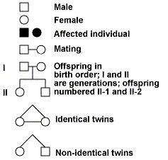
In some situations, we do not have the opportunity to perform controlled crosses. Rather we need to analyze an existing population. This is always the case when studying human genetics. Scientists have devised another approach, called pedigree analysis, to study the inheritance of genes in humans. Pedigree analysis is also useful when studying any population when progeny data from several generations is limited. Pedigree analysis is also useful when studying species with a long genration time.
A series of symbols are used to represent different aspects of a pedigree. Once phenotypic data is collected from several generations and the pedigree is drawn, careful analysis will allow you to determine whether the trait is dominant or recessive. Here are some rules to follow. For those traits exhibiting dominant gene action: 1) affected individuals have at least one affected parent 2) the phenotype generally appears every generation 3) two unaffected parents only have unaffected offspring |
|
|
|
What are the aspects of a dominant pedigree?
|
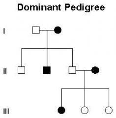
For those traits exhibiting dominant gene action:
affected individuals have at least one affected parent the phenotype generally appears every generation two unaffected parents only have unaffected offspring |
|
|
|
What are the aspects of a recessive pedigree?
|
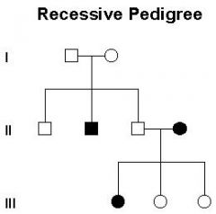
And for those traits exhibiting recessive gene action:
unaffected parents can have affected offspring affected progeny are both male and female |
|
|
|
What is a gene and a locus?
|
Gene: a gene is a stretch of DNA that codes for a trait. In molecular biology, the gene codes for a protein, which acts to bring about a trait.
Locus: location of a gene or DNA sequence on a chromosome. A gene is a unit of heredity in a living organism. It is normally a stretch of DNA that codes for a type of protein or for an RNA chain that has a function in the organism. All proteins and functional RNA chains are specified by genes. All living things depend on genes. Genes hold the information to build and maintain an organism's cells and pass genetic traits to offspring. A modern working definition of a gene is "a locatable region of genomic sequence, corresponding to a unit of inheritance, which is associated with regulatory regions, transcribed regions, and or other functional sequence regions ". Colloquial usage of the term gene (e.g. "good genes, "hair color gene") may actually refer to an allele: a gene is the basic instruction, a sequence of nucleic acid (DNA or, in the case of certain viruses RNA), while an allele is one variant of that instruction (variant meaning it could be the long hair variant of the gene or short hair variant of the gene).All organisms have many genes corresponding to many different biological traits, some of which are immediately visible, such as eye color or number of limbs, and some of which are not, such as blood type or increased risk for specific diseases, or the thousands of basic biochemical processes that comprise life. When proteins are manufactured, the gene is first copied into RNA as an intermediate product. In other cases, the RNA molecules are the actual functional products. For example, RNAs known as ribozymes are capable of enzymatic function, and other RNAs have a regulatory role. The DNA sequences from which such RNAs are transcribed are known as RNA genes. The biochemistry of the intermediate proteins determines how they interact in the cell. Therefore, biochemistry predicts how combinations of different alleles will produce varying traits. Extended expression patterns seen in diploid organisms include facets of incomplete dominance, codominance, and multiple alleles. |
|
|
|
What are Allele (single, multiple)?
|
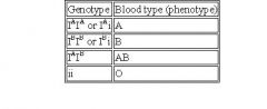
An allele is a variant of a gene. A gene may have a number of alleles. All alleles of the same gene exist at the same locus.
A cell holds 2 alleles of each gene. One allele from mom, one allele from dad. When a gene that codes for a trait has only 2 alleles, then that's the simple case we're used to seeing. This is called a single allele. For example, the trait for height in peas is governed by T and t. TT and Tt gives tall plants, and tt gives short ones. When a gene has more than 2 alleles, then that's called multiple alleles. For example, blood type is governed 3 alleles: IA, IB and i. Because a cell can only hold 2 of these alleles, the different combinations an individual can have are in the picture. An excellent example of multiple allele inheritance is human blood type. Blood type exists as four possible phenotypes: A, B, AB, & O. There are 3 alleles for the gene that determines blood type. (Remember: You have just 2 of the 3 in your genotype --- 1 from mom & 1 from dad). Notice that the allele for "O" (i) is recessive to the alleles for "A" & "B". With three alleles we have a higher number of possible combinations in creating a genotype. As you can count, there are 6 different genotypes & 4 different phenotypes for blood type. Note that there are two genotypes for both "A" & "B" blood --- either homozygous (IAIA or IBIB) or heterozygous with one recessive allele for "O" (IAi or IBi). Note too that the only genotype for "O" blood is homozygous recessive (ii). And lastly, what's the deal with "AB" blood? What is this an example of? Type AB is an example of codominance. The IA allele & the IB allele are "equal". What I mean is that neither one dominates the other. Instead, when inherited together in the genotype, they appear together in the phenotype. Wa-la ! Codominance. |
A and B Blood groups can be heterozygous, with the recessive allele being the iO, or homozygous, with IA or IB. A and B are codominant together, and are dominant over the blood antigen O. Therefore, if a man heterozygous for blood type A (AO) married a woman heterozygous for blood type B (BO), they could possibly have children with the genotypes AO, BO, OO, or AB. Therefore, all blood types are possible in this mating. However, if a question says that the man and/or woman has blood type A or blood type B, that could mean that they are heterozygous or homozygous for those blood types.
|
|
|
What is Homozygosity and heterozygosity?
|
Homozygous: when the two alleles that an individual carries are the same. For example, AA or aa.
Heterozygous: when the two alleles that an individual carries are different:. For example, Aa. |
|
|
|
What is a Wild type?
|
Wild type: the "normal" allele or phenotype for an organism. The wild-type is usually the most prevalent, although it doesn't necessarily have to be true. Wild type (or wildtype) refers to the phenotype of the typical form of a species as it occurs in nature. Wild type is the type of alleles found in the "wild" - in nature.
|
|
|
|
What is Recessiveness?
|
Recessiveness: the "weak" allele. The recessive allele is only expressed if both copies are present. Only a single copy is needed for the dominant allele. The recessive allele is usually denoted as the lower case letter, the dominant allele is usually denoted as the upper case letter. For example, blond hair is recessive. Both alleles for blond hair need to be present, otherwise the hair is dark.
|
|
|
|
What is complete dominance?
|
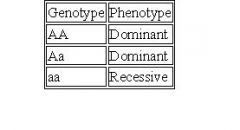
A kind of dominance wherein the dominant gene completely masks the effect of the recessive gene in heterozygous condition.
For instance, an individual carrying two alleles that are both dominant (e.g. AA), the trait that they represent will be expressed. But if the individual carries two alleles in a manner that one is dominant and the other one is recessive, (e.g. Aa), the dominant allele will be expressed while the recessive allele will be suppressed. Hence, the heterozygote (Aa) will have the same phenotype as that of the dominant homozygote (AA). This condition is called complete dominance. If one of the parents is homozygous dominant, then ALL of the offspring will have the dominant phenotype, regardless of the genotype of the other parent. |
|
|
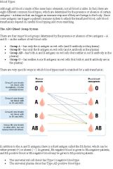
What is Codominance?
|
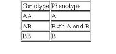
An example of co-dominance is the A and B blood type alleles. Type A cells have A antigens. Type B cells have B antigens. Type AB makes both antigens.
A condition in which the alleles of a gene pair in a heterozygote are fully expressed thereby resulting in offspring with a phenotype that is neither dominant nor recessive. |
Basically, there are 2 ways that you can donate blood. You can donate red blood CELLS or blood PLASMA. Type O blood is the universal donor of red blood CELLS because it does not have any A or B antigens on its cells. However, it would not be a good donor of plasma cells because it has anti-A and anti-B antibodies in its plasma. It would be a universal recipient of plasma cells. Type AB blood is the universal recipient of red blood CELLS because type A and type B blood has no anti-A nor anti-B antibodies, since it has A and B antigens on its red blood cells. Type AB blood would be a universal donor of plasma cells since it doesn't have any anti-A or anti-B antibodies in its plasma.
|
|
|
What is incomplete dominance?
|
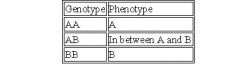
An example of incomplete dominance is the color of chickens. A cross between black chickens and white chickens give rise to bluish grey chickens.
A kind of dominance occurring in heterozygotes in which the dominant gene or allele is only partially expressed, and usually resulting in an offspring with an intermediate phenotype. In incomplete dominance, a heterozygous organism carrying two alleles wherein one is dominant and the other one is recessive, (e.g. Aa), the dominant allele will only be partially expressed. Hence, the heterozygote (Aa) will have an intermediate phenotype. A typical example is the color of the flower in which R symbolizes the dominant allele for red pigment and r is the recessive allele for no pigment. In incomplete dominance, the heterozygous plant carrying both alleles, Rr, will not be able to produce enough red pigment (since the dominant allele is only partially expressed) and therefore will appear pink. |
|
|
|
What is genetic leakage?
|
Genetic leakage is when genes flow from one species to another. This is a concern for antibiotic resistance where genes are able to cross species.
Mnemonic - genes leak from one species to another. |
|
|
|
What is penetrance?
|
Penetrance is the frequency that a genotype will show up in the phenotype. 100% penetrance means that if you have the genes for being smart, then you'll definitely be smart! Less than 100% penetrance means that you may have the genes for being smart, but you may not actually be smart. Penetrance in genetics is the proportion of individuals carrying a particular variation of a gene (allele or genotype) that also express an associated trait (phenotype). In medical genetics, the penetrance of a disease-causing mutation is the proportion of individuals with the mutation who exhibit clinical symptoms. For example, if a mutation in the gene responsible for a particular autosomal dominant disorder has 95% penetrance, then 95% of those with the mutation will develop the disease, while 5% will not. A gene is a stretch of DNA that codes for a trait. An allele is a variance of the trait and the genotype is the 2 alleles that code for the trait. The gene may have more than 2 alleles (multiple alleles), but the genotype of an individual only has 2 alleles. Common examples used to show degrees of penetrance are often highly penetrant. There are several reasons for this: 1) Highly penetrant alleles, and highly heritable symptoms, are easier to demonstrate, because if the allele is present, the phenotype is generally expressed. Mendelian genetic concepts such as recessiveness, dominance, and co-dominance are based on this principle. 2) Alleles which are highly penetrant are more likely to be noticed by clinicians and geneticists, and alleles for symptoms which are highly heritable are more likely to be inferred to exist, and then are more easily tracked down. Penetrance only considers whether individuals express the trait or not. For variation in the degree of expression of a given trait, that is expressivity. Expressivity refers to variations of a phenotype in individuals carrying a particular genotype.
|
|
|
|
What is expressivity?
|
Expressivity is to what degree a penetrant gene is expressed. Constant expressivity means that if your genes for being smart manages to penetrate (show up as a trait), then you're IQ is 120. Variable expressivity means that your IQ doesn't have to be 120, it could be somewhat lower or somewhat higher. Expressivity is a term used in genetics that refers to variations of a phenotype in individuals carrying a particular genotype. The term can be used to qualitatively or quantitatively characterize the extent of the phenotype variation given a particular genotype. The term is equivalent to the severity of a condition in clinical medicine. For example, the amount of blood ejected from the pumping heart with each contraction can be quantitated by echocardiography and is called the ejection fraction. If a specific genotype is associated with the development of congestive heart failure, the expressivity would be represented by the range of ejection fractions seen in patients that have that genotype. As a more qualitative example, the "blue" gene might have an expressivity of 25% for individuals that express the "blue" gene and appear light blue, and 75% for individuals that express the "blue" gene and appear dark blue. This differs from penetrance, which refers to the likelihood of the gene generating any phenotype at all, while expressivity refers to the influence of an expressed gene in individuals.
|
|
|
|
What occurs during meiosis II?
|
Second division of meiosis: Gamete formation (separation of sister chromatids)
Prophase 2: DNA does not replicate. Metaphase 2: Chromosomes align at the equatorial plate. Anaphase 2: Centromeres divide and sister chromatids migrate separately to each pole. Telophase 2: Cell division is complete. Four haploid daughter cells are obtained. They divide the replicated chromosomes further to produce a cell with a chromosome. One parent cell produces four daughter cells. Daughter cells have half the number of chromosomes found in the original parent cell and with crossing over, are genetically different. Meiosis differs from mitosis primarily because there are two cell divisions in meiosis, resulting in cells with a haploid number of chromosomes |
|
|
|
What occurs during meiosis I?
|
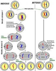
Meiosis is the process by which a single parent diploid cell (Both homologous chromosomes) divides to produce four daughter haploids cells (One homologous chromosome of the pair). Meiosis is the type of cell division by which germ cells (eggs and sperm) are produced. Meiosis involves a reduction in the amount of genetic material. Meiosis comprises two successive nuclear divisions with only one round of DNA replication. Four stages can be described for each nuclear division. Interphase: Before meiosis begins, genetic material of the diploid cell is duplicated. First division of meiosis: Separation of the tetrad (2 pairs of homologous chromosomes)
Prophase 1: Duplicated chromatin condenses. The two pairs of homologous chromosomes consists of two, closely associated sister chromatids. Crossing-over between the two pairs of homologous chromosomes occurs during this stage. Metaphase 1: Homologous chromosomes (tetrad) align at the equatorial plate. Where independent assortment occurs. Anaphase 1: Homologous pairs of the tetrad separate with sister chromatids remaining together. Telophase 1: Two daughter cells are formed with each daughter having ½ of each homologous pair of chromosomes. Each of the two daughter cells has a nucleus with a haploid set of REPLICATED chromosomes. |
|
|
|
What is a Gene pool?
|
Gene pool: all of the alleles in a species or population. A large gene pool indicates extensive genetic diversity, which is associated with robust populations that can survive bouts of intense selection. Meanwhile, low genetic diversity like inbreeding and population bottlenecks can cause reduced biological fitness and an increased chance of extinction.
|
|
|
|
What is the significance of meiosis?
|
Significance of meiosis: meiosis introduces genetic variability and increases genetic diversity by genetic recombination. Genetic recombination is the product of independent assortment and crossing-over, which introduces genetic variability. Genetic recombination is a process by which a molecule of DNA is broken and then joined to a different DNA molecule. In eukaryotes, recombination occurs in meiosis as a way of facilitating chromosomal crossover. The crossover process leads to offspring having different combinations of genes from their parents. Chromosomal crossover (or crossing over) is an exchange of genetic material between homologous pairs of chromosomes. It is one of the final phases of genetic recombination, which occurs during prophase 1 of meiosis. Crossover usually occurs when matching regions on matching chromosomes break and then reconnect to the other chromosome. Independent assortment is the process of random segregation and assortment of chromosomes during anaphase I of meiosis resulting in the production of genetically unique gametes. The Law of Independent Assortment speaks of alleles of a gene separating independently from alleles of another gene. Hence, the inheritance pattern of one trait will not affect the inheritance pattern of another. For instance, the gene for the eye color is inherited independently from the gene for hair color. That is, not all individuals with brown eyes will always have a black hair color; others may still have a different hair color. It is because the gene coding for the eye color separates independently (and randomly) from the gene coding for the hair color during formation of gametes (meiosis). Independent assortment of genes is important to produce new genetic combinations that increase genetic variations within a population. Prophase I of meiosis is where crossing over occurs, anaphase I of meosis is where independent assortment occurs.
|
|
|
|
What are the Important differences between meiosis and mitosis?
|
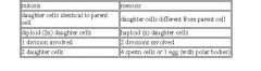
During prophase I of meiosis, DNA is exchanged between homologous chromosomes in a process called crossing over. The new combinations of DNA created during crossover are a significant source of genetic variation, and may result in beneficial new combinations of alleles. The paired and replicated chromosomes are called tetrads, which have two chromosomes and four chromatids, with one chromosome coming from each parent. A tetrad is simply the two pairs of homologous chromosomes, but it enables crossing over of each pair. The first part of meiosis involves separating the tetrad, the second part separates the sister chromatids. The homologous chromosomes is the pair that arises from each parent's chromosome that was replicated into sister chromatids. In mitosis, the two chromosomes that are replicated are already called the homologous chromosomes. Diploid organisms have two copies of each chromosome - (except the sex chromosomes), one from each parent. Each pair of chromosomes is homologous. For example, the two #7 chromosomes are homologous. The homologue to the #3 chromosome would be the other #3 chromosome. Each chromosome contributed by the father has a corresponding chromosome that was contributed by the mother. These corresponding chromosomes constitute a homologous pair. The DNA sequences of homologous chromosomes are not identical (because one's from the mom and one's from the dad). Sister chromatids are exactly identical, because they are replicated from the original chromosome. Mitosis is a form of eukaryotic cell division that produces two daughter cells with the same genetic component as the parent cell. In diploid multicellular organisms sexual reproduction involves the fusion of two haploid gametes to produce a diploid zygote. Mitotic divisions of the zygote and daughter cells are then responsible for the subsequent growth and development of the organism. In the adult organism, mitosis plays a role in cell replacement, wound healing and tumour formation and we want the daughter cells to have exactly the same copy of chromosomes as the parent. Polar bodies are by-products of the egg’s division during meiosis.
|
|
|
|
What is independent assortment when genes are segregated?
|
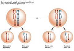
Independent assortment generates genetic variation.
A cell has 2 copies of each somatic chromosome- one from mom, one from dad (homologous chromosomes). Independent assortment shuffles these chromosomes, and then places only one copy of each into the gamete. This way, the gamete may have chromosome 1 from mom, chromosome 2 from dad, chromosome 3 from dad, ... etc. The mechanism of independent assortment is the following: During metaphase I of meiosis, homologous chromosome pair up along the metaphase line in random orientation - sometimes the mom's chromosome is on the left, sometimes it's on the right. During anaphase I of meiosis, the homologous chromosomes are pulled apart. Those on the left will be put into one daughter cell, those on the right will be put into another. |
|
|
|
What is linkage when when genes are segregated?
|
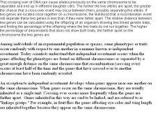
Because of independent assortment, genes on different chromosomes are randomized. However, genes on the SAME chromosome can not be randomnized by this mechanism. Only the chromosome itself is randomly assorted. Genes on the same chromosome are linked to some extent. Recombination includes both crossing over and independent assortment. Without recombination, all alleles for those genes linked together on the same chromosome would be inherited together. Recombination allows a more independent selection between the two alleles that occupy the positions of single genes, as recombination shuffles the allele content between homologous chromosomes. Crossing over is a mechanism that reduces linkage. However, crossing over is only efficient when the genes are physically apart from each other on the chromosome. When the genes are further apart on the chromosome, crossing over makes them less linked. The physically closer the genes are on the chromosome, the more linked they are. Genetic linkage is a term which describes the tendency of certain loci or alleles to be inherited together. Genetic loci on the same chromosome are physically close to one another and tend to stay together during meiosis, and are thus genetically linked. At the beginning of normal meiosis, a chromosome pair (made up of a chromosome from the mother and a chromosome from the father) intertwine and exchange sections or fragments of chromosome. The pair then breaks apart to form two chromosomes with a new combination of genes that differs from the combination supplied by the parents. Through this process of recombining genes, organisms can produce offspring with new combinations of maternal and paternal traits that may contribute to or enhance survival.
|
|
|
|
What is recombination when genes are segregated?
|
recombination: also called genetic recombination, is the process that introduces genetic diversity into the gametes during meiosis. There are 2 processes that make up recombination: independent assortment and crossing over.
|
|
|
|
What are single crossovers when genes are segregated?
|
single crossovers: results in genetic recombination. The chromatids involved in this single crossover exchange alleles at a given locus. Results in 2/4 recombinants (2 sister chromatids out of 4 sister chromatids). Crossing over occurs during Prophase I of meiosis.
|
|
|
|
Do crossovers occur for mitosis or meiosis?
|
The interchange of sections between pairing homologous chromosomes during the prophase of meiosis. Crossovers do not occur for mitosis.
|
|
|
|
What is a chromatid?
|
A chromatid is one among the two identical copies of DNA making up a replicated chromosome, which are joined at their centromeres, for the process of cell division (mitosis or meiosis). The term is used so long as the centromeres remain in contact. When they separate (during anaphase of mitosis and anaphase 2 of meiosis), the strands are called sister chromatids.
In other words, a chromatid is "one-half of a replicated chromosome". It should not be confused with the ploidy of an organism, which is the number of homologous versions of a chromosome. Ploidy is the number of complete sets of chromosomes in a biological cell. In humans, the somatic cells that compose the body are diploid (containing two complete sets of chromosomes, one set derived from each parent), but sex cells (sperm and egg) are haploid. |
|
|
|
What is the quantity of chromatids during the S phase, during Mitosis, and after Mitosis?
|
In humans, for example, there are normally 23 pairs of homologous chromosomes in each cell (N=23). However, the quantity of chromatids will be a multiple of 23. It can be either 4N, 2N or 1N. N does not refer to haploid or diploid; it refers to the number of chromatids in the cell as a multiple of the haploid number of chromosomes for the organism. For example, because a human haploid germ cell has 23 chromosomes, then "N" refers to a multiple of 23. (e.g. 2N=46 chromatids). The last is only seen in haploid gametes, with only one of each homologous chromosome pair. Such are created in gametogenesis.
4N:In a cell with 4N chromatids, there are 23 chromosome pairs (46 chromosomes), and each chromosome has 2 chromatids. Thus, there are 92 chromatids in each cell (4xN). It occurs after the S phase of interphase. 2N: Immediately after a mitosis, where a cell has divided in two, but not yet duplicated its DNA in S phase, there are still 23 chromosome pairs (46 chromosomes). However, each chromosome only has one chromatid. Thus there are 46 chromatids (2xN) 1N: Immediately after meiosis, each cell, called a gamete, only has half the number of chromosomes (23 chromosomes). Furthermore, each chromosome only has one chromatid. Thus, there are 23 chromatids (1xN) |
|
|
|
What is the difference between sister chromatids and chromosomes?
|
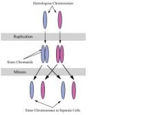
Sister chromatids are 2 identical copies of a chromosome connected by a centromere. Compare sister chromatids to homologous chromosomes, which are the two different copies of the same chromosome that diploid organisms (like humans) inherit, one from each parent. In other words, sister chromatids contain the same genes and same alleles, and homologous chromosomes contain the same genes but two copies of alleles, each of which might or might not be the same as each other. A full set of sister chromatids is created during the S subphase of interphase, when all the DNA in a cell is replicated. Identical chromosome pairs are separated into two different cells during mitosis, or cellular division. All cells (except sex cells) have the same number of chromosomes - it doesn't matter if an organism develops from 2 cells to 800 cells, each of those cells have the same number of chromosomes.
|
|
|
|
What are double crossovers?
|
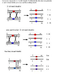
double crossovers:
Scenario 1: results in no genetic recombination. The chromatids involved in this double crossover exchange alleles at first, but then it exchanges them back, resulting in no net recombination. This is called the 2-strand double crossover. Results in 0/4 recombinants. Scenario 2: results in genetic recombination. The chromatids exchange alleles during a crossover. Then, one of the crossover chromatid exchanges with a different chromatid. This is called the 3-strand double crossover. Results in 2/4 recombinants. There are 2 options for 3-strand crossovers because the crossover could happen with one of the 2 other chromatids. Scenario 3: results in genetic recombination. The chromatids exchange, then 2 totally different chromatids on the same chromosome exchange. This is called the 4-strand double crossover. Results in 4/4 recombinants. A two-strand double crossover is the result of two the same two sister chromatids exchanging DNA. These two events cancel each other out and result in parental or non-recombinant progeny. Three-strand double crossovers result when one chromatid crosses over with two other chromatids. The result is two parental and two recombinant progeny, the same as a single crossover event. The four-strand double crossover is a single crossover between two chromatids and then a second single crossover between the remaining chromatids. The result is that all four products are recombinant. |
|
|
|
Explain the significance of very few genes on Y chromosome for sex-linked characteristics.
|
Very few genes on Y chromosome. The Y chromosome is very small and carries few genes of importance. All the sex-linked alleles are carried on the X chromosome. Sex linkage is the phenotypic expression of an allele related to the chromosomal sex of the individual. This mode of inheritance is in contrast to the inheritance of traits on autosomal chromosomes, where both sexes have the same probability of expressing the trait. Since humans have many more genes on the X than the Y, there are many more X-linked traits than Y-linked traits. X-linked traits are maternally inherited from carrier mothers or from affected father. Each son born to a carrier mother has a 50% probability of inheriting the X-chromosome carrying the mutant allele. There are a few Y-linked traits; these are inherited from the father. X-linked inherited recessive diseases occur far more frequently in males because they only have one X chromosome. Females must receive a copy of the gene from both parents to have such a recessive disease. However, they will still be carriers if they receive one copy of the gene.
|
1) X-linked recessive diseases are more common in males
2) The disease can skip generations while being transmitted through heterozygous carrier females 3) If a mother is a heterozygous carrier, her children have a 50% chance of inheriting the disease 4) Affected males transmit the gene to all their daughters, who become carriers 5) There is no father-to-son transmission 6) Males that carry the mutant gene have the disease |
|
|
What is sex determination?
|
Sex determination: XX = female, XY = male
In mammals, the female is the homozygous sex, with two X chromosomes (XX), while the male is heterozygous, with one X and one Y chromosome (XY). Genes on the X or Y chromosome are called sex linked genes. |
|
|
|
What is cytoplasmic inheritance?
|
Cytoplasmic inheritance = inheritance of things other than genomic DNA.
Inheritance of traits through DNA that is not connected with the chromosomes but rather to DNA from organelles in the cell. In eukaryote organisms the zygote normally receives the bulk of its cytoplasm from the egg cell and the male gamete contributes little more than a nucleus. Any genes contained in the cell organelles of the cytoplasm will therefore show maternal inheritance. The genes located in mitochondria are very important for proper cellular function, yet the genomes replicate independently of the DNA located in the nucleus. Genes located in the mitochondria are inherited only from the mother. The mother and father both contribute to genes that are transmitted from their nucleus. |
|
|
|
What is mitochondrial inheritance?
|
All cellular organelles, such as mitochondria, is inherited from the mother.
In sexual reproduction, mitochondria are normally inherited exclusively from the mother. The mitochondria in mammalian sperm are usually destroyed by the egg cell after fertilization. The fact that mitochondrial DNA is maternally inherited enables researchers to trace maternal lineage far back in time. |
|
|
|
What is the general concept of mutation?
|
general concept of mutation-error in DNA sequence
Mutation = change in DNA sequence by means other than recombination. |
|
|
|
What are the types of mutations (random, translation error, transcription error, base substitution, insertion, deletion, frameshift)?
|
Random mutation = random changes in DNA sequence. Can be due to radiation, chemicals, replication error ...etc.
Translation error = even if the DNA for a gene is perfect, errors during translation can cause expression of a mutant phenotype. Transcription error = even if the DNA of a gene is perfect, errors during transcription can cause expression of a mutant phenotype. Base substitution = mutation involving a base (ATGC) changing to a different base in DNA or RNA molecule. Also called point mutation. Occurs during replication. Insertion = an extra base is added/inserted into the DNA sequence. Occurs during replication. Deletion = a base is taken out of the DNA sequence. Occurs during replication. Addition/insertion and deletion mutations result in a frameshift mutation during DNA replication. A frameshift mutation is a genetic mutation caused by insertions and deletions of a number of nucleotides that is not evenly divisible by three from a DNA sequence. Due to the triplet nature of gene expression by codons, the insertion or deletion can change the reading frame (the grouping of the codons), resulting in a completely different translation from the original. The earlier in the sequence the deletion or insertion occurs, the more altered the protein produced is. A frameshift mutation will in general cause the reading of the codons after the mutation to code for different amino acids. The stop codon ("UAA", "UGA" or "UAG") in the original sequence will not be read, and a stop codon could result at an earlier or later site. The protein being created could be abnormally short or abnormally long, and will most likely not be functional. Base pair substitutions can also code for a stop codon. |
|
|
|
What are chromosomal rearrangements (inversion, translocation)?
|
Inversion: A defect in the chromosome in which a segment of the chromosome breaks off and reinserted in the same place but in the reverse direction relative to the rest of the chromosome.
Translocation: Chromosomal translocation, that is a chromosomal segment is moved from one position to another, either within the same chromosome or to another chromosome. |
|
|
|
What is advantageous versus deleterious mutation?
|
Advantageous = results in a benefit to the fitness of the organism. For example, a butterfly may produce offspring with new mutations. The majority of these mutations will have no effect; but one might change the color of one of the butterfly's offspring, making it harder (or easier) for predators to see. If this color change is advantageous, the chance of this butterfly surviving and producing its own offspring are a little better, and over time the number of butterflies with this mutation may form a larger percentage of the population.
A deleterious mutation has a negative effect on the phenotype, and thus decreases the fitness of the organism. An advantageous mutation has a positive effect on the phenotype, and thus increases the fitness of the organism. Deleterious = results in a harmful effect to the fitness of the organism. For example, a mutation that causes an organism to be sterile. Deleterious means to cause harm. |
|
|
|
What are inborn errors of metabolism?
|
inborn errors of metabolism = genetic diseases resulting in faulty metabolism. For example PKU (Phenylketonuria) is an inborn error of metabolism where people can't metabolize phenylalanine. There's no cure, but the treatment involves avoiding things containing the amino acid phenylalanine.
|
|
|
|
What is the relationship of mutagens to carcinogens?
|
Mutagen = something that causes mutation.
Carcinogen = something that causes a mutation that causes cancer. All carcinogens are mutagens. Not all mutagens are carcinogens. A mutagen is a substance or agent that causes an increase in the rate of change in genes (subsections of the DNA of the body's cells). These mutations (changes) can be passed along as the cell reproduces, sometimes leading to defective cells or cancer. Examples of mutagens include certain biological and chemical agents as well exposure to ultraviolet light or ionizing radiation. Mutagenesis is the formation of mutations. Do not confuse a mutagen with a carcinogen (a substance that causes cancer). Mutagens may cause cancer, but not always. A carcinogen is a substance that causes cancer (or is believed to cause cancer). A carcinogenic material is one that is known to cause cancer. The process of forming cancer cells from normal cells or carcinomas is called carcinogenesis. Carcinogenesis (the creation of cancer), is the process by which normal cells are transformed into cancer cells. Cell division is a physiological process that occurs in almost all tissues and under many circumstances. Under normal circumstances, the balance between proliferation and programmed cell death, usually in the form of apoptosis, is maintained by tightly regulating both processes to ensure the integrity of organs and tissues. Mutations in DNA that lead to cancer (only certain mutations can lead to cancer and the majority of potential mutations will have no bearing) disrupt these orderly processes by disrupting the programming regulating the processes. Carcinogenesis is caused by this mutation of the genetic material of normal cells, which upsets the normal balance between proliferation and cell death. The uncontrolled and often rapid proliferation of cells can lead to benign tumors; some types of these may turn into malignant tumors (cancer). |
|
|
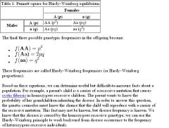
What is the Hardy-Weinberg Principle ?
|

Five Assumptions of Hardy-Weinberg: 1) Infinitely large population (no genetic drift) 2) No mutation 3) No migration 4)Random mating (no sexual selection) 5)No natural selection
The Hardy–Weinberg principle states that both allele and genotype frequencies in a population remain constant—that is, they are in equilibrium—from generation to generation unless specific disturbing influences are introduced. Those disturbing influences include non-random mating, mutations, selection, limited population size, "overlapping generations", random genetic drift and gene flow. Gene flow is the transfer of alleles of genes from one population to another. It is important to understand that outside the lab, one or more of these "disturbing influences" are always in effect. That is, Hardy–Weinberg equilibrium is impossible in nature. Genetic equilibrium is an ideal state that provides a baseline to measure genetic change against. In the simplest case of a single locus with two alleles: the dominant allele is denoted A and the recessive a and their frequencies are denoted by p and q; freq(A) = p; freq(a) = q; p + q = 1. If the population is in equilibrium, then we will have freq(AA) = p2 for the AA homozygotes in the population, freq(aa) = q2 for the aa homozygotes, and freq(Aa) = 2pq for the heterozygotes. Maintained gene flow between two populations can also lead to a combination of the two gene pools, reducing the genetic variation between the two groups. It is for this reason that gene flow strongly acts against speciation, by recombining the gene pools of the groups, and thus, repairing the developing differences in genetic variation that would have led to full speciation and creation of daughter species. |
|
|
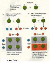
What is a Testcross (backcross; concepts of parental, F1, and F2 generations)?
|
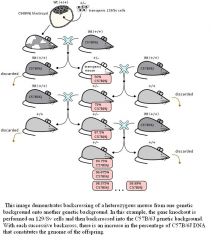
Test cross: so you have something with dominant phenotype. It could either be Aa or AA. To find out, you cross it with the homozygous recessive aa. If Aa, half the offspring will express the recessive phenotype. If AA, no offspring will express the recessive phenotype.
Back cross = mating between the offspring and the parent = preserve parental genotype. Backcrossing is a crossing of a hybrid with one of its parents or an individual genetically similar to its parent, in order to achieve offspring with a genetic identity which is closer to that of the parent. It is used in horticulture, animal breeding and in production of gene knockout organisms. A hybrid carries two different alleles of the same gene. Parental generation = P = generation of the parent. On a pedigree, the is the row that represents the parents F1 generation = Filial 1 = children. On a pedigree, this is the row below the parents, and represents the children of the parents. F2 generation = Filial 2 = grandchildren. On a pedigree, this is the row below the F1, and represents the children of the F1 and grandchildren of the parents. |
|
|
|
What is the fitness concept?
|
Fitness is defined as the ability to pass your genes on, or reproductive success.
The classical trick question gives you an individual who is strong, healthy, long-living, but does not reproduce. In this case, no matter how good the other traits are, if the individual does not reproduce, then it has a fitness of zero. |
|
|
|
What is selection by differential reproduction?
|
Individuals who reproduce more viable offspring are selected for.
Individuals who reproduce less viable offspring are selected against. |
|
|
|
What are is directional selection and natural selection?
|
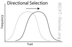
Natural selection = survival of the fittest. Natural selection is the process by which certain heritable traits—those that make it more likely for an organism to survive and successfully reproduce —become more common in a population over successive generations. It is a key mechanism of evolution. Natural selection acts on the phenotype, or the observable characteristics of an organism, but the genetic (heritable) basis of any phenotype which gives a reproductive advantage will become more common in a population. Over time, this process can result in adaptations that specialize populations for particular ecological niches and may eventually result in the emergence of new species. In other words, natural selection is an important process (though not the only process) by which evolution takes place within a population of organisms.
Directional selection: selects for a trait on one extreme. For example, selection for height of canopy trees in a rainforest: trees compete for sunlight, so selection favors trees to become higher and higher. |
|
|
|
What is stabilizing selection?
|
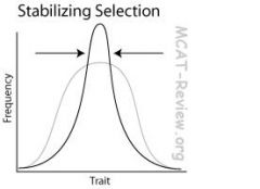
Stabilizing selection: selects for a trait that is moderate, and selects against the extremes. For example, birthweight: too low birthweight means that the baby is premature, too high birthweight means that the mom will have a hard time delivering, so there's a "just right" birthweight that is selected for.
|
|
|
|
What is disruptive selection?
|
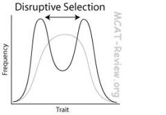
Disruptive selection: selects for the extremes. For example, birds occupying a habitat with 2 distinct niches (eating berries for a living and eating seeds for a living): small beaks are selected for eating berries, large beaks are selected for cracking seeds, medium beak is left out.
|
|
|
|
What is group selection?
|
Group selection = natural selection acting on the group, not the individual. Group selection refers to the idea that alleles can become fixed or spread in a population because of the benefits they bestow on groups, regardless of the alleles' effect on the fitness of individuals within that group.
Group selection describes natural selection operating between groups of organisms, rather than between individuals. This would produce adaptations that benefit the group, rather than the individual. Darwin's theory of evolution was based upon individual selection, and he rejected the idea of group selection. Animals sometimes restrain their reproduction. The idea was brought to prominence in 1962 by the British biologist Wynne-Edwards, who argued that animals restrain their reproduction in order not to over-eat the local food supply. If all the individuals in a group reproduce at the maximum rate, their offspring might over-eat the food supply, and the group would then go extinct. This could be avoided by collectively restraining their reproduction. Natural selection on individuals does not favor reproductive restraint. An individual that increases its reproduction will be favored relative to individuals that produce fewer offspring. Within a group, if some individuals produce more offspring than others, the former will proliferate. For Wynne-Edward's theory to be correct, selection on groups must be weaker than selection on individuals. |
|
|
|
Explain how evolutionary success is an increase in percent representation in the gene pool of the next generation.
|
If the frequency of an allele increased, then that's evolutionary success for that allele.
If the frequency of alleles of an individual increased in a population, then that's evolutionary success for that individual. |
|
|
|
What is the definition of species?
|
Three conditions for biological species
Be able to interbreed. Be able to produce fertile, viable offspring. Does this naturally. A dog and a cat can't interbreed, so they don't belong the the same species. A horse and a donkey can interbreed, but their offspring, the mule, is sterile. So horses and donkeys aren't the same species. Some species of flowers can cross pollinate to produce fertile offspring. However, this never occurs in nature because one is bee-pollinated and the other is bird-pollinated. Thus, they are different species even though they can potentially produce fertile offspring. |
|
|
|
What is polymorphism?
|
Polymorphism is just a fancy word for different forms of alleles. Polymorphism means having multiple alleles of a gene within a population, usually expressing different phenotypes. In other words, the occurrence of more than one form or morph. In order to be classified as such, morphs must occupy the same habitat at the same time and belong to a population with random mating. Polymorphism is common in nature; it is related to biodiversity, genetic variation and adaptation; it usually functions to retain variety of form in a population living in a varied environment. Polymorphism results from evolutionary processes, as does any aspect of a species. It is heritable, and is modified by natural selection.
|
|
|
|
What is adaptation and specialization?
|
Adaptation is the genetic change in a population caused by natural selection. Adaptation is the evolutionary process whereby a population becomes better suited to its habitat. This process takes place over many generations.By using the term adaptation for the evolutionary process, and adaptive trait for the bodily part or function (the product), the two senses of the word may be distinguished.
Adaptation is caused by Darwin's natural selection, not by Lamarck's ideas. According to Lamark, certain traits of a creature could come about via use or disuse. When a trait was used many times, the organs which the trait was dependent on were strengthened, and the trait became commonplace. If a trait fell into disuse the organ was weakened, and the trait was not carried on to its offspring. If a trait was strong within the parent, its offspring would also have the same characteristics. The adaptation theory says that a giraffe's neck is long because long necks increase the survival rate, so more long-necked giraffes survive to reproduce, and over many generations, the population evolved long necks. The wrong idea by Lamarck is that the giraffe had to reach for higher leaves on trees, so it stretched itself a longer neck. Specialization = adaptation of traits to better fill a niche. Those animals that have adapted traits over time, fill the niche better. |
|
|
|
What are the concepts of ecological niche, competition?
|
A species' ecological niche is what resources the species uses to survive in its environment. Two species can avoid competition, and better use the environment's resources by occupying different niches. As long as two species occupy different niches, there's no competition because they use different resources. When niches overlap, there's competition. Specialization occurs to better occupy a particular niche. A niche is a term describing the relational position of a species or population in its ecosystem to each other. The ecological niche describes how an organism or population responds to the distribution of resources and competitors (e.g., by growing when resources are abundant, and when predators, parasites and pathogens are scarce) and how it in turn alters those same factors (e.g., limiting access to resources by other organisms, acting as a food source for predators and a consumer of prey). Competition is an interaction between organisms or species, in which the fitness of one is lowered by the presence of another. Limited supply of at least one resource (such as food, water, and territory) used by both is required. Species less suited to compete for resources should either adapt or die out. According to evolutionary theory, this competition within and between species for resources plays a critical role in natural selection.
|
|
|
|
What is the concept of population growth through competition?
|
Population growth is checked by competition. When resources get scarce, competition increases, which slows down population growth. Competition within a species can force members within the species to occupy different niches, which drives speciation. Competition is a negative interaction that occurs among organisms whenever two or more organisms require the same limited resource. All organisms require resources to grow, reproduce, and survive. Organisms, however, cannot acquire a resource when other organisms consume or defend that resource. Therefore, competitors reduce each other's growth, reproduction, or survival. The competitive exclusion principle: species cannot coexist if they have the same niche. The word "niche" refers to a species' requirements for survival and reproduction. These requirements include both resources (like food) and proper habitat conditions (like temperature, pH ). The idea is that if two species had identical niches (required identical resources and habitats) they would attempt to live in the exact same area and would compete for the exact same resources. If this happened, the species that was the best competitor would always exclude its competitors from that area. Therefore, species must at least have slightly different niches in order to coexist. Speciation is the evolutionary process by which new biological species arise.
|
|
|
|
What is inbreeding?
|
Inbreeding is mating between relatives. Inbreeding increases the frequency of homozygotes, decreases heterzygotes, and decreases genetic diversity. Inbreeding depression occurs because of the increase in the frequency of homozygous recessive detrimental alleles. Inbreeding depression is reduced fitness in a given population as a result of breeding of related individuals. It is often the result of a population bottleneck (an evolutionary event in which a significant percentage of a population or species is killed or otherwise prevented from reproducing). In general, the higher the genetic variation within a breeding population, the less likely it is to suffer from inbreeding depression.
Some species (naked mole rats) naturally inbreed because: 1) They stay in one small area and don't migrate much. 2) Detrimental homozygous recessive alleles are eliminated because of many generations of natural selection due to the small gene pool. Breeding between closely related individuals, called inbreeding, may on one hand result in more recessive deleterious traits manifesting themselves, because the genomes of pair-mates are more similar: recessive traits can only occur in offspring if present in both parents' genomes, and the more genetically similar the parents are, the more often recessive traits appear in their offspring. Consequently, the more closely related the breeding pair is, the more homozygous deleterious genes the offspring may have, resulting in very unfit individuals. For alleles that confer an advantage in the heterozygous and/or homozygous-dominant state, the fitness of the homozygous-recessive state may even be zero (meaning sterile or unviable offspring). |
|
|
|
What is outbreeding?
|
Outbreeding is mating with non-relatives, which is just the opposite of inbreeding.
Outbreeding increases heterozygosity. |
|
|
|
What are bottlenecks and genetic drift?
|
A bottleneck is a severe reduction in population size. This can be caused, for example, by a natural disaster that wipes out a majority of the population.
Genetic drift is the random changes in allele frequencies. The effect of genetic drift increases as population size decreases. Bottlenecks increase the effect of genetic drift. Genetic drift—along with natural selection, mutation, and migration—is one of the basic mechanisms of evolution. Genetic drift is an important evolutionary process, which leads to changes in allele frequencies over time. It may cause gene variants to disappear completely, and thereby reduce genetic variability. In contrast to natural selection, which makes gene variants more common or less common depending on their reproductive success, the changes due to genetic drift are not driven by environmental or adaptive pressures, and may be beneficial, neutral, or detrimental to reproductive success. The effect of genetic drift is larger in small populations, and smaller in large populations. |
|
|
|
What is divergent evolution?
|
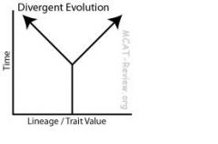
Divergent evolution means the same lineage, evolving apart to be more different.
For example, bats and horses. Both share the same lineage as mammals, but the limb of the bat became wings while the horse developed hooves. Divergent evolution produces homologous structures (bat's wing and horse's hoof). A body part with the same basic structure and embryonic origin as that of another organism, though not necessarily sharing the same function. An important consequence of divergent evolution is speciation, the divergence of one species into two or more descendant species. Divergent evolution refers to the independent development of dissimilar characteristics in tow or more lineages sharing a common ancestry. If a bat, a human, an alligator, and a penguin all evolved from a common ancestor, then they should share common anatomical traits. In fact, they do. Compare the forelimbs of the human, the bat, the penguin, and the alligator. Find the humerus, radius, ulna, and carpals in each forelimb. Though the limbs look strikingly different on the outside and though they vary in function, they are very similar in skeletal structure. More significantly, they are derived from the same structures in the embryo. Structures that are embryologically similar, but have different functions, are called homologous structures. Though these animals look different, a comparison of homologous structures indicates that they are quite similar. This suggests that these animals evolved from a common ancestor. |
|
|
|
What is parallel evolution?
|
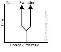
Parallel evolution
Same lineage, evolving closer together to be similar, using similar mechanisms. Parallel evolution is the development of a similar trait in different not closely related species, but descending from the same ancestor. Parallel evolution is sometimes difficult to distinguish from convergent evolution. Parallel evolution occurs when different species start with similar ancestral origins, then evolve similar traits over time. This kind of thing happens because the two different species, though they don't necessarily share a common ancestor, experience similar kinds of environmental pressures and survive only by undergoing similar adaptations. When both descendants are similar in a particular respect, evolution is defined as parallel if the ancestors considered were also similar, and convergent if they were not. An example of parallel evolution is coloration that serves as a warning to predators and for mating displays have evolved in many different species. |
|
|
|
What is convergent evolution?
|
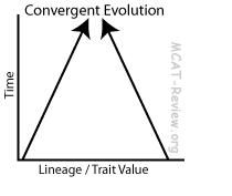
Convergent evolution
Different lineage, evolving closer together to be similar, using different mechanisms. For example, bats and butterflies. Both have wings, but they came from totally different lineages, evolved through different mechanisms/mutations. Convergent evolution produces analogous structures (bat's wing and butterfly's wing). Convergent evolution describes the acquisition of the same biological trait in unrelated lineages. The opposite of convergent evolution is divergent evolution, whereby related species evolve different traits. Analogous structures are features of different species that are similar in function but not necessarily in structure (bat's wing and butterfly's wing don't have the same kind of structure) and which do not derive from a common ancester and which evolved in response to a similar environmental challenge. Structures that are embryologically similar, but have different functions, are called homologous structures. Though these animals (for example a bat, human, alligator, and penguin) look different, a comparison of homologous structures indicates that they are quite similar and share a common ancester. |
|
|
|
What is parasitism?
|
Relationship where one benefits (parasite), and the other is harmed (host).
For example, worms living inside animal intestines. |
|
|
|
What is commensalism?
|
Relationship where one benefits, and the other is not affected.
For example, some plants seeds disperse by sticking to animal fur. |
|
|
|
What is mutualism?
|
Relationship where both species benefit.
For example, lichens are made from a mutualistic relationship between fungi and algae. The fungus provides anchor/absorption, and the alga provides photosynthesis. |
|
|
|
What is the Relationship between ontogeny and phylogeny?
|
Ontogeny = development through the life of an organism.
Phylogeny = development through evolutionary time of lineages/species. Phylogeny is the evolutionary history of species. Ontogeny describes the origin and the development of an organism from the fertilized egg to its mature form. Phylogenetics is the study of evolutionary relatedness among various groups of organisms (for example, species or populations). While developmental or ontogenetic processes can influence subsequent evolutionary or phylogenetic processes, individual organisms develop (ontogeny), while species evolve (phylogeny). |
|
|
|
What is primordial soup and how does it relate to the origin of life?
|
Oparin and Haldane proposed that conditions during the early years of Earth's existence favored the abiotic synthesis of organic molecules. Atmospheric gases and the oceans bonded together in various ways and accumulated, forming a primordial soup of organic precursor molecules. The energy for the formation of these bonds was supplied by a number of sources, including the sun and lightning. Miller tested this hypothesis in the laboratory by mixing gases circulated past a source of lightning. Organic molecules were formed, including simple amino acids. The experiment has been modified and able to generate all 20 amino acids, many lipids, and the 5 nitrogenous bases of DNA and RNA. It is postulated that organic monomers were abiotically synthesized in a similar way on primitive Earth, and as they accumulated, they were able to react and form polymers. Abiotic components are non-living chemical and physical factors in the environment.
|
|
|
|
What is the RNA world hypothesis of the origin of life?
|
The question "How do simple organic molecules form a protocell?" is largely unanswered but there are many hypotheses. Some of these postulate the early appearance of nucleic acids ("genes-first") whereas others postulate the evolution of biochemical reactions and pathways first ("metabolism-first"). The RNA world hypothesis describes an early Earth with self-replicating and catalytic RNA but no DNA or proteins. Factors supportive of an important role for RNA in early life include its ability to act both to store information and catalyse chemical reactions (as a ribozyme); its many important roles as an intermediate in the expression and maintenance of the genetic information (in the form of DNA) in modern organisms; and the ease of chemical synthesis of at least the components of the molecule under conditions approximating the early Earth. Relatively short RNA molecules which can duplicate others have been artificially produced in the lab. Such replicase RNA, which functions as both code and catalyst provides a template upon which copying can occur. If these were present, Darwinian selection would favour the proliferation of such self-catalysing structures, to which further functionalities could be added. Several models reject the idea of the self-replication of a "naked-gene" and postulate the emergence of a primitive metabolism which could provide an environment for the later emergence of RNA replication.
|
|
|
|
Explain the concept Evolutionary time as measured by gradual random changes in genome.
|
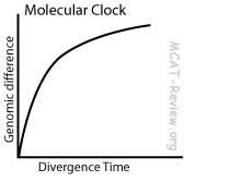
Random changes in frequencies of alleles (genetic drift) that are not acted on by natural selection occur at a constant rate. These mutations are neutral because they are not acted on by natural selection.
By measuring the amount of these neutral mutations, you can find out how much time has passed. You can compare genome differences between two species to find out how long ago they diverged. Another name for this concept is the Molecular Clock. It is used to estimate the time of occurrence of events called speciation. The rate of evolutionary change of any specified protein was approximately constant over time and over different lineages. The molecular data used for such calculations is usually nucleotide sequences for DNA or amino acid sequences for proteins. |
|
|
|
Explain how prokaryotes fit in with the autotrophic and heterotrophic theories concerning the origin of life.
|
The autotrophic hypothesis on the origin of life asserts that the first living beings (prokaryotes) on earth were producers of their own food, just like plants. The ancestors of cyanobacteria (blue-green algae) is believed to have thrived on the earth and been involved in photosynthesis where light energy was converted to chemical energy. The formation oxygen would then transform the Earth's atmosphere and pave the way for it's biodiversity. According to the heterotrophic hypothesis the first living beings were very simple heterotrophic organisms, i.e., not producers of their own food, which emerged from the gradual association of organic molecules into small organized structures. The heterotrophic hypothesis is the most accepted hypothesis.
|
|
|
|
What are protocells in regards to the origin of life?
|
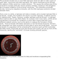
The theoretical protocell is made up of only two molecular components, a RNA replicase and a fatty acid membrane. An extremely pared down and simple version of a cell, the protocell is nonetheless capable of growth, replication, and evolution. Although a working version of a protocell has not yet been achieved in a laboratory setting, the goal appears well within reach.
|
|
|
|
Explain how eukaryotes were involved in the origin of life?
|
According to the most accepted hypothesis aerobic eukaryotic cells emerged from the association of aerobic prokaryotes engulfed by primitive anaerobic eukaryotic cells. This would have been the origin of mitochondria that thus would have primitively been aerobic bacteria engulfed by eukaryotic anaerobes. This hypothesis is called the endosymbiotic hypothesis on the origin of mitochondria. For the reason that mitochondria are the center of the aerobic cellular respiration, the powerhouse of the eukaryotic cell, it is supposed that they were once aerobic prokaryotes. The endosymbiotic hypothesis to explain the emergence of aerobic is further strengthened by the following evidence: mitochondria have their own DNA, similar to bacterial DNA; mitochondria reproduce asexually by binary division, like bacteria do; both organelles have ribosomes and synthesize proteins.
These two organisms would have a mutually beneficial relationship, but would benefit the incorporation. The endosymbiotic theory suggests that mitochondira are descendents of prokaryotes that were engulfed by endocytosis into a vesicle lined with a membrane derived from the cell membrane of the eukaryote host. This is the outer membrane. The inner membrane is the plasma membrane of the endosymbiotic prokaryote. The cell wall of bacteria does not resemble a plasma membrane, it just protects the outside of the cell from dessication and is used for structural support. |
|
|
|
What is the chordate and what are the notochord and the dorsal nerve cord?
|
Chordate = one of the phylums in the kingdom Animalia. Chordates (phylum Chordata) are animals which are either vertebrates or one of several closely related invertebrates. They are united by having, for at least some period of their life cycle, a notochord, a hollow dorsal nerve cord, and pharyngeal pouches/brachial arches.
Chordate features :1) Notochord = the "backbone" of the embryo, except that it's not made of bone. In vertebrates, bones will replace the notochord to form the vertebrae. The notochord is a flexible, rod-shaped body found in embryos of all chordates. It is composed of cells derived from the mesoderm and defines the primitive axis of the embryo. 2) Dorsal nerve cord = forms the nervous system. In higher chordates, the nerve cord develops into the brain and spinal cord. The dorsal nerve cord is a hollow cord dorsal to the notochord. It is formed from a part of the ectoderm that rolls, forming the hollow tube. Dorsal means the "back" side, as opposed to the belly side (ventral). |
|
|
|
What are pharangeal pouches and brachial arches?
|
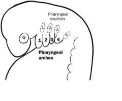
DEVELOPMENT OF BRACHIAL ARCHES ‑ structures which develop in embryo; are comparable to gills of fish; reflect fact that ontogeny (development of individual) resembles phylogeny (evolution of species); are important in understanding final structure and innervation of head and neck. Week 4 of development will form brachial arch components (head and neck). Neural crest cells invade future head and neck region of embryo; these cells enter the pharangeal pouches and brachial arches where they contribute to the development of the head and neck. Brachial arch are covered by ectoderm externally; lined internally by endoderm; core of arch formed by mesenchyme; mesenchyme will form muscles, arteries, connective tissue, cartilage and parts of skeleton. Pharangeal pouches are lined with an endodermal with outpocketings and these pouches are located between adjacent brachial arches. FATE OF BRACHIAL ARCHES ‑ contribute to formation of face, neck, mouth, larynx, and pharynx. In fish, the brachial arches give rise to gills. FATE OF Pharangeal POUCHES: forms auditory tube and tympanic membrane; tonsils, parathyroid, and thymus gland.
|
|
|
|
What is Vertebrate phylogeny and how are they related to each other? What was there in the beginning?
|
Fish: In the beginning, there was fish. Jawless (Agnatha): The very first fish was jawless, slimy, eel-like.
The 7 living classes of vertebrates are distinguished mostly on the basis of their skeletal system, general environmental adaptation, and reproductive system. The 7 living classes include: Agnatha, Chondrichthyes, Osteichthyes, Amphibia, Reptilia, Aves, and Mammalia. The characteristics of each class: Agnatha consists of jawless fish that do not have scales which are the lampreys and hagfish. Chondrichthyes are vertebrates that have rough cartilage rather than bone, such as sharks and rays. Osteichtyes are bony fish, such as tuna, bass, and salmon. Amphibia spend part of their time on land and part of their time in water. Examples of amphibia are frogs, toads, and salamanders. Reptilia such as snakes and lizards have lungs to breathe on land and skin that does not need to be kept wet. Reptiles can produce amniotic eggs. Aves includes all birds who also produce amniote eggs and have forelimbs modified as wings. Mammalia are all mammals that conceive young who are nourished with milk from mammary glands. |
|
|
|
What was the Cartilaginous (Condrichthyes/sharks/rays) vertebrate class?
|
Cartilaginous (Condrichthyes/sharks/rays): Then some developed jaws and a skeleton. Condrichthyes has a skeleton made of cartilage.
|
|
|
|
What was the Bony (Osteichthyes/food fish) vertebrate class?
|
Bony (Osteichthyes/food fish): Osteichthyes has a skeleton made of bone.
Amphibians: the Bony Fish came onto land because their bony skeleton is strong enough to support their weight. Reptiles: Can penetrate further onto land because they don't dry out like amphibians do. Similar to amphibians, the reptiles lay eggs. Birds: First to branch off from the reptiles. Like the reptiles, birds lay eggs. (which came first, the chicken or the egg? Ans: the egg, because the chicken is a bird, and reptiles laid eggs before birds even existed.) Mammals: Next to branch off from the reptiles. Unlike the reptiles and birds, mammals don't lay eggs. |
|
|
|
What do the vertebrates comprise of?
|
Vertebrates are members of the subphylum Vertebrata, chordates with backbones or spinal columns. About 58,000 species of vertebrates have been described. Vertebrata is the largest subphylum of chordates, and contains many familiar groups of large land animals. Vertebrates comprise cyclostomes, bony fish, sharks and rays, amphibians, reptiles, mammals, and birds.
All vertebrates are built along the basic Chordate body plan: A stiff rod running through the length of the animal (vertebral column or notochord), with a hollow tube of nervous tissue (the spinal cord) above it and the gastrointestinal tract below. In all vertebrates the mouth is found at or right below the anterior end of the animal, while the anus opens to the exterior before the end of the body. The remaining part of the body continuing aft of the anus forms a tail with vertebrae and spinal cord, but no gut. The defining characteristic of a vertebrate is the vertebral column, in which the notochord (a stiff rod of uniform composition) has been replaced by a segmented series of stiffer elements (vertebrae) separated by mobile joints (intervertebral discs, derived embryonically and evolutionarily from the notochord). Women have lower bone density than men. Women and men have the same number of vertebrae. |
|
|
|
What is the taxonomic levels of living organisms in order from the groups consisting of the least closely related to the most closely related?
|
The taxonomic levels are:
Kingdom, phylum, class, order, family, genus, and species. KPCOFGS. "Keep Putting Cake Out For Goodness Sakes!" Distantly related organisms may be in the same kingdom, but as one descends the hierachy the taxonomic groups consist of smaller and smaller numbers of more and more closely related organisms. Individual species are named according to their genus name and their species name, like Homo sapiens. Individual species can be uniquely named using this combination of names because these are the two lowest levels of the taxonomic series. Other mneumonic is : "King Phillip Came Over From Great Spain" |
|
|
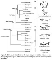
What is the traditional classification of vertebrates?
|
Conventional classification has living vertebrates grouped into seven classes based on traditional interpretations of gross anatomical and physiological traits.
Class Agnatha (jawless fish) Class Chondrichthyes (cartilaginous fishes) Class Osteichthyes (bony fishes) Class Amphibia (amphibians) Class Reptilia (reptiles) Class Aves (birds) Class Mammalia (mammals) |
|
|
|
What is normality?
|
Normality refers to compounds that have
multiple chemical functionalities, such as sulfuric acid, H2SO4: a 1M solution of H2SO4 will contain only one mole of H2SO4 in 1 liter of solution, but if you titrate the solution with base, you will find that it contains two moles of acid. This is because a single molecule of H2SO4 contains two acidic protons. Thus, a 1M solution of H2SO4 will be 2N. Normality = molarity x n (where n = the number of protons exchanged in a reaction). There is a relationship between normality and molarity. Normality can only be calculated when we deal with reactions, because normality is a function of equivalents. Normality measures the number of equivalents per liter of solution. The definition of an equivalent will depend on the type of reaction taking place in the solution. The only time normality is likely to appear on the MCAT is with an acid-base reaction. In an acid-base reaction an equivalent is defined as the mass of acid or base that can furnish or accept 1 mole of protons. So a 1 molar solution of H2SO4 would be called a 2 normal solution because it can donate 2 protons for each mole of H2SO4. |
|
|
|
What is parts per million?
|
This is a way of expressing very dilute concentrations of substances. Just as per cent means out of a hundred, so parts per million or ppm means out of a million. Usually describes the concentration of something in water or soil. One ppm is equivalent to 1 milligram of something per liter of solution (mg/l) or 1 milligram of something per kilogram of solution (mg/kg).
In terms of percents, 1 ppm equals 0.0001 percent. For Solids 1 kilogram (kg) = 1 million milligrams (mg) so: 1 mg/kg = 1 part per million Parts per million can also be expressed as: 1,000,000 times grams of solute divided by grams of solution. Parts per billion is 1000 greater than parts per million. Parts per million is 10^6 times the ratio of the mass of the solute to the total mass of the solution. Parts per million is NOT number of solute molecules per million molecules, it is the mass of the solute per mass of solution times one million. |
|
|
|
How do you calculate weight percent (X%)
|
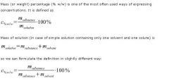
Weight percentage is the only percentage concentration that is always unambiguous. Note that it is expressed in % units (as opposed to % w/v).
|
|
|
|
How do you calculate weight/volume percent?
|
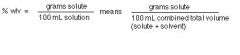
In Latin "per cent" means "for every 100 hundred." When we express the concentration of a solute in % (w/v), we are reporting the mass of solute for every 100 mL of solution.
For example, a 4% (w/v) solution of NaCl in water is made by dissolving 4 g NaCl in enough water to give a total final volume of 100 mL. Percent weight/volume does have units. It's not unitless like Percent weight/weight |
|
|
|
What is buffering capacity?
|
Buffers reduce the variation in
the pH of an end-product. Buffer capacity = No.of moles of acid or base added per lit. solution / change in pH. The buffer capacity of a buffer is maximum when acid to salt or base to salt ratio is equal to 1 i.e., it contains equal number of moles of acid (or base) and the salt. All buffer solutions remain effective over a small pH range: this pH-range is characteristic of the buffer and is termed as the buffer-range. Buffer capacity of an acidic buffer is maximum if 1) PH = pKa 2) [Salt] = [weak acid] Buffer capacity of a basic buffer is maximum if 1) POH = pKb 2) [Salt] = [weak base] |
|
|
|
How do you calculate pH of a Weak Acid?
|
Question:
What is the pH of a 0.01 M benzoic acid solution? Given: benzoic acid Ka= 6.5 x 10⁻⁵ Solution: C6H5COOH → H+ + C6H5COO- The formula for Ka is Ka = [H+][B-]/[HB] where [H+] = concentration of H+ ions [B-] = concentration of conjugate base ions [HB] = concentration of undissociated acid molecules for a reaction HB → H+ + B- Benzoic acid dissociates one H+ ion for every C6H5COO- ion, so [H+] = [C6H5COO-]. Let x represent the concentration of H+ that dissociates from HB, then [HB] = C - x where C is the initial concentration. Enter these values into the Ka equation: Ka = x · x / (C - x) Most weak acids barely dissociate in solution. Values for C - x then, would be very close to C to seem unchanged. If we substitute C for (C - x) in the Ka equation, Ka = x²/(C - x) Ka = x²/C Find pH pH = -log[H+] pH = -log(x) Solve for x Benzoic acid dissociates in water as |
|
|
|
How do you calculate the pH of a weak base?
|
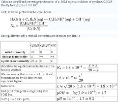
|
|
|
|
What is imprinting?
|
In imprinting, the perception of an object is enhanced in some way during a critical period early in development. The term was first used to describe the behavior of ducklings when they tend to follow any large moving object they encounter during this critical period after hatching. Habituation is a form of non-associative learning in which the response to a stimulus decreases over time, perhaps due to some kind of neural fatigue. Conditioning is a form of associative learning. In classical conditioning, for instance, an animal may come to associate the sound of a bell with the presentation of food.
|
|
|
|
What is erythroblastosis fetalis?
|
|
|