![]()
![]()
![]()
Use LEFT and RIGHT arrow keys to navigate between flashcards;
Use UP and DOWN arrow keys to flip the card;
H to show hint;
A reads text to speech;
15 Cards in this Set
- Front
- Back

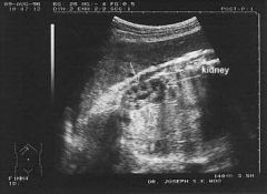
1. This is a _____________ of the __________ (organ).
2. The WHITE material seen in the centre of the image represents the ____ of the ______ ________ and which is present in every person (exception = pathology). |
1. ULTRASOUND KIDNEY
2. FAT RENAL PELVIS |
|
|
1. ULTRASOUNDS are USEFUL in RENAL EXAMS because they good at showing _________ & __________ which contain ___________.
2. However not all _________ contain __________. Some contain ________ ACID which cannot be seen on ULTRASOUND.
3. If there was an OBSTRUCTION to A KIDNEY one would expect to see a "_________ __" KIDNEY
|
1. WATER STONES which contain CALCIUM
2. STONES CALCIUM URIC
3 BALLOONED UP |
|
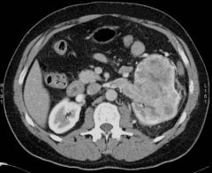
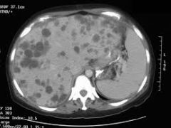
1. This image is a __________ showing a normal ____ KIDNEY but a __________ on the ________ KIDNEY. Diagnosis = _______ ____ _________.
2. In this image the _________ is invading the _______ ________ which is quite common for this form of disease. |
1. CT SCAN RIGHT KIDNEY = NORMAL TUMOUR on LEFT KIDNEY RENAL CELL CARCINOMA
2. TUMOUR INVADING RENAL VEIN |
|
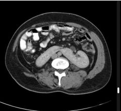
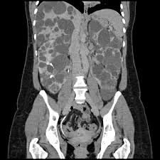
1. This is a _______ with _________ image.
2. The condition present is a _________ __________
3. People with this condition have their ________ located ________ up in the abdomen and more _________.
4. This makes people in these cases more at risk of _______ from a small injury as the organs are usually protected by the ______ ________. |
1. CT with CONTRAST
2. HORSESHOE KIDNEY
3. KIDNEYS LOCATED HIGHER SUPERFICIAL
4. BLEEDING PSOAS MUSCLE |
|
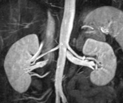
1. This is a ________ of the RENAL ________ and __________.
2. _____'s are useful for assessing _______ ________ and pathology related to them. |
1. MRI RENAL VESSELS AORTA
2. MRI's BLOOD VESSELS |
|
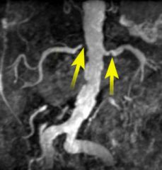
1. In this MRI of the RENAL VESSELS there is _______ RENAL ARTERY ________.
2. This is likely to have been caused by SEVERE _________ which can be seen on the _____ also. |
1. BILATERAL RENAL ARTERY STENOSIS
2. ATHEROSCLEROSIS AORTA
|
|
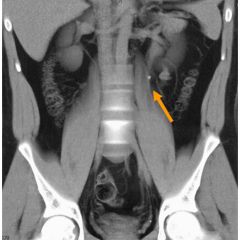
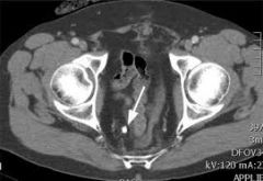
1. In this ____ there is a small _____ stone in the ______ __________.
2. Smaller stones are more likely to cause ________ as they can travel down the ________ compared to ___ ones which hardly move and cannot get into the _________.
3. If there is a blockage in the piping this can cause a ________ of URINE back to the KIDNEY.
4. It can also cause _________ in the URETER which is a condition known as _____________. |
1. CT CALCIUM LEFT URETER
2. PAIN URETER BIG URETER
3. REFLUX
4. DISTENTION HYDROURETER |
|
1. ______________ are useful for Fetal Development as can see whether RENAL development is normal.
2. In particular attention is paid to whether ____ SPOTS are seen in the KIDNEY as these represent _____ ______. If these cant be seen there is an abnormality.
|
1. ULTRASOUNDS
2. DARK SPOTS RENAL PYRAMIDS |
|
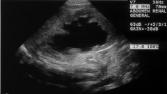
1. ULTRASOUND can also be useful in seeing whether the KIDNEY is full and _________ as seen in this case due to an ___________.
2. An ____________ in the URETER or further down can cause __________ which can result in more fluid in the KIDNEY and cause DISTENTION and damage to the KIDNEY tissues = ______________. |
1. DISTENDED OBSTRUCTION
2. OBSTRUCTION REFLUX HYDRONEPHROSIS |
|
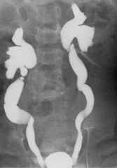
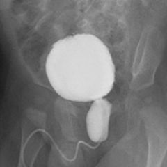
1. This image is of a MCU _________ ___________ which shows a ______-______ ________ caused by ________ _______ _________. This is evident as the kidneys, ureters, and bladder all appear _______ and blown up. |
1. MICTURATING CYSTOURETHROGRAM VESICO-URETERIC REFLUX POSTERIOR URETHRAL VALVES DISTENDED |
|
1. Another MCU _________ __________ that shows the URETHRA and ________ as _______ and Blown up causing ______-_______ REFLUX.
2. This is caused by ________ _________ _________.
3. This condition occurs in neo-nates and is due to problems in development that result in remnant membranes affecting the ________ __________. |
1. MICTURATING CYSTOURETHROGRAM BLADDER DISTENDED VESICO-URETERIC REFLUX
2. POSTERIOR LEAFLET VALVES
3. PROSTATIC URETHRA |
|
This image shows a _______ _______.
If the stone is bigger than _____ then they will not be able to ______ it out so will need to see a _________ for __________. |
RENAL COLIC 6MM PASS UROLOGIST INTERVENTION |
|
? |
ADULT POLYCYSTIC KIDNEY |
|
|
ADULT POLYCYSTIC KIDNEY |
|

|
TUBEROUS SLCEROSIS |