![]()
![]()
![]()
Use LEFT and RIGHT arrow keys to navigate between flashcards;
Use UP and DOWN arrow keys to flip the card;
H to show hint;
A reads text to speech;
72 Cards in this Set
- Front
- Back
- 3rd side (hint)
|
The three parts of the lymphatic system: |
A network of lymphatic vessels (lymphatics)
Lymph
Lymph nodes |
|
|
|
Lymphatic System functions |
Returns interstitial fluid and leaked plasma proteins back to the blood Together with lymphoid organs and tissues, they provide the structural basis of the immune system |
|
|
|
Once interstitial fluid enters lymphatics, it's called... |
Lymph |
|
|
|
Lymphatic System diagram |
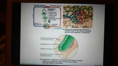
Lymphatic capillaries are blind-ended tubes in which adjacent endothelial cells overlap each other, firming flaplike minivalves. |
|
|
|
Where does lymph flow? |
Lymphatic vessels are a one-way system Lymph flows toward the heart |
|
|
|
Lymphatics (lymph vessels) include: |
Lymphatic capillaries Lymphatic collecting vessels Lymphatic trunks and ducts |
|
|
|
Lymphatic capillaries |
Similar to blood capillaries Except, They are very permeable (take up cell debris, pathogens, and cancer cells) Endothelial cells overlap to form one-way minivalves and are anchored by collagen filaments, preventing collapse of capillaries |
|
|
|
Where are lymphatic capillaries absent in the body? |
Bones Bone marrow Teeth CNS |
|
|
|
Lacteals and their function |
Specialized lymph capillaries present in intestinal mucosa They absorb digested fat and deliver fatty lymph (chyle) to the blood |
|
|
|
Lymphatic collecting vessels |
Similar to veins
Except
They have thinner walls, with more internal valves
Anastomes appear more frequently |
|
|
|
Collecting vessels in the skin travel with... |
Superficial veins |
|
|
|
Deep vessels travel with... |
Arteries |
|
|
|
Lymphatic collecting vessels nutrients are supplied from.... |
Branching vasa vasorum |
|
|
|
Lymphatic trunks |
Are formed by the union of the largest collecting ducts: Paired lumbar Paired bronchomediastinal Paired subclavian Paired jugular trunks A single intestinal trunk |
|
|
|
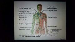
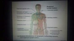
Lymphatic ducts |
Lymph is delivered into one of two large ducts: The right lymphatic duct drains the right upper arm and the right side of the head and thorax The thoracic duct arises from the cisterna chyli and drains the rest of the body Each empties lymph into venous circulation at the junction of the internal jugular and subclavian veins on its own side of the body |
|
|
|
Lymphatic ducts diagram |
|

|
|
|
What propelles lymph? |
Pulsations of nearby arteries Contraction of smooth muscle in the walls of the lymphatics |
|
|
|
Lymphoid cells (lymphocytes) |
Lymphocytes are the main warriors if the immune system |
|
|
|
What are the two main varieties of lymphocytes? |
T cells (T lymphocytes) B cells (B lymphocytes) |
|
|
|
What do T cells and B cells protect against? |
Antigens Anything that the body perceives as foreign: Bacteria and their toxins; viruses Mismatched RBCs or cancer cells |
|
|
|
T cells |
Manage the immune response Attack and destroy foreign cells |
|
|
|
B cells |
Produce plasma cells, Which secrete antibodies |
|
|
|
Other lymphoid cells |
Macrophages phagocytize foreign substances and help activate T cells Dendritic cells capture antigens and deliver them to lymph nodes Reticular cells produce stroma that supports other cells in lymphoid organs |
|
|
|
Other lymphoid cells diagram |
|
|
|
|
Lymphoid tissue |
Houses and provides a proliferation site for lymphocytes
Furnishes a surveillance vantage point |
|
|
|
What are the two main types of lymphoid tissue? |
Diffuse lymphatic tissue Lymphatic follicles |
|
|
|
Diffuse lymphatic tissue |
Comprises scattered reticular tissue elements in every body organs Larger collections in the lamina propria of mucous membranes and lymphoid organs |
|
|
|
Lymphatic follicles (nodules) |
Are solid, spherical bodies of tightly packed reticular elements and cells The germinal center is composed of dendritic and B cells May form part of larger lymphoid organs |
|
|
|
Lymph nodes |
Principal lymphoid organs of the body Embedded in connective tissue, in clusters along lymphatic vessels Near the body surface in inguinal, axillary, and cervical regions of the body |
|
|
|
Lymph nodes diagram |
|
|
|
|
Lymph node functions |
Filter lymph - Macrophages destroy microorganisms and debris Immune system - Lymphocytes are activated and mount an attack against antigens |
|
|
|
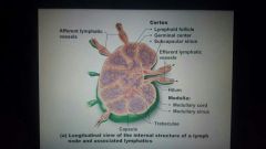
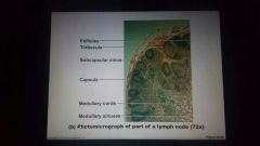
Structure of a lymph node |
Bean shaped
External fibrous capsule
Trabeculae extend inward and divide the node into compartments
Two histologically distinct regions:
Cortex Medulla |
|
|
|
The cortex of a lymph node contains... |
Follicles with germinal centers, heavy with dividing B cells |
|
|
|
What nearly encapsulate the follicles? |
Dendritic cells |
|
|
|
What houses T cells in transit? |
The deep cortex |
|
|
|
T cells circulate continuously among the... |
Blood
Lymph nodes
Lymphatic stream |
|
|
|
Lymph node cortex diagram |
|
|
|
|
Medullary cords extend _____ from the cortex and contain ____, _____, and ______ |
Inward
B cells, T cells, and plasma cells |
|
|
|
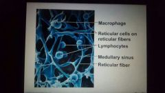
Lymph sinuses contain.... |
Macrophages |
|
|
|
Lymph node microscope image |
|
|
|
|
Circulation in the Lymph Nodes |
Lymph enters via afferent lymphatic vessels, Travels through large subcapsular sinus and smaller sinuses, And exits the node at the hilus via efferent vessels Fewer efferent vessels, causing flow of lymph to stagnate, allowing lymphocytes and macrophages time to carry out functions |
|
|
|
Lymph node diagram |

|
|
|
|
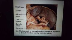
Spleen |
Largest lymphoid organs
Served by splenic artery and vein, which enter and exit at the hilus |
|
|
|
Spleen functions |
Site of lymphocyte proliferation and immune surveillance and response
Cleanses the blood of aged cells and platelets and debris |
|
|
|
Spleen diagram |
|
|
|
|
Spleen facts |
Stores breakdown products of RBCs (ex. Iron) for later reuse Stores blood platelets Site of fetal erythrocyte production (normally ceases after birth) Has a fibrous capsule and trabeculae Contains lymphocytes, macrophages, and huge numbers of erythrocytes |
|
|
|
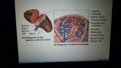
Structure of the spleen |
Two distinct areas: White pulp around central arteries Mostly lymphocytes on reticular fibers and involved in immune functions Red pulp in venous sinuses and splenic cords |
|
|
|
Red pulp |
Is rich in macrophages for disposal of worn-out RBCs and bloodborne pathogens |
|
|
|
Spleen histology diagram |
|
|
|
|
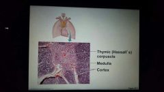
Thymus |
Size with age:
I'm infants, it is found in the inferior neck and extends into the mediastinum, where it partially overlies the heart
Increases in size and is most active during childhood
Stops growing during adolescence and then gradually atrophies |
|
|
|
Thymic lobes contain |
Contain an outer cortex and inner medulla |
|
|
|
Thymus cortex contains |
Contains densely packed lymphocytes and scattered macrophages |
|
|
|
Thymus medulla contains |
Contains fewer lymphocytes and thymic (Hassall's) corpuscles involved in regulatory T cells development |
|
|
|
Thymus diagram |
|
|
|
|
How does the thymus differ from other lymphoid organs? (In important ways) |
It functions strictly in T lymphocyte maturation It does not directly fight antigens The stoma of the thymus consists of star-shaped epithelial cells (not reticular fibers) |
|
|
|
Thymocytes |
Provide the environment in which T lymphocytes become immunocompetent |
|
|
|
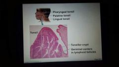
Tonsils |
Simplest lymphoid organs Form a ring of lymphatic tissue around the pharynx |
|
|
|
List the lymphatic tissues forming a ring around the pharynx |
Palatine tonsils Lingual tonsils Pharyngeal tonsil Tubal tonsils |
|
|
|
Palatine tonsils location |
At posterior end of the oral cavity |
|
|
|
Lingual tonsils location |
Grouped at the base of the tongue |
|
|
|
Pharyngeal tonsil location |
In posterior wall of the nasopharynx |
|
|
|
Tubal tonsils location |
Surrounding the openings of the auditory tubes into the pharynx |
|
|
|
Tonsils facts |
Contain follicles with germinal centers Are not fully encapsulated Epithelial tissue overlying tonsil masses invaginates, forming tonsillar crypts |
|
|
|
Tonsil crypts |
Trap and destroy bacteria and particulate matter |
|
|
|
Tonsil diagram |
|
|
|
|
Peyer's patches |
Clusters of lymphoid follicles In the wall of the distal portion of the small intestine Similar structures are also found in the appendix |
|
|
|
Peyer's patches and the appendix |
Destroy bacteria, preventing them from breaching the intestinal wall Generate "memory" lymphocytes |
|
|
|
Peyer's patch Diagram |

|
|
|
|
MALT |
Mucosa-associated lymphatic tissue
Including:
Peyer's patches, tonsils, and the appendix (digestive tract)
Lymphoid nodules in the walls of the bronchi (respiratory tract) |
|
|
|
MALT function |
Protects the digestive and respiratory system from foreign matter |
|
|
|
Development of the lymphatic system |
Beginnings of the lymphatic vessels and main clusters of lymph nodes are apparent by the 5th week of embryonic development
These arise from the budding lymph sacs from developing veins
Lymphatic organs (except the thymus) arise from the mesoderm Except for the spleen and tonsils, lymphoid organs are poorly developed at birth |
|
|
|
Development of the thymus |
The thymus (endodermal origin) forms as an outgrowth of the pharynx |
|