Reading...
![]()
Play button
![]()
Play button
![]()
Use LEFT and RIGHT arrow keys to navigate between flashcards;
Use UP and DOWN arrow keys to flip the card;
H to show hint;
A reads text to speech;
106 Cards in this Set
- Front
- Back
- 3rd side (hint)
|
What are the histological layers of tubular organs?
|
Tunica mucosa
Tunica submucosa Tunica muscularis Tunica serosa or adventitia |
|
|
|
What layers make up the tunica mucosa of tubular organs?
|

Lamina epithelialis mucosa
Lamina propria mucosa Lamina muscularis mucosa |
|
|
|
Describe the tunica mucosa of tubular organs
|

The tunica mucosa is made up of three layers, the lamina epithelialis mucosa, lamina propria mucosa and lamina muscularis mucosa.
Lamina epithelialis mucosa and lamina propria mucosa are loose CT. They are vascular with glands, lymph vessels and occasional nodules. Lamina muscularis mucosa has an inner circular layer and outer longitudinal layer of smooth muscle. |
|
|
|
Describe the tunica submucosa of tubular organs
|
Dense, irregular fibroelastic connective tissue. Glands may be present. Blood, lymph vessels, submucosal (Meissner’s) plexus (ganglia). The motility of mucosa and activity of glands are most prominent in the gastrointestinal tract.
|
|
|
|
Describe the tunica muscularis of tubular organs
|
The thick muscular layer. Associated with peristaltic activity in the gastrointestinal tract, moving ingesta along the digestive tract. Smooth muscle (except in esophagus) with an inner circular layer, outer longitudinal layer. Nerves and vessels (both lymph and blood) are seen between the muscle layers. Myenteric (Auerbach’s) plexus (ganglia) is between the layers, regulating tunica muscularis.
|
|
|
|
Compare and contrast tunica serosa and tunica adventitia
|

Tunica serosa covers organs within body cavities, like intrathoracic or intraperitoneal. These are mesothelial cells.
Tunica adventitia would cover the stuff outside of the body cavities (like most blood vessels), blending in with surrounding connective tissue and fascia |
|
|
|
What is the lamina muscularis mucosa and is it always present?
|

It is the deepest layer of tunica mucosa. It may or may not be present, but when it is, it is made up of smooth muscle.
|
|
|
|
Which is thicker, the tunica muscularis or lamina muscularis mucosa?
|

The tunica muscularis is much thicker. The lamina muscularis mucosa may or may not even be present.
|
|
|
|
What is lamina propria submucosa? What does it indicate?
|

The blended layer of lamina propria mucosa and tunica submucosa. It indicates that the lamina muscularis mucosa is missing.
|
|
|
|
What are the three main parts of the respiratory system?
|
Conduction, transition and respiratory.
|
|
|
|
Where does conduction take place in the respiratory system?
|
Outside and within the lungs. Most of the respiratory system has the primary function of conducting air into the lungs.
It is made up of the nasal cavity, paranasal sinuses, nasopharynx, larynx, and trachea. This portion of the respiratory system also has the task of filtering, humidifying, and warming or cooling the inspired air that eventually goes to the respiratory portion of the lungs. And also is involved in phonation. |
|
|
|
What is the transition zone of the respiratory system?
|
A section of lungs that has both conduction and respiratory functions.
|
|
|
|
What is the respiratory portion of the lung primarily responsible for?
|
Exchange of gases - O2 for CO2
|
|
|
|
What are different tasks of the conducting system of the respiratory tract.
|
Phonation, filtering, humidifying, and warming or cooling the inspired air that eventually goes to the respiratory portion of the lungs.
|
|
|
|
What is an example of phonation?
|
Clucking.
Phonation is the creation of the sounds characteristic of a species. It happens in the conduction of the respiratory system. |
|
|
|
What parts of the respiratory system are involved in conduction?
|
Nasal cavity, paranasal sinuses, nasopharynx, larynx, trachea, primary, secondary, tertiary bronchi, bronchioles, terminal bronchioles.
|
|
|
|
What are nares?
|
The start of the nasal cavity making up the initial opening to the environment (air) and ends with the choana (funnel) that leads into the nasopharynx.
What a bunch of hooey to say nostrils. |
|
|
|
What are vibrissae?
|
Short stiff hairs filtering large dust particles out of the anterior portion of the nasal cavity (vestibule).
|
|
|
|
What sort of epithelium lines the vestibules of the nasal cavities?
|
Keratinized stratified squamous epithelium.
|
|
|
|
What sort of glands are found in the dermis?
What function to they serve? |
sebaceous, sweat, serous, mixed glands.
These glands help humidify the inspired air. |
|
|
|
What is the transition from the skin lining the vestibule to the mucous membrane?
|
ciliated pseudostratified columnar epithelium
|
|
|
|
What is a mucocutaneous junction?
|
The junction between cutaneous skin to mucus membrane.
|
|
|
|
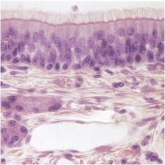
Describe the respiratory epithelium of the nasal cavity.
|
The respiratory epithelium is a ciliated, pseudostratified columnar epithelium with goblet cells – humidifies and cleanses the inspired air. The lamina propria contains many branched, tubuloalveolar, mixed glands (nasal glands) that are mostly serous. Erectile tissue (highly vascularized) may also be present that may become engorged with blood thus warming or cooling the air by heat transfer.
|
|
|

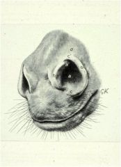
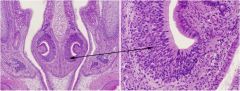
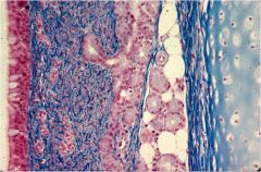
Identify the olfactory and respiratory epithelium on the nasal cavity in this picture.
|
This is the head of a rat showing a combination of olfactory and respiratory epithelium.
Olfactory epithelium on the dorsal, medial, and lateral surfaces of the nasal cavity. Respiratory epithelium on the ventral surfaces of the nasal cavity. |
|
|
|
What are the three cell types of the olfactory region?
|
Sustentacular, basal, and olfactory
|
|
|
|
Describe sustentacular cells.
|
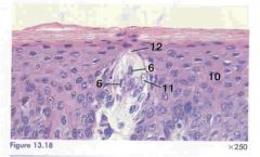
Sustentacular cells are tall with broad apices and narrow bases, an apically oriented round to oval nucleus and granules. Give electrical insulation, support and nourishment to olfactory cells
|
|
|
|
Describe the basal cells of the olfactory region.
|
Basal cells are short cells that do not extend to the surface and have a high proliferative capacity. The basal cells are the cells from which both sustentacular and olfactory cells are derived.
|
|
|
|
When does a nares become a vestibule, and a vestibule a choana?
|
We aren't 100% on this, but think that the nares is the external portion of the nasal cavity, the vestibule is right inside that - the opening, and it narrows into the choana.
|
|
|
|
What type of neurons are in olfactory cells?
|
Modified bipolar neurons. Their basal processes extend to the brain as axons of the olfactory nerve. The apex is a bulbous projection from which modified non-motile cilia project.
|
|
|
|
What is the purpose of the olfactory glands? In what organizational level of respiratory cells are they located? What type of glands are they?
|
The olfactory glands cleanse the olfactory surface and solubilize odor-producing substances.
They are found in the lamina propria and are tubuloalveolar serous glands. |
|
|
|
T/F Olfactory epithelium have goblet cells.
|
False. Respiratory epithelium have lots of them.
|
|
|
|
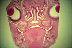
What are the vomeronasal organs?
|

Paired, tubular structure parallel to the base of the rostral nasal septum.
Portions of this organ are lined by respiratory and olfactory epithelia, the thick portion being pointed to is the olfactory epithelium, up top is the respiratory epithelium |
|
|

Are vomeronasal organs lined with respiratory epithelium, olfactory epithelium, or both?
|
Both. The thick outer "C" is the olfactory portion, the smaller interior bump is the respiratory epithelium.
|
|
|
|
Are the olfactory epithelium of the vomeronasal organs lined with microvilli or cilia?
|
Microvilli
|
|
|
|
Are the olfactory epithelium of the vomeronasal organs lined with microvilli or cilia?
|
Microvilli
|
|
|
|
What sort of glands does the lamina propria of the vomeronasal organ contain?
|
Serous, mucous, and seromucoid.
|
|
|
|
What two cavities do vomeronasal organs connect?
|
The oral and nasal cavities.
|
|
|
|
What sort of epithelial cells line the paranasal sinuses?
|
Cuboidal, squamous, or thin pseudostratified ciliated columnar epithelia.
|
|
|
|
Where is the nasopharynx and what does it connect?
|
The nasopharynx is the portion of the respiratory tract that is dorsal to the soft palate. It connects the nasal cavity to the oropharynx.
|
|
|
|
What is the predominant epithelial lining of the nasopharynx?
|
Ciliated psuedostratified columnar epithelium with goblet cells.
|
|
|
|
What epithelium is the soft palate on the oral side of the nasopharynx composed of?
|
stratified squamous epithelium.
|
|
|
|
What does the larynx connect
|
The larynx connects the pharynx and the trachea.
|
|
|
|
What type of mucosal epithelium lines the larynx?
|
Stratified squamous or ciliated pseudostratified columnar epithelium
|
|
|
|
What cells wrap around the epiglottis from one side to another?
|
a. Taste buds
b. Stratified squamous epithelium |
|
|
|
What is the function of the larynx?
|
It is the position for phonation of most mammals.
Striated muscle in this area helps with swallowing. |
|
|
|
start with slide 20
|
start with slide 20
|
|
|
|
What is the trachea an extension of?
|
The larynx.
|
|
|

What type of cells are the mucosa of the trachea?
|

Ciliated pseudostratified columnar epithelium with some goblet cells.
|
|
|

Describe the lamina propria-submucosa of the trachea.
|

The lamina propria-submucosa of the trachea has prominent elastic fibers. There are branched, coiled, and tubuloalveolar glands (both mucous and serous) between and peripheral to the tracheal cartilage.
|
|
|

Describe the tunica muscularis of the trachea
|
The tunica muscularis of the trachea is smooth muscle. This muscle may be interrupted by glands.
|
|
|

Describe the tunica adventitia of the trachea.
|

The tunica adventitia of the trachea blends with the surrounding cervical fascia.
|
|
|
|
Is the tunica adventitia of the trachea in the thoracic or pleural cavity?
|
Trick question! It isn't in either cavity, it is in the mediastinum.
|
|
|
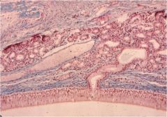
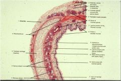
This is a slide of the trachea. You should be able to see the ciliated pseudostratified columnar epithelium. You should be able to see the goblet cells and the glands in the propria-submucosa and tracheal cartilage. You should be able to see elastic fibers.
|

This is a special stain of a section of the trachea showing the ciliated pseudostratified columnar epithelium with goblet cells, and the large numbers of glands in the lamina propria-submucosa adjacent to the tracheal cartilage. Blue is the elastic fibers.
|
|
|
|
What kind of cells line the carina?
|
Ciliated pseudostratified columnar epithelium (respiratory epithelium).
|
|
|
|
What does the lung secrete?
|
CO2.
|
|
|
|
What purpose do the elastic fibers in the lung serve?
|
The elasticity is beneficial to the exchange of CO2 to O2.
|
|
|
|
Describe the change in cartilage shape from the primary to tertiary bronchi.
|
The cartilage rings change from the horseshoe shape to a plaque-like shape and then the cartilage is lost all together. Bronchioles have no cartilage.
|
|
|
|
Do goblet cells become more or less abundant as you go down the respiratory tubes?
|
As a generalization, they decrease in the smaller bronchii. They decrease to reduce fluid accumulation.
|
|
|
|
The mucosa of the bronchi is the same as in the trachea, and the lamina propria also contains elastic fibers, but the lamina muscularis has a different arrangement, what is it?
|
The muscle is arranged in a helical pattern.
|
|
|
|
What is bronchial fluid a secretion of? What is it made up of?
|
Bronchial fluid is a secretion of all laryngobronchial cells and is a mixture of secretory and transudative materials including mucins, serum proteins, lactoferrin, sIgA, IgM, and glycoproteins.
|
|
|
|
What is the function of sIgA?
|
Surface protection - it's secreted into the bronchial fluid by the laryngobronchial cells.
|
|
|
|
What does the mucosa of the Bronchioles consist of?
|
The mucosa of the bronchioles consists of simple columnar or cuboidal cells and is devoid of goblet cells.
|
|
|
|
At what level do cilia stop in the respiratory system?
|
The lining is ciliated in the primary bronchioles, but the cilia diminish into the respiratory bronchioles.
|
|
|
|
Do cilia or glands extend further into the bronchioles?
|
Cilia extend farther into the respiratory tree than do glands.
|
|
|
|
Where cilia and glands end in the bronchioles, what begins?
|
Clara cells - they pick up the protection.
|
|
|
|
What sort of fibers do the bronchioles contain?
|
Collagenous and elastic fibers.
|
|
|
|
Is the lamina muscularis of the bronchioles continuous or discontinuous?
|
The lamina muscularis of the bronchioles is continuous.
|
|
|
|
Is cartilage present in the bronchioles?
|
No. No cartilage is present in the bronchioles.
|
|
|
|
What are the primary conducting pathways into the secondary lobule?
|
The terminal bronchioles.
|
|
|
|
What are respiratory bronchioles?
|
After the terminal bronchioles, there should be alveoli, but in some species there are respiratory bronchioles that look like terminal bronchioles that have out-pockets of gas exchange alveoli.
|
|
|
|
What are Clara cells?
|
Clara cells are granular secretory vesicles that produce protective secretions containing glycoproteins, which degrade toxins in inhaled air via the cytochrome p450 pathway. They produce surfactant-like material in the bronchioles, and divide to regenerate the bronchiolar epithelium.
|
|
|
|
What is the function of respiratory bronchioles?
|
If respiratory bronchioles are present, they both conduct and exchange gases.
|
|
|
|
What do cells of the tunica mucosa of the respiratory bronchioles consist of?
|
The mucosa of respiratory bronchioles consists of cuboidal epithelium that may or may not be ciliated. The cuboidal cells are interrupted by alveolar outpouching from the bronchiolar walls.
|
|
|
|
Where do respiratory bronchioles terminate?
|
Alveolar ducts.
|
|
|

What are R and A in this picture?
|
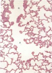
R is an alveolus outcropping of the respiratory bronchioles.
A is the terminus of the respiratory bronchiole, the alveolar duct. |
|
|
|
Is APUD:
a) the guy that runs the convenience store on the Simpsons. b) an acronym for automatically puckering uber derriere c) an acronym for amine precursor uptake and decarboxylation. These are small granular cells occuring singly or in small clusters. Some are innervated. These cells are intimately associated with the conducting airway smooth muscles and the pulmonary capillaries of the bronchioalveolar portals. D) All of the above |
C)
|
|
|
|
What are the exchange components of the lung?
|
Alveolar ducts, saccules, and alveoli.
|
|
|
|
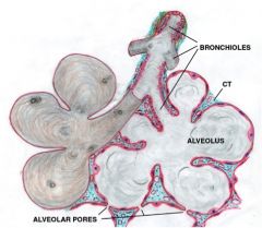
What are alveolar ducts are lined by?
|
Alveoli. They may have smooth muscle at the apical borders between alveoli or saccules.
|
WTF? I have a question I don't get this question....
|
|
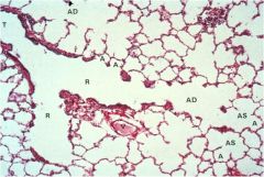
What are AD, AS, and A?
|

AD - Alveolar ducts
AS - Alveolar saccules A - Alveoli |
|
|

What is the middle of saccule called?
|
The atrium.
|
|
|
|
What are the primary lining cells of the alveoli?
|
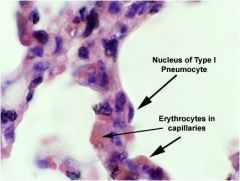
Type I Pneumocytes.
|
|
|

What type of epithelium is a Type I Pneumocyte?
|

Type I Pneumocytes are simple squamous epithelial cells.
|
|
|

What is the function of Type I Pneumocytes?
|

Type I Pneumocytes have well developed basement membranes and form tight junctions between cells, effectively preventing extracellular fluid from seeping into the alveolar lumen. Their primary function is to maintain an interface between the air and blood to allow passage of gases.
|
|
|
|
What is the function of Type II Pneumocytes?
|
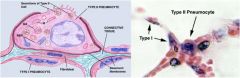
Type II Pnuemocytes are the secretory cell of the alveolar lining. They secrete a surfactant reducing surface tension on the wall of the alveolus.
As these cells secrete surfactant continuously, they are also responsible for breaking the surfactant down along with pulmonary alveolar macrophages (PAMs). |
|
|
|
Is a PAM?
a) a character on The Office. b) a map of backwards land. c) a breaker downer of surfactant along with Type II Pneumocytes. |

c) a breaker downer of surfactant along with Type II Pneumocytes.
|
|
|
|
What would "picket fences" of Type II Pneumocytes mean?
|

Major repair of the respiratory cells.
|
|
|
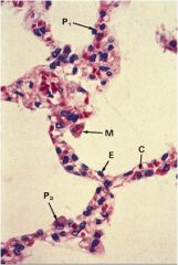
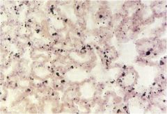
What are P1, P2, and M in this picture?
|

P1 - type I pneumocytes
P2 - type II pneumocytes M - PAMs |
|
|
What is the effect on cilia of the PAMs?
|

Cilia speed the trek of the PAMs upward to be swallowed or expectorated.
Mmm, tasty. |
|
|

What are heart failure cells?
|
The macrophages may be filled with phagocytized, extravasated red cells, which then break down producing hemosiderin.
|
|
|
|
What makes up the Blood-Air Barrier?
|

Type I pneumocytes.
|
|
|
|
What secretes the septal space?
|
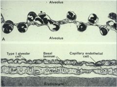
Both the pneumocytes and the endothelial cells.
|
|
|
|
What is ventilation in comparison to perfusion?
|
Ventilation is the oxygenation of blood within the pulmonary capillaries. Perfusion is the movement of blood through the capillaries.
They must be matched for normal lung function. The exchange of gases within the lung is not only important to proper oxygenation of tissues, but also acts to modulate acid-base balance (usually determined by blood pH). |
|
|
|
What vessels provide the nutritional blood supply to the lung?
|
Bronchial arteries
|
|
|
|
Are there lymphatics in the interalveolar septa?
|
No
|
|
|
|
what are the effects of the parasympathetic or sympathetic systems on the lungs?
|
Parasympathetic stimulation causes bronchoconstriction.
Sympathetic stimulation causes bronchodilation. |
|
|

Where is the syrinx in this pictures?
What is the syrinx? |
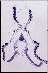
It's the purple triangle.
It's similar to the carina, in that it's at the crotch of the trachea. It's function is mainly phonation. |
|
|
|
In the avian lung, where does the secondary bronchi branch off of?
|
The secondary bronchi comes off the mesobronchus, or primary bronchus.
|
|
|
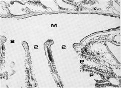
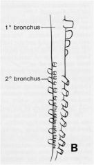
Identify M, P, and 2 in this picture.
|

M - mesobronchus - the primary bronchi
P - parabronchi - the tertiary bronchi 2 - the secondary bronchi |
|
|

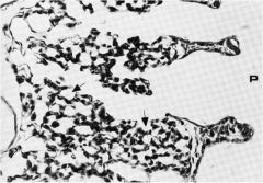
Where is the vestibulum in this picture of an avian lung?
|

The vestibulum is the widened portion of the mesobronchus, similar to it, but that it lacks cartilage.
I'm actually not sure if it's in this picture, but I can't figure out how to remove it! |
|
|
|
Does the vestibulum of the avian lung have cartilage?
|
No.
|
|
|
|
What epithelium lines the secondary bronchi in the avian lung?
|
A columnar to cuboidal epithelium devoid of goblet cells.
|
|
|
|
What avian lung structure is analgous to the respiratory bronchioles in mammals?
|
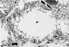
Parabronchi and air vesicles.
P and V in the picture. |
|
|
|
What structures are off of the vesicles of the avian lung?
|
Air capillaries. Next to every air capillary will be a blood capillary (the dark spots).
|
|
|
|
Describe the counter current gas exchange of the avian lung.
|
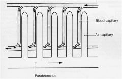
The parabronchi are perpendicular to the blood capillary axis within the lung. This permits air exchange independent of flow direction. The mixed venous blood enters a blood capillary and flows adjacent to and in the opposite direction of the flow within the air capillary. Capillary flow exits at the point where the parabronchial air enters enhancing gas exchange and equilibration between the blood and air capillaries.
|
|
|
|
What are the two units of the avian lung?
|

The paleopulmon and the neopulmon.
|
|
|
|
How does the bidirectional/unidirectional system of respiration work in avian lungs?
|
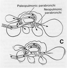
Air moves both bidirectional and unidirectional during inspiration and expiration – unidirectional through the paleopulmonic parabronchi and bidirectional through the neopulmonic parabronchi. This allows birds to extract more O2 from a specific volume of inspired air than mammals can.
|
|