![]()
![]()
![]()
Use LEFT and RIGHT arrow keys to navigate between flashcards;
Use UP and DOWN arrow keys to flip the card;
H to show hint;
A reads text to speech;
49 Cards in this Set
- Front
- Back
|
are lysosomes membrane enclosed? |
yes |
|
|
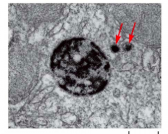
TEM of lysosome stained to reveal the location of acid phosphatase - a lysosomal enzyme -red arrows indicate vesicles from the Golgi delivering enzymes to the lysosome |
|
|
what are acid hydrolases? |
hydrolytic enzymes that are delivered from the Golgi to LYSOSOMES |
|
|
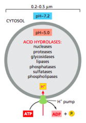
Lysosomes. The acid hydrolases are hydrolytic enzymes that are active under acidic conditions. An H+ ATPase in the membrane pumps H+ into the lysosome, maintaining its lumen at an acidic pH |
|
|
How does the low pH of lysosomes protect the rest of the cell from lysosomal enzymes in case the lysosome breaks? |
lysosomal enzymes are acid hydrolases with an optimal pH of 5.0
cytosol is pH 7.2, so the lysosomal enzymes would inactive in the cytosol if they accidentally spilled out |
|
|
plant and fungal cells have large-fluid filled lysosomes called ___________
What are they used for?? |
plant and fungal cells have large-fluid filled lysosomes called vacuoles
-used for storage of nutrients and wastes -regulation of osmotic pressure and size -degradation |
|
|
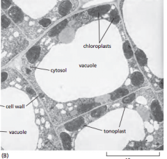
TEM of plant vacuole
This electron micrograph of cells in a young tobacco leaf shows the cytosol as a thin layer, containing chloroplasts, pressed against the cell wall by the enormous vacuole. |
|
|
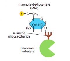
how are hydrolases delivered to lysosomes from the Golgi? |
Have mannose-6-phosphate(M6P) -unique marker added to N-linked oligosaccharides on lysosomal hydrolases -M6P is added in the golgi |
|
|
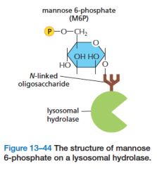
The structure of mannose 6-phosphate on a lysosomal hydrolase. |
|
|
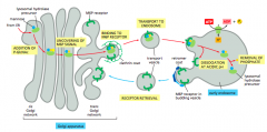
The transport of newly synthesized lysosomal hydrolases to endosomes The sequential action of two enzymes in the cis and trans Golgi network adds mannose 6-phosphate (M6P) groups to the precursors of lysosomal enzymes (see Figure 13–46). The M6P-tagged hydrolases then segregate from all other types of proteins in the TGN because adaptor proteins (not shown) in the clathrin coat bind the M6P receptors, which, in turn, bind the M6P-modified lysosomal hydrolases. The clathrin-coated vesicles bud off from the TGN, shed their coat, and fuse with early endosomes. At the lower pH of the endosome, the hydrolases dissociate from the M6P receptors, and the empty receptors are retrieved in retromercoated vesicles to the TGN for further rounds of transport. In the endosomes, the phosphate is removed from the M6P attached to the hydrolases, which may further ensure that the hydrolases do not return to the TGN with the receptor.
|
|
Transport of hydrolases from Golgi to lysosome (hint: look at this pic) |
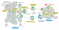
1) N-linked oligosaccharides added to hydrolases in ER, then transported to the Golgi (not in diagram) 2) enzymes within Golgi recognize a signal patch on the hydrolase and phosphorylate mannose on the N-linked oligosaccharide in a two-step process: 2a) phosphorylated N-acetyl glucosamine (P-GlcNAc) is added 2b) GlcNAc is cleaved to uncover the M6P signal 3) M6P receptor in the trans-Golgi binds M6P on the hydrolase and clathrin-coat adaptor proteins 4) clathrin-coated vesicles bud off the Golgi. When the coats are lost, the vesicles fuse with endosomes 5) because the endosomal lumen is acidic, M6P signal is released by the receptor 6) M6P dephosphorylated; keeps hydrolase in lysosome 7) receptors recycle back to the trans-Golgi |
|
|
The transport of newly synthesized lysosomal hydrolases to endosomes |
1. addition of P-GlcNAc 2. Uncovering of M6P signal 3. Binding to M6P receptor 4. transport to endosome 5. Dissociation at Acidic pH 6. Removal of phosphate 7. Receptor retrieval
|
|
|
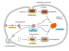
four pathways for degradation in lysosomes?? (recall: enzymes are delivered from the Golgi to endosomes) |
1. Endocytosis 2.Phagocytosis 3.Autophagy 4.Pinocytosis |
|
|
Four pathways to degradation in lysosomes. Materials in each pathway are derived from a different source. Note that the autophagosome has a double membrane. **In all cases, the final step is the fusion with lysosomes.*** |
|
|
Endocytosis |
budding from the plasma membrane carry substances into the cell for degradation • fusion of endosome + vesicles carrying hydrolytic enzymes from Golgi = lysosome |
|
|
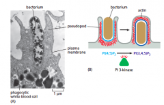
phagocytosis |
= “cell eating”
entire cells and large molecules are engulfed by the plasma membrane to form a phagosome
phagosome + lysosome = new lysosome
• in protozoa: for feeding • in animal cells: used by specialized phagocytes (e.g. neutrophils and macrophages) scavenge dead cells and defend against invaders
CARGO-triggered-particles bind to receptors this activates signals inside the cell that lead to actin polmerization and extension of pseudopods |
|
|
autophagy |
= “self-eating” - cytosol and old organelles are engulfed by a double membrane to form an autophagosome • fusion of autophagosome + preexisting lysosome = new lysosome |
|
|
pinocytosis |
="cell drinking" -fluids and portions of the plasma membrane are ingested as vesicles -fusion of vesicles with endosomes +vesicles carrying hydrolytic enzymes from Golgi = lysosome
occurs all the time = constitutive
most are clathrin-coated
|
|
|
2 types of endocytosis?? |
1. phagocytosis = "cell eating" 2. pinocytosis = "cell drinking"
|
|
|
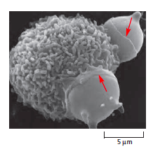
SEM of a mouse macrophage phagocytosing two red blood cells (red arrows) |
|
|
TEM of a neutrophil phagocytosing a bacterium |
|
|
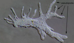
pseudopods:are temporary projections of eukaryotic cell membranes or unicellular protists |
|
|
|
Receptor mediated endocytosis |
special type of pinocytosis in which specific molecules are taken up from extracellular fluid
molecules (ligands) bind to transmembrane receptors on the cell surface and are then taken up by clathrin-coated vesicles
allows small amounts of ligands outside the cell to become concentrated inside the cell |
|

|
receptor-mediated endocytosis |
|
|
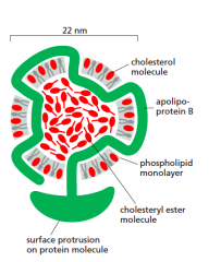
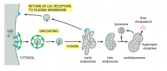
Cholesterol transport |
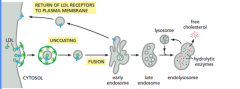
transported in the blood as a low density lipoprotein (LDL)
when cholesterol is needed for membrane synthesis, cells insert LDL receptors into their plasma membranes
1) LDL binds to the receptors, becomes concentrated in a clathrin-coated pit and then taken into the cell by endocytosis
2) the vesicles fuse with an endosome; low pH in the endosome causes LDL to be released from receptors
3) endosome fuses with vesicles containing hydrolytic enzymes, resulting in the release of cholesterol from LDL particles
some LDL receptors get degraded in the lysosome, while others are recycled back to the plasma membrane |
|
|
low density lipoprotein (LDL) particle |
|
|
The receptor-mediated endocytosis of LDL |
|
|
what is the typical lifespan of an LDL receptor? |
20 hours!
it takes about 10 min for the receptor to make a roundtrip from the plasma membrane to the endosome and back
a typical receptor makes several hundred trips |
|
|
what happens if an LDL receptor is defective? |
high lvls of LDL
atheriosclerosis, cholesterol deposits on artery walls
strokes, heart attacks
individuals with defective genes encoding for LDL receptors develop atherosclerosis prematurely |
|
|
recycling endosomes |
serve as an intracellular storage site for receptors and transporters that can be rapidly conveyed to the membrane when needed |
|
|
glucose transporters in fat and muscle cells |
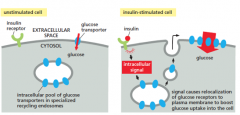
(recycling endosomes)
in unstimulated cells, a pool of glucose transporters, which mediate glucose uptake, are stored in recycling endosomes
when insulin binds to its receptor on the cell surface, it stimulates vesicles to rapidly bud off the endosome and deliver transporters to the plasma membrane
INCREASES GLUCOSE UPTAKE |
|
|
constitutive secretory pathway
(EXOCYTOSIS TYPE 1) |
operates continuously in all cells
supplies plasma memb. w/ newly synthesized proteins and lipids
releases secreted prtns and small molec. to the extracellular space |
|
|
regulated secretory pathway
(EXOCYTOSIS TYPE 2) |
occurs in specialized secretory cells
cargo (ex. hormones, neurotransmitters) stored in secretory vesicles from the Golgi
released rapidly on demand to signals |
|
|
Transport from the Trans-Golgi |
1. pathway to lysosomes - NEEDS M6P signal
2. Regulated secretion pathway- need signal to concentrate and package cargo into secretory vesicles... signal is unknown
3. constitutive secretory pathway- occus by default, no signal needed, protein that leave the golgi w/ no signal displayed are AUTOMATICALLY exocytosed at plasma memb.
|
|
|
TEM of insulin released from pancreatic beta cells |
|
|
Regulated secretion |
signal:
1. chemical: a hormone 2. electrical: an action potential
|
|
|
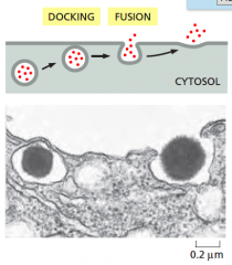
Regulated secretion at Neuromuscular Junction (NMJ) |
Rapid bc: 1. Complexin blocks SNARES from completely paring, and prevents fusion 2. action potential, Ca2+ enters cell, binds to synaptotamin, removes complexin 3. SNAREs lock into place, vesicle and plasma memb. fuse |
|
|
What does complexin do? |
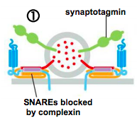
blocks SNARES from completely paring, and prevents fusion
(CYTOSOL) |
|
|
What is the role of Ca2+ in a neuromuscular junction? |
It binds to to synaptotagmin, which removes complexin blocker |
|
|
how do lysosomes maintain their acidic pH? |
they have proton pumps (V-type ATPases) that pump protons into the lumen to acidify it |
|
|
what are lysosomes' hydrolytic enzymes called? |
(acid) hydrolases |
|
|
vacuoles (and their function) |
-lysosomes of plant and fungal cells -functions: store nutrients and wastes, regulate osmotic pressure and size, degradation |
|
|
Compare and contrast the different kinds of endocytosis
(note that the textbook considers endocytosis different than phagocytosis or pinocytosis or autophagy, but these three processes are also viewed as kinds of endocytosis) |
Compared: -All kinds of endocytosis have a final step of fusion resulting in a lysosome
Contrasted: -pinocytosis is the only kind of endocytosis where fusion involves "endosomes + vesicles carrying hydrolytic enzymes from Golgi = lysosome," rather than "endosome/phagosome/autophagosome + preexisting lysosome = new lysosome" -phagocytosis involves large molecules/microorganisms whereas pinocytosis involves liquids/small molecules -phagocytosis requires the cargo to bind to receptors on the phagocyte surface, whereas (most kinds of) pinocytosis does not require any trigger; receptor-mediated endocytosis is a special kind of pinocytosis that does require a cargo-trigger |
|
|
Give an example of each kind of exocytosis |
phagocytosis: white blood cell engulfs a bacterium
autophagy: dead mitochondria along with cytosol are engulfed by a double-membrane, which fuses with a preexisting lysosome
pinocytosis (not receptor mediated): loose amino acids in the extracellular space are brought into the cell
receptor-mediated endocytosis (a kind of pinocytosis): cholesterol is needed for membrane synthesis and is taken from LDL from eaten food |
|
|
What's an example of the use of recycling endosomes? |
glucose transporters in fat and muscle cells
(see the appropriate slide) |
|
|
What's the fastest kind of exocytosis? |
phagocytosis |
|
|
compare and contrast the two different exocytotic pathways
(remember that the definition of exocytosis is secretion to outside the cell) |
compared: -both begin at the trans Golgi and end in the extracellular space (or close to it) -both involve secretion of proteins to outside the cell
contrasted:
constitutive secretory pathway: -does not require a signal -occurs in all cells continuously -directs membrane proteins and membrane lipids, as well as proteins that are to be secreted outside the cell
regulated secretory pathway: -requires a signal -occurs in specialized cells -secretes non-membrane proteins |
|
|
describe the three pathways of protein sorting in the trans Golgi network |
transport to lysosomes via endosomes -already discussed -proteins with the M6P trigger signal travel to lysosomes via endosomes in clathrin-coated transport vesicles
constitutive secretory pathway -already discussed -in unpolarized cells, the protein (which we know is a membrane protein) has no signal and is directed to a random position on the membrane, since it has no specialized function -in polarized cells (e.g. intestinal cells), secreted and plasma membrane proteins possess signals and are thus selectively directed to a position of the membrane (i.e. apical or basolateral) |
|
|
why is secretion at the neuromuscular junction so rapid? |
1) vesicles are already docked at the presynaptic membrane via SNAREs that are only partially paired (complexin blocks SNAREs from completely pairing and prevents fusion)
2) upon arrival of the action potential, calcium ions enter the cell and bind to synaptotagmin (a calcium ion sensor), which removes the complexin block
3) SNAREs lock into place and the vesicle and plasma membrane rapidly fuse to exocytose the neurotransmitter |