Reading...
![]()
Play button
![]()
Play button
![]()
Use LEFT and RIGHT arrow keys to navigate between flashcards;
Use UP and DOWN arrow keys to flip the card;
H to show hint;
A reads text to speech;
48 Cards in this Set
- Front
- Back
- 3rd side (hint)
|
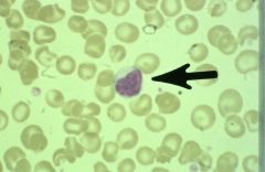
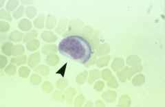
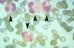
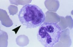
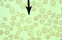
platelet
|
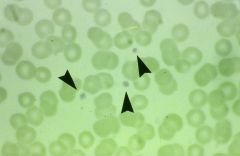
what are the arrows pointing to?
|
|
|
|
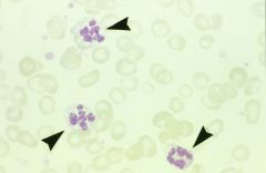
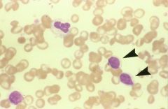
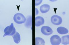
giant platelets
Post spleenectomy is most common reason to find them Also mb dt: May-Hegglin, leukemic myelofibrosis. |
what are the arrows pointing to?
|
|
|
|
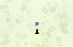
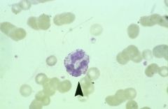
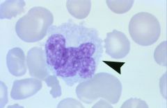
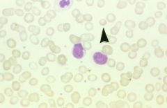
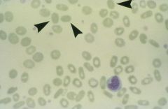
segmented neutrophil
|
what is the arrow pointing to?
|
|
|
|
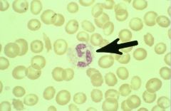
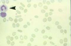
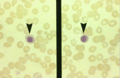
usually only one seen in an RBC. DNA nuclear fragments.
Found in megaloblastic anemia, sickle-cell anemia, and post splenectomy. |
Erythrocyte Howell-Jolly bodies:
|
|
|
|
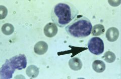
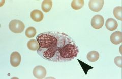
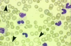
segmented neutrophils
|
what is the arrow pointing to?
|
|
|
|
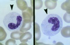
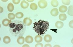
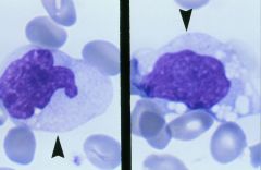
Band
The indentation is greater than 1/2 of the width of the hypothetical round nucleus. Opposite edges of nucleus become parallel giving horseshoe appearance. |
what is the arrowing pointing to?
|
|
|
|
Band
|
what is this? (arrow)
|
|
|
|
Neutrophilic bands constitute what percentage of WBCs in a normal peripheral blood smear?
|
1-5%
|
|
|
|
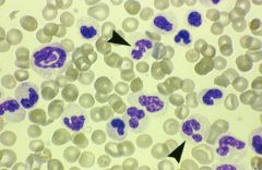
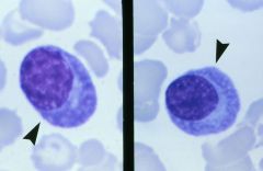
the seg is on the left, the band on the right
|
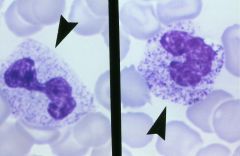
which is the segmented neutrophil and which is the neutrophilic band?
|
|
|
|
Dohle bodies, RER residual aggregates, but may increase in infectious dz, burns, cytotoxic chemicals, poisons.
|
what are the pale blue inclusions in these cells' cytoplasm?
|
|
|
|
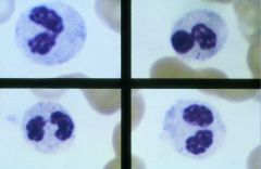
Hypersegmented neutrophils (5 or more lobes)
Due to decreased B12 and Folate. |
what type of WBCs are these? what causes them to look like this?
|
|
|
|
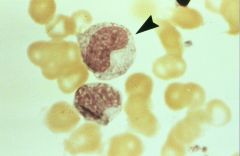
Peutz-Huet anomaly.
Hereditary anomaly characterized by hypolobulation of the nucleus of granulocytes. Pince-nez (bottom left). |
I.D. these cells
|
|
|
|
neutrophilic metamyelocyte.
slightly indented nucleus, small pinkish-blue granules. Rarely seen in normal peripheral blood. |
what is this WBC?
|
|
|
|
lymphocytes
|
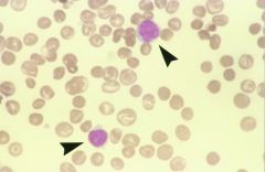
what are these (arrows)?
|
Clumped nuclear chromatin, dark stained.
|
|
|
what is the diameter of a lymphocyte?
|
small: 7-10 micron range.
|
|
|
|
lymphocyte
|
what is this?
|
Clumped nuclear chromatin, dark stained.
|
|
|
lymphocytes
|
what are the arrows pointing to?
|
Clumped nuclear chromatin, dark stained.
|
|
|
lymphocyte (small, mature)
|
what is this?
|
Clumped nuclear chromatin, dark stained.
|
|
|
lymphocytes
|
what are the arrows pointing to?
|
Clumped nuclear chromatin, dark stained.
|
|
|
infectious mononucleosis
|
what is this showing?
|
Plasmalike cells with indented nuclei and “early” loose nuclear structure.
|
|
|
basophil
|
what is this?
|
|
|
|
these WBCs have grains of histamine and heparin, are NOT phagocytic and normally are less than 1 per 100 in peripheral cells
|
basophil
|
|
|
|
neutrophil
|
what is this?
|
|
|
|
these prominent purplish and blue-black granules are associated with what?
|
Toxic granulation: prominent purplish and blue-black granules, associated with severe infxn and other toxic states.
|
|
|
|
eosinophil
|
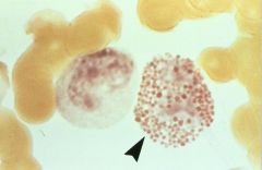
what is this?
|
|
|
|
how do you identify an eosinophil?
|
Granulocytes characterized by acid stain eosin readily as pink/red granules.
|
|
|
|
what make up eosinophilic granules? what are they toxic to?
|
major basic protein & eosinophilic cationic protein. they are toxic to several parasites.
|
|
|
|
eosinophilia
|
what is going on here?
|
|
|
|
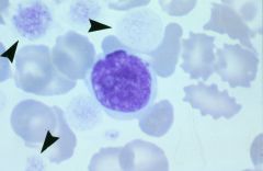
monocyte
|
what is this?
|
deeply indented nucleus, grey-blue cytoplasm “ground-glass” of lightly stained numerous fine granules.
|
|
|
classic horse-shoe shaped monocyte
|
what is this?
|
ground glass appearance
|
|
|
atypical monocyte
|
what is this?
|
|
|
|
monocyte
|
what is this?
|
|
|
|
bone marrow: monoblast
|
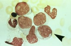
what's this? (arrow)
|
Monocytic leukemia: fine chromatin, visible nucleoli, and no cytoplasmic granules.
|
|
|
Plasma cells. Maybe seen in young children, viral infx, herpes, EBV. Not seen in peripheral blood of healthy adult.
|
what are these cells and when are they seen?
|
Non-granular cyto stains a dark blue, no vacuoles, pale cyto adjacent to nucleus “peri-nuclear clear zone”
|
|
|
macrocytes
|
what are the arrows pointing to?
|
|
|
|
when will you see macrocytes?
|
B12 and folate deficiencies
|
|
|
|
macrocytes
|
what are the arrows pointing at?
|
|
|
|
presence in the blood of erythrocytes with excessive variation in size
|
anisocytosis
|
|
|
|
spherocytes
|
identify these cells.
|
red blood cells with no area of central pallor like a normal red blood cell.
|
|
|
hypochromic microcytic
|
identify these rbcs
|
|
|
|
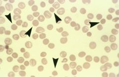
teardrop (dacrocyte)
|
identify the rbc (arrow)
|
|
|
|
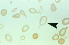
target cell (codocyte)
|
identify this cell
|
|
|
|
when will you see a target cell?
|
Common in thalassemia, sickle-cell anemia, Hb-S thalassemia, other hemoglobinopathies
|
|
|
|
target cells
|
identify these cells
|
|
|
|
ovalocytes/elliptocytes. A few is normal, but small #’s are seen in: iron deficiency, thalassemia, other hemoglobinopathies
|
identify these cells and when are they seen?
|
|
|
|
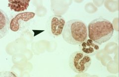
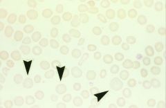
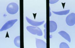
thin elongated eythrocytes with a point at each end. Schistocytes of all types may be found. Found in Hb-S Thal and Sickle Cell.
|
Drepanocytes
|
|
|
|
nucleated RBCs. Pernicious anemia & related B12-folic acid deficiency diseases. Usually see oval macrocytes and microcytes, and teardrops.
|
identify these cells and when they are seen.
|
|
|
|
sickle cells/drepanocytes
|
identify these cells
|
|