![]()
![]()
![]()
Use LEFT and RIGHT arrow keys to navigate between flashcards;
Use UP and DOWN arrow keys to flip the card;
H to show hint;
A reads text to speech;
30 Cards in this Set
- Front
- Back
|
General Knee Anatomy |
Comprised of 3 bones: - femur - tibia - patella
True knee joint is the femur & tibia - Tibiofemoral joint |
|
|
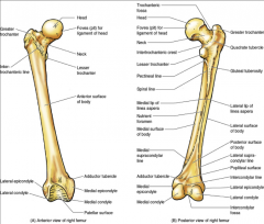
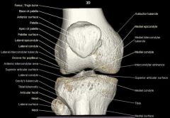
Femur |
- largest bone in body
- knee is comprised of the distal femur
- hip is comprised of the proximal femur |
|
|
Femoral Shaft (Linea Aspera) |
seperates the lateral & medial surfaces posteriorly |
|
|
Femoral Shaft (Supracondylar Lines) |

begin where the femoral shaft widens medially & laterally |
|
|
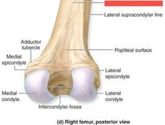
Bony Landmarks of Distal Femur (Medial & Lateral Condyle) |

|
|
|
Bony Landmarks of Distal Femur (Intercondylar Notch/Fossa) |

|
|
|
Bony Landmarks of Distal Femur (Patellar Groove) |

|
|
|
Bony Landmarks of Distal Femur (Medial & Lateral Epicondyle) |

|
|
|
Bony Landmarks of Distal Femur (Adductor Tubercle) |
above medial epicondyle |
|
|
Bony Landmarks of Distal Femur (Popliteal Surface) |
|
|
|
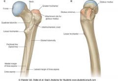
Bony Landmarks of Proximal Femur (Lesser & Greater Trochanter) |

|
|
|
Bony Landmarks of Proximal Femur (Intertrochanteric Line - anterior) |

|
|
|
Bony Landmarks of Proximal Femur (Intertrochanteric Crest) |

|
|
|
Medial & Lateral Tibial Plateaus |

flat area of the tibia |
|
|
Intercondylar Eminence |
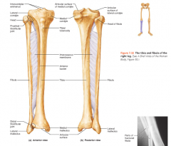
roughened area between medial & lateral plateaus |
|
|
Medial & Lateral Tibial Condyles |
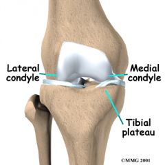
|
|
|
Gerdy's Tubercle |

IT band inserts here
located laterally |
|
|
Fibular Articular Facet |
located laterally & posteriorly of tibial condyle
where fibula inserts
|
|
|
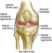
Patella |
- sesamoid bone & short bone
- improves efficiency of knee extension during the last 30 degrees
- convex anterior aspect
- triangular posterior border - surface is lined with hyaline cartilage (smooth surface) - total of 5 facets (articular surfaces)
- can function without patella
- base of patella is proximal
- apex of patella is distal
|
|
|
Tibiofemoral Joint |
- largest joint in the body
- modified hinge joint - flexion - extension
- synovial joint |
|
|
Patello-Femoral Joint |
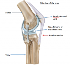
- articulation between patella & trochlear groove of the femur
- during extension, patella glides cranially
- during flexion, patella glides caudally |
|
|
Medial Retinaculum |
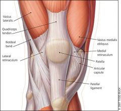
- fibrous band which attaches the medial border of the patella
- holds the patella medially |
|
|
Bursas (Suprapatellar) |

above the knee between the quadriceps & femur |
|
|
Bursas (Infrapatellar) |

between the skin & patellar tendon |
|
|
Bursas (Deep Infrapatellar) |

between the tendon & fat pads |
|
|
Bursas (Prepatellar) |

anterior between skin & patella |
|
|
Bursas (Pes Anserine) |
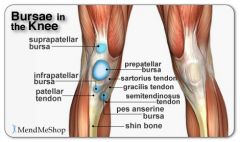
tibia & pes anserine insertion |
|
|
Iliotibial Band |
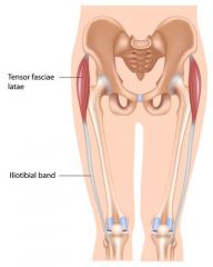
- thickened fascia that extends distally from tensor fascia latae to Gerdy's Tubercle
- gives the lateral aspect of the knee increased stability |
|
|
Plica |
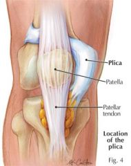
- remnants of embryonic tissue that appears in the knee as folds in the joint lining
- usually thin, elastic, pliable tissues
- can become thickened areas of scar tissue .... therefore inelastic with repetitive stress |
|
|
Infrapatellar Fat Pad |
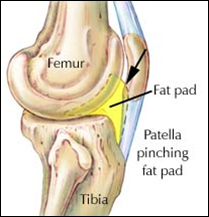
- from the lower pole of the patella to the tibia, posterior to the patellar tendon
- shock absorber & nutrition source for the tendon |