Reading...
![]()
Play button
![]()
Play button
![]()
Use LEFT and RIGHT arrow keys to navigate between flashcards;
Use UP and DOWN arrow keys to flip the card;
H to show hint;
A reads text to speech;
31 Cards in this Set
- Front
- Back
|
Coronal Plane
|
yields a front and back piece
|
|
|
Sagittal Plane
|
yields a left and right piece
|
|
|
Horizontal Plane
|
yields a top and bottom piece
|
|
|
The 4/5 anatomical lobes
|
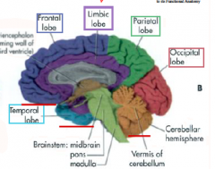
Frontal, Parietal, Occipital, Temporal, and (limbic)
|
|
|
Central Sulcus
|
separates the frontal and parietal lobe
|
|
|
Lateral Sulcus (Sylvian fissure)
|
separates the frontal/parietal lobe with the temporal lobe. It's particularly deep (deep sulci are called fissures)
|
|
|
parietooccipial sulcus
|
separates the parietal and occipital lobes (seen in the medial view)
|
|
|
Gyrus
|
a ridge in the brain
|
|
|
Sulcus
|
a groove between the ridges
|
|
|
Corpus Callosum
|
a bundle of axons joining the two cerebral hemispheres together
|
|
|
Major fossae of the cranial cavity
|
anterior cranial fossa (holds the frontal lobe),
middle cranial fossa (holds the temporal lobe)... deeper posterior cranial fossa (holds the cerebellum and brianstem)...largest and deepest |
|
|
Tentorium cerebelli
|
an infolding of dura. creates a tent over cerebellum.
separates the occipital lobe (above) from the cerebellum (below) |
|
|
Anterior Cranial Fossa (bones)
|
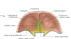
Frontal bone (red)
Ethmoid bone (green) Sphenoid bone (yellow) |
|
|
Middle cranial fossa (bones)
|
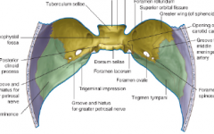
Sphenoid bone (yellow)
Temporal bone (green) |
|
|
Posterior cranial fossa (bones)
|
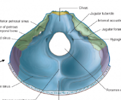
Sphenoid bone (yellow)
Temporal bone (green) Occipital bone (blue) |
|
|
Cerebral Cortex
|
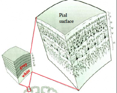
the layers of neurons that reside along the outer surface of the cerebrum. Most of the human cortex is a six layer cortex (neocortex) (each layer is a neuron), numbered from the outside (pial surface) in.
"gray matter" -->(6 layers) neuronal cell bodies (outside... some subcortical). Stains purple via Nissl stain. "white matter" -->where the (myelinated fatty) axons reside (inside) |
|
|
Corona radiata (specific white matter region)
|

radiating white matter immediately deep to the cortex that fans out like a "crown"
|
|
|
Internal capsule (Specific white matter region)
|
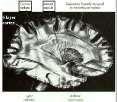
-deep to the corona radiata
-deep white matter tracts that course between nuclei of the basal ganglia and thalamus |
|
|
The five anatomical regions of the internal capsule
|
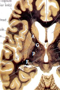
-Anterior limb
-Posterior limb -Genu ("at the bend") -Retrolenticular -Sublenticular |
|
|
Organization of the cerebral cortex
|
- the cerebral cortex is highly organized
- information is first processed in primary sensory cortices and then travels to association cortices (higher order cortical areas) where integration occurs. |
|
|
Diencephalon (includes)
|
epithalamus, thalamus, hypothalamus
and subthalamus. |
|
|
Thalamus
|
-bilateral and highly organized
-comprised of many nuclei -"gateway" to the cerebral cortex: majority of sensory and motor pathways relay through the thalamus before reaching the cerebral cortex. -many cortical regions also send projections back to the thalamus. |
|
|
Hypothalamus
|
-Important in maintaining the internal environment in a physiological range (promotes maintenance of homeostasis).
-Like the thalamus, the hypothalamus is comprises of many different nuclei, each with a specific function. |
|
|
Cerebellum
|
-The cerebellum receives extensive sensory input
-The cerebellum projects to subcortical structures and (indirectly) to cortical regions. -The cerebellum influences motor, cognitive and behavioral functions. |
|
|
Brainstem
|
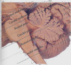
Three divisions: Midbrain (most rostral) ,Pons, Medulla (most caudal)
-a lot of nuclei -White matter tracts also travel through the brainstem (ie. from the spinal cord to the cerebral cortex). |
|
|
Spinal cord
|
-The rostral spinal cord is continuous with the brainstem at the caudal medulla
-Spinal nerves emerge from the spinal cord to form peripheral nerves that carry sensory/motor information to/from the CNS. |
|
|
Ipsilateral (homolateral)
|
same side (red/green)
|
|
|
Contralateral
|

opposite side (red/green)
|
|
|
Bilateral
|
both sides (two green)
|
|
|
The Ventricles
|
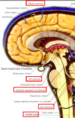
A series of continuous
spaces deep in the brain that contain cerebrospinal fluid (CSF) © Lateral ventricle (2) © 3rd ventricle (1) - the space between the right and left thalamus (holds spinal fluid) © Cerebral aqueduct (1) -between the midbrain © 4th ventricle (1) - posterior to the pons, anterior to the cerebellum © Central canal of the spinal cord (1) |
|
|
Layers of Meninges
|
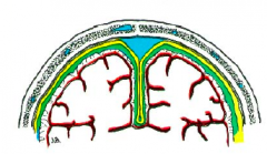
Dura Mater (yellow)- external meningeal layer. Thick.
Arachnoid (green)- cover the general surface of the brain. Intermediate meningeal layer. Thin membrane. Pia Mater (red)- internal meningeal layer. Adheres to surface of brain; the only layer to dip in sulci. |