![]()
![]()
![]()
Use LEFT and RIGHT arrow keys to navigate between flashcards;
Use UP and DOWN arrow keys to flip the card;
H to show hint;
A reads text to speech;
77 Cards in this Set
- Front
- Back
|
Bacteria Ribosome structure and importance |
16 S
Targeted by many antibiotics because different size than Eukaryotes |
|
|
Defining feature of clostridium |
form spores that are visible under microscope |
|
|
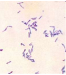
corynebacterium sp. |
|
|
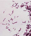
Clostridium tetani |
|

|
Staphylococcus sp. |
|
|
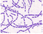
Bacillus anthracis |
|
|
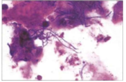
Dermatophilus Congolensis |
|

|
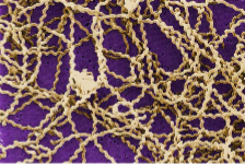
Streptococcus pyogenes |
|
|
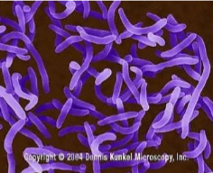
Campylobacter sp |
|
|
Vibrio sp. |
|
|
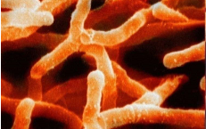
Actinomyces sp |
|

|
Streptococcus sp |
|
|
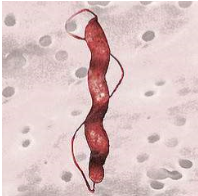
Leptospira sp |
|

|
Diplococci |
|

|
Steptococci |
|

|
Staphylococci |
|

|
Bacilli |
|

|
vibrio |
|

|
Actinomyces |
|
|
function of bacterial capsule |
attachment to surfaces protection from phagocytosis some killing and digestion nutrient reserve protection against desiccation |
|
|
composition of capsule |
polysaccharides and some polypeptides |
|
|
function of gram-positive cell wall |
prevent osmotic lysis provides rigidity and shape |
|
|
function of gram-negative cell wall |
prevents osmotic lysis provides rigidity and provides permeable outer membrane LPS has many functions |
|
|
composition of gram-positive cell wall |
peptidoglycan with teichoic acids |
|
|
composition of gram-negative cell wall |
peptidoglycan surrounded by phospholipid protein-lipopolysaccharide |
|
|
significance of peptidoglycans in cell wall |
only found in bacterial cells so can be targeted by antibiotics |
|
|
composition of plasma membrane |
phospholipid and protein |
|
|
lysozyme |
found in tears, saliva, and macrophages natural defense against bacteria lysozome sensitive bonds found cell wall |
|
|
Functions of LPS |
causes very strong immune response Acts as an endotoxin increases structural integrity protects against chemical attach increases negative charge of cell membrane stabilizes membrane structure |
|
|
Function of teichoic acid in gram positive cell wall |
provide rigidity by attracting cations (Mg and Na) |
|
|
staining with crystal violet |
both gram positive and gram negative appear light blue |
|
|
staining with iodine |
both gram positive and gram negative appear dark blue |
|
|
staining with decolorize |
gram positive appears dark blue gram negative appears unstained |
|
|
staining with basic fuchsin |
gram positive appear purple gram negative appear pink |
|
|
components of an acid fast cell wall |
lipid layer over peptidoglycan |
|
|
staining acid fast bacteria with fuschin and methylene blue |
acid fast bacteria appear pink not-acid fast appear blue |
|
|
surface components are used as: |
permeability barriers adhesins protection against phagocytosis enzymes for cell surface reactions sensing proteins that respond to environmental conditions |
|
|
significance of a lack of cell wall |
affects function of antibiotics cannot survive in the environment Often intracellular |
|
|
Detection of motility |
Flagellar stain Mobility agar Direct microscopic observation |
|
|
Peritrichous Flagella |
flagella come off all around the cell |
|
|
polar flagella |
flagella only come off either end of the cell (can be off only one or both) |
|
|
tactic behaviour |
ability to move in response to environment stimuli |
|
|
chemotaxis |
a bacterium can sense the quality and quantity of certain chemicals and swim towards or away from them |
|
|
flagella of leptospira |
single flagella causes bacteria to swim in a spiral pattern |
|
|
fimbriae (pili) |
rodlike surface appendages mediates attachment to host tissue confer antigenic specificity many different types |
|
|
capsule of S. pneumoniae |
composed of polysaccharide determinant of virulence protects against phagocytosis in the lung |
|
|
capsule of B anthracis |
composed of poly D-glutamic acid protects against phagocytosis and complement-mediated lysis in blood |
|
|
capsule of S. pyogenes |
composed of hyaluronic acid antigenic disguise the prevents recognition by phagocytes |
|
|
biofilm creation |
cells excrete a sticky extracellular matrix coating that make them hard to remove |
|
|
some examples of fast growing bacteria |
E. coli bacillus streptococcus staphylococcus lactobacillus |
|
|
some examples of slow growing bacteria |
rhizobium japonicum mycobacterium treponema pallidum |
|
|
basic steps of cell division |
cell elongates and DNA replicates Cell wall and plasma membrane begin to divide Cross wall forms completely around divided DNA cells separate |
|
|
species that elongate at low temperatures |
E. coli Salmonella enteritidis |
|
|
species that elongates and both high and low temperatures |
Listeria |
|
|
Species that elongate in an acid environment |
Listeria Bacillus Clostridium Lactobacillus |
|
|
Species that elongate in the human body conditions
|
Shigella Salmonella |
|
|
Steps of sporulation |
1) spore septum begins to isolate replicated DNA and portion of cytoplasm 2) Plasma membrane starts to surround section isolated in step one 3) spore septum surrounds isolated portion forming forespore 4) peptidoglycan layer forms between membranes 5) spore coat forms 6) endospore is freed from cell |
|
|
survival features of spores |
core wall of peptidoglycans surround cortex core is dehydrated |
|
|
difference of surface coats b/w vegetative and endpspores |
vegetative - usually gram-positive murein cell and wall polymer Endospore - thick spore coat, cortex and core wall |
|
|
differences of appearance b/w vegetative and endospore |
vegetative - nonrefractile endospres - refractile |
|
|
differences in cytoplasmic water activity b/w vegetative and endospore |
vegetative - high endospore - low |
|
|
differences in heat resistance b/w vegetative and endospore |
vegetative - low endpsore - high |
|
|
differences in chemical and acids b/w vegetative and endospore |
vegetative - low endospore - high |
|
|
chemically defined media |
exact amount of every ingredient is knows |
|
|
basic nutritive media |
for less fastidious bacerteria |
|
|
enriched media |
for growth of fastidious bactera |
|
|
selective media |
agar media with inhibitory substance Eliminates growth of species other than one of interest |
|
|
Indicator media |
detects biochemical reactions characteristic of certain bacteria |
|
|
significance of plasmids |
antibiotic resistance toxins and virulence bacteriocins transposons |
|
|
3 methods of genotypic variants |
mutation recombination transposition |
|
|
recombination |
exchange of DNA between bacteria |
|
|
transposition |
relocation of portions of DNA in the genome |
|
|
three mechanisms of recombination |
transformation transduction conjugation |
|
|
competence |
ability to bind DNA ability to translocate across cell envelope ability to integrate DNA in own chromosome different steps mediated by specific proteins |
|
|
requirements for gene transfer |
appropriate source of free DNA competent recipient cells |
|
|
steps in phage DNA transfer |
1) phage injects its DNA 2) phage enzymes degrade host DNA 3) cell synthesizes new phages that incorporate phage and host DNA 4) phage injects donor DNA 5) donor DNA is incorporated into recipient's chromosome through recombination |
|
|
conjugative transfer |
donor cell latches on with pilus and forms mating bridge plasmid copy is transferred donor must have a plasmid that codes for ability to be a donor |