Reading...
![]()
Play button
![]()
Play button
![]()
Use LEFT and RIGHT arrow keys to navigate between flashcards;
Use UP and DOWN arrow keys to flip the card;
H to show hint;
A reads text to speech;
24 Cards in this Set
- Front
- Back
- 3rd side (hint)
|
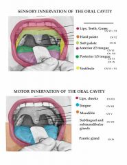
A. What is the sensory innervation of:
1. Lips, teeth, gums 2. Hard palate 3. Soft palate 4. Anterior 2/3 tongue 5. Posterior 1/s tongue 6. Vestibule B. What is the motor innervation of: 1. Lips, cheeks 2. Tongue 3. Mandible 4. Sublingual and submandibular glands 5. Parotid |
SEE DIAGRAM
|
Sensory
T, V = 32 Motor T = T S = S |
|
|
Describe:
A. the sensory limb (afferent limb) and B. central connections of the salivatory reflex? |
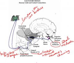
A. The receptors for the salivatory reflex are taste buds, groups of cells in surface of the tongue and oral cavity. Food taken into the mouth goes into saliva and the food molecules wash over the receptive taste buds and:
1. activate the afferent neurons of the lingual branch of V3. 2. The processes of these neurons travel centrally, joining the chorda tympani and then the facial nerve. 3. The afferent neurons then terminate in the rostral medulla in the nucleus of the solitary tract and their cell body is in the geniculate ganglion. B. In the central relay of the reflex, the cells of the nucleus of the solitary tract project rostrally into the pons, where they terminate in the superior salivatory nucleus. |
|
|
|
Describe the 2 neuron relays of the motor (efferent) limb of the salivatory reflex?
|

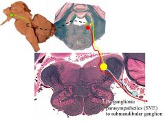
1. The first is the axons of the superior salivatory nucleus which exit the brainstem in the facial nerve and retrace the path through the chorda tympani and lingual branch of V3, terminating in the submandibular ganglion. These are preganglionic parasympathetics.
2. The second relay are the cell bodies of the submandibular ganglion send axons which end on the secretory cells of the sublingual and submandibular glands, causing secretion of saliva. This latter relay are postganglionic parasympathetics. |
LOOK AT NETTER PAGE 127
|
|
|
Describe the experimental procedure of classical conditioning conducted by Pavlov and why was it significant?
|
-Ivan Pavlov was investigating gastric secretions in the dog and he made an opening to the stomach so he could collect and measure the amount of these secretions when the dog was fed meat powder. From time to time he noticed that gastric secretion occurred even when the dog expected the meat, but the powder was not physically present.
-He made a similar experimental setup to collect salivary secretions and he first measured the amount of saliva secreted when meat powder was placed on the dog's tongue. Next, he sounded a tone whenever the meat powder was about to be presented. Finally, he sounded a tone without following it up with delivering the meat powder. In this case, saliva continued to be secreted. -This showed that repeated pairing of the auditory stimulus to the gustatory stimulus could lead to substitution of the former for the latter in evoking the salivatory response. |
|
|
|
Describe:
1. unconditioned stimulus/response 2. conditioned stimulus/response |
1. A tastant contacting the taste buds of the tongue which elicits a reflex secretion of saliva is called an unconditioned stimulus, and the secretion itself is called the unconditioned response.
2. Following an unconditioned response, the tone is known as the conditioned stimulus and the subsequent secretion of saliva is the conditioned response. Extended presentation of the conditioned stimulus without a periodic delivery of a reward (the unconditioned stimulus) leads to extinction of the conditioned stimulus/conditioned response sequence. |
|
|
|
Where does classical conditioning occur in the brain?
|
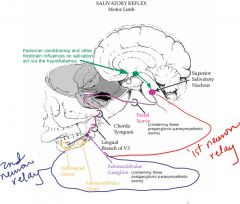
It occurs in the amygdala, a structure in the temporal lobe anterior to the hippocampus. The amygdala sends strong projections to the hypothalamus, which in turn projects to the preganglionic nuclei of the brainstem and spinal cord, including the salivatory nuclei of the pons.
ASIDE: The green projection above diagrams how the higher central centers are in a position to modify the function of the unconditioned reflex circuitry. |
|
|
What does this represent?
|

The pathway of the afferent limb and the efferent limb of the salivatory reflex. The salivatory reflex is an autonomic reflex and it involves wetting of the food.
|
|
|

What does this illustrate?
|
The green projection above diagrams how the higher central centers are in a position to modify the function of the unconditioned reflex circuitry. Therefore the Green = influence of hypothalamus on preganglionic sympathetic and thus influencing it.
|
|
|
|
Describe the somatomotor reflex of the oral cavity?
|
This is the process of mastication. “After food enters the mouth, it is usually broken down by the grinding action of the teeth and is mixed with saliva. The food is repeatedly passed between the opposing teeth as the result of the movements of the tongue and the ’trampoline-like’ action of the buccinator muscles of the cheeks.” The wetted mass may be formed into a shape (the bolus) for delivery to and passage along the gastrointestinal tube.
NOTES: In the oral cavity we chew food to make it small enough to swallow without choking It is to crush and wet the food before it enters the pharynx. Tongue muscles tell us when the food is. Buccinator muscles tell us where the food is and where we need to move it to keep on chewing it. In chewing there is touch of the tongue, cheeks and roof of the mouth. There is a lot of activity involving touch and movement to crush the food. |
|
|
|
What nerves are involved in the process of mastication?
|
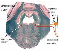
The tongue, lips, etc are innervated by CN V. The afferent neurons come into the chief sensory nucleus of V in the pons. Medial to it is the motor nucleus of V causing reflex in muscles of mastication. This is a stimulus response pathway for chewing.
|
|
|
|
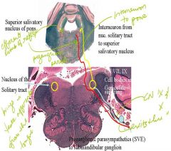
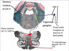
Compare the sensory nucleus of CN V to the touch receptors and state the role of motor nucleus of CN XII (hypoglossal).
|
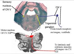
Analogous pathway for touch receptors
Blue afferent is the same as prev slide. Open medulla at the bottom Hypoglossal motor nucleus sends axons ventrally across med leminiscus and out to CN XII, which innervate the muscles of the tongue. |
|
|
|
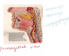
Describe the sensory and motor innervation of the pharynx?
|
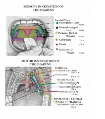
The pharynx has sensory and motor components to pull food back and down thru esophagus and to the stomach
They are innervated mainly by CN IX. Motor innervation are levator veli palatini; uvula; and pillars seen in colored outline. They bring food back and dump it into the stomach A lot of CN X is brought in and styloglossus in CN XII. Posterior and lateral walls of pharynx also have a muscle |
|
|
|
Describe the 2 facts about the pharynx?
|
1. First, it is a space used in common by two very different systems-the respiratory and alimentary, both of which must be kept functionally separate. Food must traverse the pharynx and by forwarded into the esophagus without entering the nasal cavity or larynx, else asphyxiation may occur. Likewise, air must pass from the nasal or oral cavities through the pharynx and into the larynx, also without interruption.
2. Second, the pharyngeal space is a right angle, so food must be re-directed from a horizontal movement to a vertical movement. Swallowing is, for the most part, carried out involuntarily, and in a rigid sequence of activated structures. |
|
|
|
What is the role of the styloglossus muscle?
|
After the food is mixed and turned into a bolus on the dorsum of the tongue, the styloglossus muscle now contracts in order to push the bolus upward and backward against the undersurface of the hard palate by pulling the root of the tongue upward and
backward. NOTES: Intrinsic tongue muscles shape the food, narrows it and elongates it. It is slippery from saliva Anterior tongue raises up to hard palate and in a sweeping motion, with contraction of posterior tongue muscles, moves the food back into the pharynx. Styloglossus muscle consecutively contracts to push the bolus into the pharynx As bolus touches mucosa, hard palate and soft palate, they evoke the stimulus response reflexes by way of the trigeminal and glossopharyngeal nerve There is a series of stimulus response reflexes as the bolus goes more posteriorly. |
|
|
|
Illustrate the sensory and motor component of the syloglossus muscle?
|
Anterior part of reflex is CN V
XII pushes bolus back into pharynx |
|
|
|
Describe the role of the palatoglossus muscles?
|
The contraction of the palatoglossus muscles now squeezes the
bolus backward into the oral part of the pharynx. The process of swallowing is an involuntary act from this point onward. Therefore, Palatoglossus jerks tongue up to dump food into pharynx. |
|
|
|
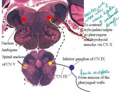
Describe the innervation of palatoglossus muscle after the bolus gets in the pharynx?
|
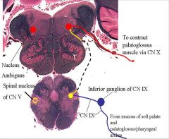
When in pharynx food sensation goes from CN IX to spinal nucleus of V (touch from pharynx). Interneuron travels rostrally from spinal nucleus of V to nucleus ambiguus– cannot see them as on this level they are scattered in the medulla. From nucleus ambiguus it goes to CN X
Motor structures in pharynx in order to contract palatoglossus muscle is mainly the CN X innervation. |
|
|
|
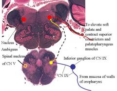
How is the nasal part of the pharynx separated from the oral part of the pharynx?
|
The nasal part of the pharynx is shut off from the oral part of the pharynx by the elevation of the soft palate, the pulling
forward of the posterior pharyngeal wall by the upper fibers of the superior constrictor muscle, and the contraction of the palatopharyngeus muscle. Likewise, When uvula is touched it contracts along with attached soft palate The other side also constrict and they meet to close off nasopharynx so food doesn’t go there Is a mechanism of the nervous system to seal off the nasal cavity so food only goes downwards. |
|
|
|
Illustrate the innervation of the superior constrictors and palatopharyngeus muscles?
|
SEE DIAGRAM
|
|
|
|
Describe the roles of these muscles:
Palatopharyngeus Salpingopharyngeus Thyrohyoid Styropharyngeus |
The larynx and laryngeal part of the pharynx are now pulled
upward by the contraction of the stylopharyngeus, salpingo- pharyngeus, thyrohyoid, and palatopharyngeus muscles. The main part of the larynx is thus elevated to the posterior surface of the epiglottis, and the entrance into the larynx is closed. The bolus moves downward over the epiglottis, the closed entrance into the larynx, and reaches the lower part of the superior, middle and inferior constrictor muscles.Finally, the lower fibers of the inferior constrictor muscle relax, and the bolus enters the esophagus. |
|
|
|
Discuss the pathway of innervation of these muscles?
Palatopharyngeus Salpingopharyngeus Thyrohyoid Styropharyngeus |
SEE PICTURE
|
|
|
|
Describe the concept of feed - forward inhibition?
|
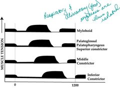
If you measure tension of all these muscles and evoked a swallow u get first bump
With bolus there is a reflex contraction– Muscles do not contract immediately when stimulated by the sensation of the bolus; rather when initial stimulus hits the sensory surface there is a neural system that goes to next muscle in sequence and inhibits them from contracting (feed-forward inhibition)– allows bolus to move caudally. |
|
|
|
Why don't we gag when we swallow?
|
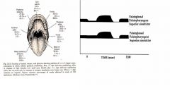
Gagging is a contraction and indicated are muscles activated by the bolus as it passes through the oral cavity. In the diagram to the right, normal is at the top and when you touch the anterior pillar and cause contraction you have not had feed-forward inhibition so there is immediate contraction because they have not gone through the process and you get gagging.
|
|