Reading...
![]()
Play button
![]()
Play button
![]()
Use LEFT and RIGHT arrow keys to navigate between flashcards;
Use UP and DOWN arrow keys to flip the card;
H to show hint;
A reads text to speech;
30 Cards in this Set
- Front
- Back
|
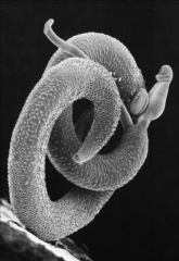
1. What species are Trematodes?
2. What species are Cestodes? 3. What species are Nematodes? |
1. Flukes - Schistosoma
2. Tapeworms - Taenia and Echinococcus 3. Roundworms - WANTED Loa Loa 3a. Wuchereria bancrofti 3b. Ascaris Lumbricoides 3c. Necator Americanus 3d. Trichuris trichiura 3e. Enterobius vermicularus 3f. Drancunculus medinensis 3g. Loa loa |
|
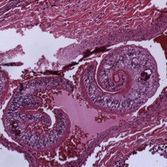
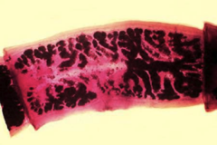
1. What species is pictured here and what category of helminth is this?
2. Describe the morphology. 3. Where are they found geographically? 4. Where are they found in the body? |
1. Schistosome - Trematode - Blood Fluke
2. Cylindrical bodies with 2 muscular suckers (1 oral, 1 attachment) 3. Africa - 2nd most prevalent tropical disease in the world 4. Found in blood vessels - OBLIGATE INTRAVASCULAR |
|
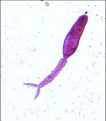
1. What is pictured here, what species is it, what form is it?
2. What is the function of this form of the helminth? 3. From what does this mature and in what intermediate host? 4. What symptom does it cause in humans? |
1. Trematode - Schistosoma - Blood Fluke - Cercariae Larva
2. Swims and penetrates the skin of the human host 3. Matures from a Sporocyst in the Snail (intermediate host) 4. Swimmer's itch |
|

1. What species is this, what form is it in?
2. Describe the morphology 3. Where do these particular ones infect? 4. What happens if this gets trapped in tissues during migration? |
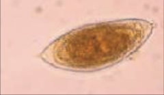
1. Trematode - Schistosome - Blood Fluke - Schistosoma Haematobium Egg
2. S. Haematobium Eggs have terminal spines 3. Venous Plexus - Urinary Bladder and Ureters 4. Granulomatous reaction - T-cells, Macs, Eosinophils |
|
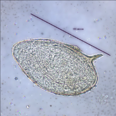
1. What species is this, what form is it in?
2. Describe the morphology 3. Where do these particular ones infect? 4. What happens if this gets trapped in tissues during migration? |

1. Trematode - Schistosome - Blood Fluke - Schistosoma Japonicum and Mansoni Egg
2. S. japonicum and S. mansoni eggs have lateral spines 3a. S. japonicum - Infects Superior Mesenteric Vein 3b. S. mansoni - Infects Inferior Mesenteric Vein 4. Granulomatous reaction - T-cells, Macs, Eosinophils |
|
|
1. What happens if a Schistosome Egg gets trapped in tissues during migration and what is the morphology?
2a. What disease from Schistosomes causes fever, hives, weight loss, cough, and headache? 2b. What causes the disease in 2a? |
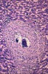
1. Granulomatous reaction - T-cells, Macs, Eosinophils - Eosinophilia, Microascess
2a. Katayama Fever 2b. Initial Schistosomulae larva release |
|
Describe the life cycle and pathogenesis of the Schistosomes
|
1. Eggs hatch into MIRACIDIA (larvae) in water
2. Eggs penetrate Intermediate Hosts (Snails) 3. Become SPOROCYTST in the snails 4. Mature into CERCARIAE (larvae) 5. Leave the snail and swim into a human via skin --> Swimmer's Itch dermatitis (allergy) 6. Cercariae lose their tail and become SCHISTOSOMULAE 7. Schistosomulae migrate thru tissues to Veins 8. Mature into ADULTS in the veins (JMH) --> Rarely pathologic (Coat themselves with Host Self substances) 9. Females deposit eggs in small venules 10. Eggs dig into the intestinal lumen, bladder, uterus using Tissue Destruction Enzymes 11. Eggs are eliminated via feces or urine |
|
1. What are complications of heavy worm burden with Schistosomes?
2. What is the non-heavy worm burden possibility? 3. What are the diagnostic techniques for Schistosomes? 4. How is this treated? |
1a. Cercarial Dermatitis (Swimmer's Itch) at entry point
1b. GI - Portal Hypertension, Fibrosis, Risk of Hep B and C, Dysentery, Ascites 1c. UTI - S. Haematobium ONLY - Renal Failure (Pyelonephritis), Glomerular Nephritis, Dysuria, Urinary Frequency, Terminal Hematuria 2. Asymptomatic Carrier 3a. Microscopy of stool or urine for Eggs 3b. Tissues biopsy of rectal (ALL) or bladder (S. haematobium ONLY) 4. Prizaquantel - causes contraction of parasite --> Poop it out |
|
1. What species is pictured here and what category of helminth is this?
2. Describe the morphology. |

1. Cestode - Tapeworms - Taenia Solium/Saginata
2a. Flat and Ribbion-like 2b. 4 muscular, cup-shaped suckers and a crown of hooklets on the Scolex 2c. Strobila body (chain of proglottids) |
|

1. What species is pictured here and what category of helminth is this?
2. Describe the morphology. 3. Where in the world is this found? 4. What is the reservoir? |
1. Cestode - Tapeworms - Taenia Solium/Saginata
2. Flat and Ribbion-like with 4 muscular, cup-shaped suckers and a crown of hooklets on the Scolex and a Strobila body (chain of proglottids) 3. Mexico, Central America, South America, Adia, Africa, India 4. Reservoir = PIGS!!! and Cattle (not usually infecting humans) |
|
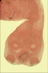
1. What is pictured and what genus and species is this from?
2. Describe the pathogenesis of this in pigs. 3. Describe the pathogenesis of this in humans. |
1. Proglottid from Taenia Solium (Cestodes - Tapeworms)
2a. Pig ingests EMBRYONATED EGG or PROGLOTTID 2b. ONCOSPHERE hatches and penetrates intestinal wall 2c. Goes into circulation 2d. Becomes CYSTICERCUS in muscle 3a. Humans eat UNDERCOOKED PIG MEAT 3b. Incubation --> 2 months --> ADULT WORM 3c. Adults produce PROGLOTTIDS 3d. Proglottid becomes GRAVID and detaches 3e. Proglottid migrates to anus or passes in stool 3f. Eggs are eaten (fecal-oral) and ONCOSPHERES --> Intestinal wall --> Blood --> Tissues 3g. LARVAE die --> Release Antigen --> Inflammatory Rxn --> Fever and Eosinophilia |
|
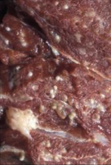
1. What species is pictured here and what category of helminth is this?
2. What are the symptoms of Taeniasis? 3. What are the symptoms of Cysticercosis? 4. What are the possible complications? |
1. Taenia Solium Cysticercus (Cestode - Tapeworm) in skeletal muscle of a pig
2. Asymptomatic or Mild nausea, low apetite, constipation or diarrhea, dizziness weakness --> 50% have eosinophilia 3. Cysticercosis - Extraintestinal Encysted Larvae --> Neurocysticercosis (gets into the CNS 60-90% of time) 4. Complications - Appendicitis, Obstruction of Panc and/or Bile Ducts |
|
|
1. What is Taenia Solium/Saginata?
2. How is Taenia Solium/Saginata diagnosed? 3. How is it treated? |
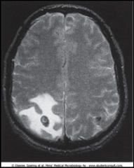
1. Cestode - Flat, Ribbon-like Roundworm with a Strobila of Proglottids, 4 suckers and a hooklet crown on the scolex
2a. Check stool for Ova and Parasites (NEVER SEE ADULTS) 2b. Imaging for Neurocysticercosis (pictured) 2c. Serology with Anti-cysticeral Antibodies 3. Preziquantel - contracts the parasite so it will detach and you can poop it out |
|
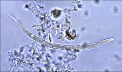
1. What organism is pictured here and what it it's classification among helminths?
2. Describe its morphology. 3. Where is this found mostly? |
1. Nematodes - Roundworms - Enterobius Vermicularis (PINWORM)
2. Small, Cylindrical, Unsegmented, White worm 3. Found worldwide and Most common helminthic infection in North America |
|
1. What organism is pictured here and what it it's classification among helminths?
2. Describe its morphology. 3. Describe the pathogenesis of this. |
1. Nematode - Roundworm - Enterobius Vermicularis (PINWORM)
2. Small, Cylindrical, White, Unsegmented worm 3a. Ingestion of Embryonated Eggs (fecal-oral) 3b. Hatch in the Small Intestine 3c. Mature in Large Intestine 3d. Fertilization of female by male 3e. Eggs Laid in Perianal Folds AT NIGHT --> Anal Itching 3f. Eggs become infectious w/in 6-8 hours |
|
1. What organism is pictured here and what it it's classification among helminths?
2. Describe its morphology. 3. What are the clinical symptoms of this organism? |
1. Enterobius Vermicularis Eggs (PINWORM) - Nematodes - Roundworms
2. Small, White, Cylindrical, Unsegmented worms 3a. Most are asymptomatic 3b. Anal Itching (Pruritis) Nocturnally or Early AM 3c. Ab/Pelvic Pain, Restless, Irritable, VAGINAL ITCHING 3d. Reinfection is common |
|
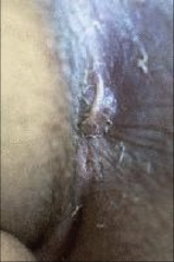
1. What is pictured here?
2. How is this diagnosed? 3. What is the treatment for it? |
1. Enterobius Vermicularis - Pinworm - Laying an Egg in perianal folds
2. Scotch tape test and Microscopy 3. Mebendazole |
|
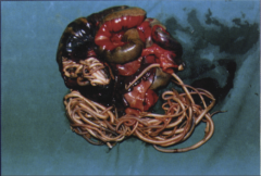
1. What organism is pictured here and what it it's classification among helminths?
2. Describe its morphology. 3. Where is it prevalent? 4. What about this organism causes clinical symptoms? |
1. Cestode - Roundworm - Ascaris Lumbricoides
2. Largest of the Intestestinal Nematodes 3. Found where there is Poor Sanitation - MOST COMMON HELMINTHIC INFECTION IN THE WORLD 4. Symptoms caused by movement of the worm |
|
1. What organism is pictured here and what it it's classification among helminths?
2. Describe its morphology. 3. Describe the pathogenesis of this organism. |
1 Ascaris Lumbricoides - Nematode - Roundworm - in the intestine
2. Largest of all Intestinal Nematodes 3a. Eggs located in feces (fecal-oral) 3b. 2-cell stage 3c. Advanced cleavage stage 3d. Eggs in external environment 3e. Develop in 10-14 days 3f. Ingestion by Humans 3g. Egg releases LARVAL WORM in Duodenum 3h. Worm --> duodenal wall --> Blood --> Liver, Heart --> Pulmonary Circulation (Loeffler's Syndrome - Pulmonary Eosinophilia 3i. LARVAE break free in Alveoli 3j. Coughed up and swallowed 3k. Fertilization of female in Small Intestine 3l. Egg production --> Shows up in feces 60-75 days after initial infection --> Repeat cycle |
|
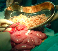
1. What organism is pictured here and what it it's classification among helminths?
2. Describe its morphology. 3. What are the symptoms of Ascariasis? |
1. Ascaris Lumbricoides - Nematode - Roundworm
2. Largest intestinal nematode 3a. Asymptomatic if egg innocculum is low 3b. GI issues --> Malnutrition --> Iron Def Anemia 3c. Growth/Cognition impairment 3d. NON-PRODUCTIVE COUGH, wheezing, Dyspnea 3e. Biliary and intestinal Obstruction (PICTURED) can become gangrenous |
|
1. What organism is pictured here and what it it's classification among helminths?
2. Describe its morphology. 3. How do you test for this? 4. Why can you not analyze stool for this during the Larval Phase? 5. What is the treatment? |
1. Ascaris Lumbricoides - Nematode - Roundworm
2. Largest of Intestinal Nematodes 3a. CBC for Eosinophilia --> Loeffler's Pulm. Eosinophilia 3b. CXR for infiltrates 3c. Larvae in sputum of pulmonary migration phase 3d. Eggs in stool in Adult Phase 3e. Abdominal Radiography 4. NO EGGS IN THE STOOL DURING LARVAL PHASE 5. Removal of Gangrenous clogged organs (PICTURED) |
|
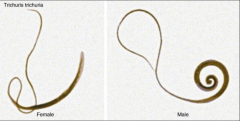
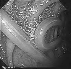
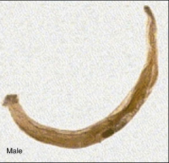
1. What organism is pictured here and what it it's classification among helminths?
2. Where is it located geographically? 3. How is it transmitted? |

1. Nematode - Roundworm - Trichuris Trichiura - WHIPWORM
2. Worldwide 3. Fecal-oral |
|

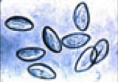
1. What organism is pictured here and what it it's classification among helminths?
2. What are the clinical symptoms of this infection? 3. Who is disease most common in? 4. How is this diagnosed and what is the treatment? |
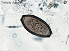
1. Egg of Trichuris Trichiura - WHIPWORM - Nematode, Roundworm
2a. Most are Asymptomatic 2b. Rectal Prolapse, Dysentery, Developmental Deficits 3. Children 4. Fecal Smears - Mebendazole |
|
1. What organism is pictured here and what it it's classification among helminths?
2. Describe its transmission and pathogenesis. 3. |

1. Nematode, Roundworm - Necator Americanus - HOOKWORM
2a. Penetrates the skin 2b. Migrates through Venous Circulation --> Lungs 2c. Coughed up and swallowed 2d. ADULTS in the Small Intestine 2e. Remains in Small Intestine for years |
|
1. What organism is pictured here and what it it's classification among helminths?
2. What are the symptoms that this causes? 3. How is this diagnosed? 4. How is it treated? |
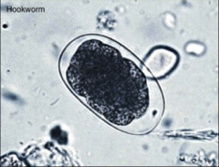
1. Necator Americanus - HOOKWORM - Nematode, Roundworm
2. Intestinal Blood Loss - Iron Deficiency Anemia 3. Find Eggs (PICTURED) in the feces 4. Mebendazole |
|
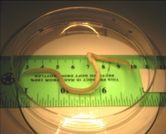
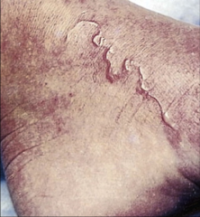
1. What organism causes this and what it it's classification among helminths?
2. Where is it located geographically? 3. How it is transmitted? 4. What causes the above symptomology? |
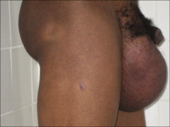
1. Wuchereria Bancrofti - Nematode, Roundworm
2. Tropical Areas, Africa, Asia, Latin America 3. Vector - Mosquito injects LARVAE 4. Adults block lymphatic vessels --> Lymphedema |
|
1. What organism causes this and what it it's classification among helminths?
2. What clinical symptoms does it cause? 3. How is this diagnosed? |
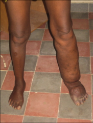
1. Wuchereria Bancrofti - Nematode, Roundworm
2. Elephantitis, Hydrocele, Lymphadenitis 3. Nocturnal Collection of Blood Demonstrates Larvae |
|
1. What organism causes this and what it it's classification among helminths?
2. Where is this located geographically? 3. How is it transmitted? |

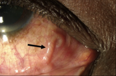
1. Loa Loa - Nematode, Roundworm
2. Western and Central Africa 3a. Infected fly bites you and injects the LARVAE 3d. Larva becomes ADULT and migrates to subconjunctiva (PICTURED) |
|
1. What organism causes this and what it it's classification among helminths?
2. How is this diagnosed? 3. How is it treated? |

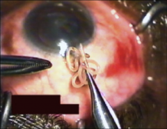
1. Loa Loa - Nematode, Roundworm
2a. Person returns from endemic area with local swellings. 2b. Visualization of adult worm beneath conjunctiva 3. Surgical Removal (Pictured) |
|
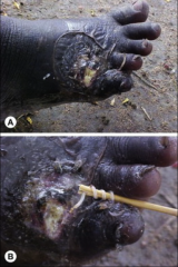
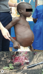
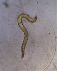
1. What is pictured above?
2. What is the transmission and pathogenesis? 3. How do they become visible? 4. How is it diagnosed and treated? |
1. Dracunculus Medinensis - Nematode, Roundworm
2a. Drink water containing COPEPODS 2b. Copepods release LARVAE 2c. Larvae --> Small intestine and Develop into ADULTS 2d. 1 year to mature 2e. Adults migrate to body surface and cause ULCERATIONS AND ERUPTIONS 3. Become visible thru ulceration 4. Diagnosed thru worm eruption - Treated with a stick |