![]()
![]()
![]()
Use LEFT and RIGHT arrow keys to navigate between flashcards;
Use UP and DOWN arrow keys to flip the card;
H to show hint;
A reads text to speech;
120 Cards in this Set
- Front
- Back
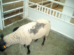
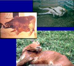
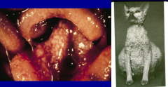
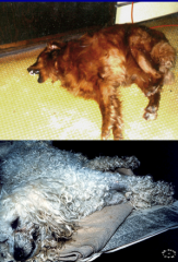
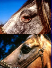
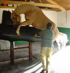
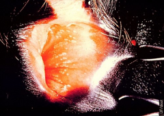
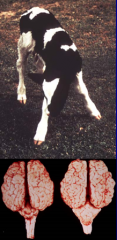
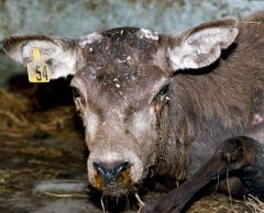
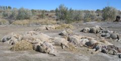
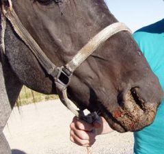
what are the clinical signs of this disease? |
scrapie in sheep non‑febrile, chronic, and fataldisease, causes pruritis- rubbing and biting flank |
|
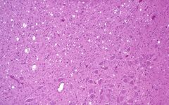
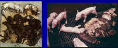
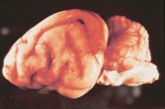
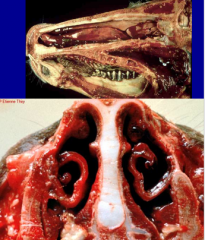
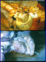
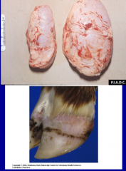
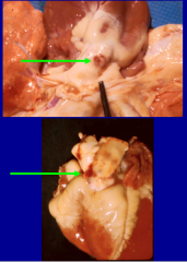
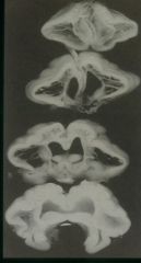
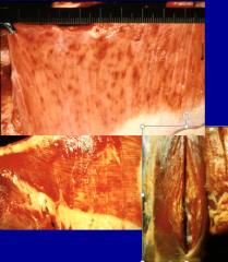
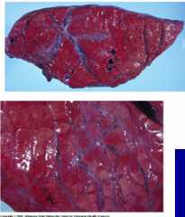
what is seen here and which disease is this? |
spongiform encephalopathy holes give brain a spongy appearance |
|
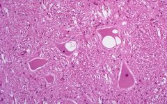
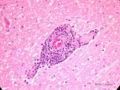
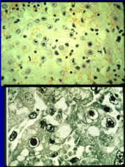
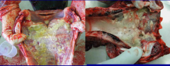
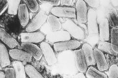
what is seen here? which disease |
spongiform encephalopathy vacuoles in neurons |
|
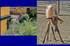
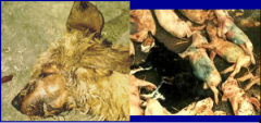
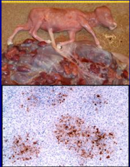
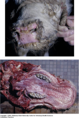
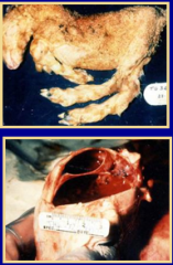
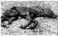
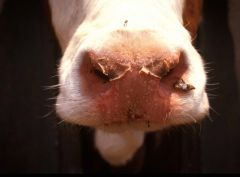
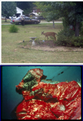
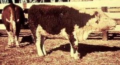
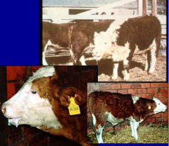
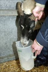
which disease is this? what are the clinical signs and who has been diagnosed? |
CWD- elk and deer rangingmule deer, black-tailed deer, white-tailed deer, Mountain elk, and Shira’smoose teethgrinding, abnormal behavior, excessive water intake, and marked loss of weight |
|
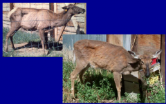
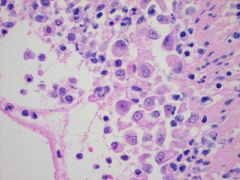
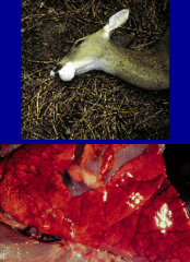
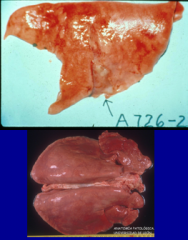
what disease is this |
CWD |
|
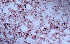
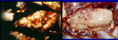
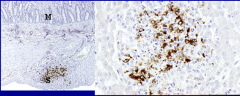
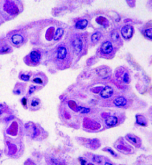
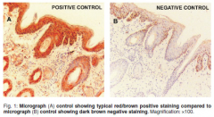
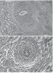
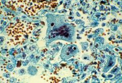
what disease is this? what is this process (Diagnosis)? |
BSE dyed with immunoperoxidase after being treated with proteinase K to digest normal PrPc |
|
|
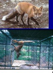
what are the reservoirs of rabies? what is the course of disease in these animals? what is the pathogenesis of a rabies infection? what is the virus type and family? |
fox, skunk, raccoon, bat, coyote rabies infects these animals, will eventually die but take longer to develop clinical signs rhabdoviridae genus lyssavirus |
|
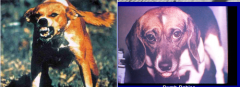
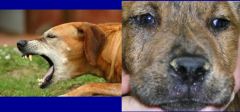
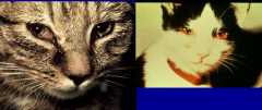
what are the clinical signs of rabies in a dog? what vaccines are available? what other viruses cause CNS dz in dogs? |
prodromal: change in behavior furious: abnormal aggressive behavior without provocation, change in voice, salivation paralytic: paraplegia, ascending paralysis killed and attenuated products distemper, rabies, pseudorabies, infectious canine hepatitis, non-respiratory parainfluenza virus, equine encephalitides, powassan, st. louis encephalitis, la crosse virus |
|
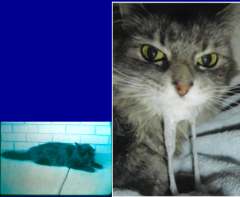
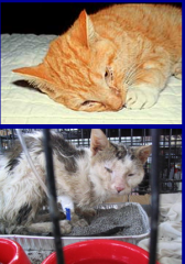
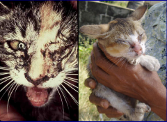
what are the clinical signs of rabies in cats? what are the other viruses that cause CNS disease in cats? |
change in behavior and temperment, meowing rabies, pseudorabies, panleukopenia, FIP, FeLV, FIV, spongiform encephalopathy |
|
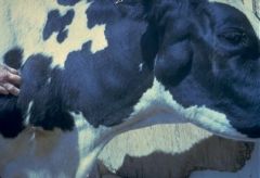
what are the clinical signs of rabies in cattle and what are the other CNS diseases? |
bellow, "choke" suspect Rabies, pseudorabies, bovine sporadic encephalopathy, bovine herpesvirus-5, malignant catarrhal fever, equine encephalitides, bluetongue virus, bovine viral diarrhea virus, akabane virus |
|
what are the clinical sign of rabies in horses? what are the differentials for CNS dz? |
lameness equine viral encephalitides, west nile, rabies, pseudorabies, equine herpesvirus-1, powassan virus, main drain virus, St. louis encephalitis virus, la crosse virus |
|
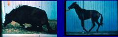
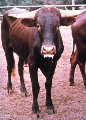
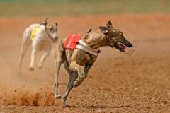
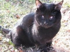
what disease is this? what is the most telling sign of infection? |
rabies, most likely to transmit virus when they become ill behavior change most telling sign |
|
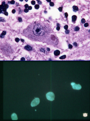
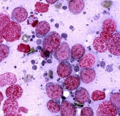
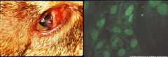
what is seen and what disease is present? what test is on the bottom? is it seen in all cases? |
negri bodies seen with rabies but not all cases FA test inclusions in neurons |
|
what process is seen here? what diseases cause this? |
SMEDI- stillborn, mummified, embryonic death and infertility pseudorabies/suid herpesvirus 1 |
|
which disease is seen? what are the clinical signs? what is the virus family and type? |
pseudorabies in dog and cat: intense pruritis usually in head region, salivate, no aggressive behavior is noted howling, meowing, vomiting and diarrhea herpesviridae, alphaherpesvirus (suid herpesvirus type 1) |
|
what disease process of FeLV and FIV is seen here? what type of virus and family are both of these? |
immunosuppression FeLV: retroviridae, gammaretrovirus FIV: retroviridae, lentivirus |
|
what disease process is seen here and what infection in cats does it accompany? what is seen with this condition? |
thymic or mediastinal lymphosarcoma- thymic -sually in cats less than 3 yrs of age 80-90% of cases associated with FeLV pleural effusion, dyspnea, regurg malignant T cells (large lymphoblasts) |
|
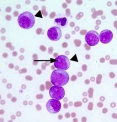
what disease process is seen here and what virus in cats is it seen with? if you wanted to detect virus in cells what test would you use? what is the process? |
acute lymphoblastic leukemia seen with FeLV IFA test: mouse Ab specific for p27, rabbit antimouse Ab tagged with fluorescent molecule |
|
|
what type of vaccine is available for FeLV? how is it administered? |
recombinant poxvirus vaccine (pureVax) intradermally |
|
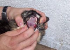
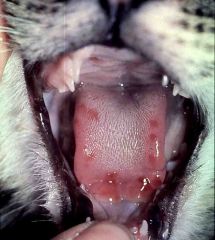
what disease process is seen? which disease is it common with? what causes these lesions? |
gingivitis and stomatitis FIV FIV immunosuppresses the cat, allows FCV to start replicating in mucosal epi and causes lesions |
|
what disease process is seen and what infection is it associated with? |
lymphocytic plasmacytic stomatitis FIV infection |
|
what disease is this? what are the presenting clinical signs? what type of virus and family? |
FIP, anorexia and icteric caused by Feline enteric coronavirus, coronaviridae |
|
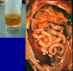
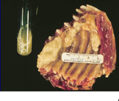
what are the characteristics of the fluid seen? what is the disease? |
FIP low cellularity, high protein <3.5 A:G ratio |
|
what virus? |
FIP |
|
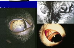
what disease process is seen? what virus is this seen with? |
severe fibrin deposits and flare due to precipitates anterior uveitis: severe iritis and keratic precipitates |
|
what disease process is seen here? what virus is it seen with and what form? what clinical signs are seen? |
pyogranulomas in brain dry form of FIP ocular lesions, behavioral changes and granulomatous lesions |
|
what lesion is seen here and what disease is seen here? |
perivascular granulomas or pyogranulomas with systemic vasculitis or thrombovasculitis FIP |
|
what is seen here in the SI and what disease is it seen with? |
pyogranulomas, macrophages predominate FIP |
|
what test is this? what is seen here and what disease does it cause? |
immunohistochem shows FCoV antigen, makes up pyogranulomas seen in FIP |
|
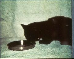
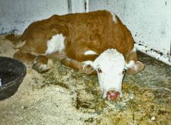
what virus and what disease is this? what is the typical posture of these animals? what is acute disease characterized by? what is not seen? |
feline panleukopeni, caused by feline parvovirus type-1, parvoviridae head over water bowl vomiting and dehydration seen acutely with severe leukopenia caused by neutropenia diarrhea is not seen acutely |
|
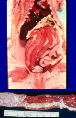
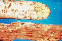
what is this lesion seen in a cat and what disease causes it? where does this virus replicate? |
turgid hyperemic small intestine, severe necrotic enteritis when intestines open virus infects crypt cells, leads to secondary bacterial infection |
|
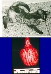
what condition is seen and what virus causes it? when was this kitten infected? what are the clinical signs? |
cerebellar hypoplasia caused by feline parvovirus type 1 (panleuk) infected in utero, ataxia and incoordination at 3 wks |
|
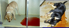
what is this virus? what is it associated with? what are the other causes of diarrhea in dogs? |
canine parvovirus-2, bloody diarrhea, anorexia, and dehydration CPV-2, CPV-1, distemper virus |
|
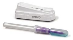
what does this test detect? |
detects antigen via monoclonal Ab |
|
|
what type of virus is canine parvovirus? |
parvovirus from parvoviridae |
|
what disease is seen here and what are the clinical signs? what does mild infection resemble? |
canine distemper virus, mucopurulent nasal discharge, sunken eyes, ocular discharge, moist productive cough, vomit and diarrhea, CNS signs (Seizure and myoclonus) indistinguishable from other respiratory disease: CAV-1 |
|
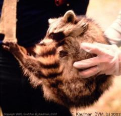
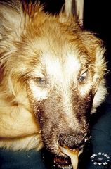
what disease is seen in this neurological raccoon? what is the mortality |
distemper virus, high mortality, cycles every 7-10 years |
|
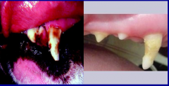
what condition is seen here and what virus causes it? |
enamel hypoplasia seen with distemper |
|
what are these seizures seen with CDV described as? when are these observed? |
chewing gum fits seen 2-4wks after the dog seemingly recovers from infection |
|
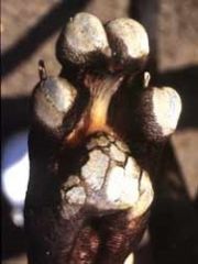
what is this condition of CDV known as? what are they more likely to develop? |
hard pad disease, hyperkaratosis of foot pads, more likely to develop CNS signs |
|
where are these inclusions seen with CDV? |
IN and IC IB seen, generally only see IC IB except in bladder wall and brain where both are present |
|
|
what type of virus and family is CDV? |
morbillivirus, paramyxoviridae |
|
what disease are these enlarged lymphnodes associated with? what if there are echymotic hemorrhages on mucosa? |
infectious canine hepatitis tonsils: canine distemper or CAV-1 |
|
what IB are seen with infectious hepatitis and where? what does ICH cause in foxes? what type of virus is ICH? |
canine adenovirus-1, adenoviridae (Ds DNA virus) IN IB seen in kuffer cells see CNS signs in foxes |
|
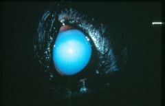
what is this condition and what virus causes it? what is the result this condition has in vaccination? is this dog blind? |
blue eye: keratitis caused by binding of antibody to cells of the cornea no blindness seen with CAV-1, vaccinate with CAV-2 (infections tracheobronchitis) |
|
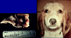
what is this infection in this 1 wk old puppy? how would you prevent this disease in the other pups? |
canine herpesvirus-1 hyperimmune serum and increase body temp |
|
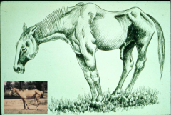
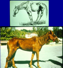
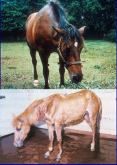
what disease is present in this recurrently infected horse? what are the clinical signs? what type and family is this virus? |
Equine infectious anemia retroviridae lentivirus acute: fever, anorexia, ataxia, weakness, thrombocytopenia, petechial hemorrhages, rapid weight loss, edema of legs and abdomen recurrent: fever, anemia, weakness, emaciation, ventral edema, thrombocytopenia, hypergammablobulinemia |
|
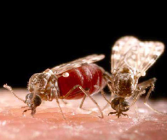
what is the pathogenesis and diagnosis of this virus? |
pathogenesis: blood sucking flies, infects macrophages/monocytes, antibodies in 2-3wks but provirus remains in macrophages which constantly undergoes antigenic drift (recurring episodes when no longer detected by Abs), signs usually caused by proinflamm mediators diagnosis: C-ELISA detects antibodies to p26, if positive confirm with Coggins (AGID) detects abs to p26 |
|
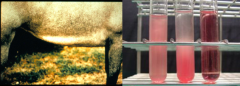
what is the basis for the ventral edema seen in EIA? what other virus causes ventral edema? what is the pathogenesis of the anemia seen? |
vasculitis/increased capillary permeability due to immune response, EVA also causes ventral edema anemia due to complement mediated hemolysis but mainly due to erythrophagocytosis |
|
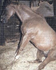
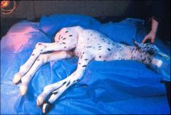
what virus causes this posterior paresis? what is this condition known as? |
equine herpesvirus type 1 also known as abortion virus equine herpesvirus myeloencephalopathy which is characterized by ataxia, posterior paresis and eventual quadriplegia |
|
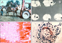
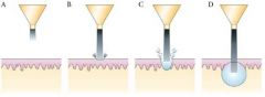
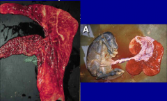
what are these lesions/conditions seen with EHV-1? what does B lesions lead to? |
A: encephalomyelopathy horse in sling B: hemorrhagic lesions in spinal cord: lead to ataxia, posterior paresis and hind quarter paralysis C: swollen axons and hemorrhage in spinal cord D: thrombo-occlusive hemorrhage in spinal cord |
|
what two viruses cause equine abortion? which one is most important? |
EHV-1 and EVA EHV-1 most important |
|
what other condition does EHV-1 cause? |
respiratory disease in young foals at weaning or a few months later |
|
what is the vaccination protocol to prevent EHV-1 abortion? |
MLV vaccines (2 dose) and inactivated vaccine (must be given frequently) goal to prevent abortion and encephalitis |
|
what virus causes this respiratory disease and conjunctivitis? what is the common name and clinical signs? where does the virus replicate? |
equine viral arteritis common name pinkeye severe conjunctivitis, depressed horse with ventral edema, nasal and ocular discharges, abortion in mares replicates in endothelial cells of blood vessels and causes arteritis (edema) |
|
what is this virus? how is it transmitted? what type of horses are more susceptible to infection? |
EVA aerosol transmission of respiratory disease venereally (Semen) and mucosal contact with infected tissue seen in standardbreds |
|
|
what is the most common way EVA is introduced onto a farm? what type and family is this virus? |
brought onto farms via semen or infected stallions arteriviridae, arterivirus |
|
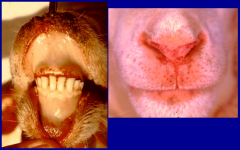
what is this virus? what is another name for this disease? what is the respiratory form called? what does it cause? what is not seen? |
bovine herpesvirus 1- red nose IBR is the respiratory form necrosis of nasal epi and turbinates NO MOUTH LESIONS |
|

what two viruses can cause open mouth breathing in cattle? |
IBR caused by BHV-1 and bovine respiratory syncytial virus |
|
what condition is seen? what virus/disease is this? what is the family/type? |
necrotic tracheitis infectious bovine rhinotracheitis virus via Bovine herpesvirus 1 herpesviridae, alphaherpesvirus |
|
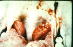
what disease is seen here (vulva of cow)? what virus causes it and when is it seen? |
infectious pustular vulvovaginitis BHV-1 2-3d post coitus |
|
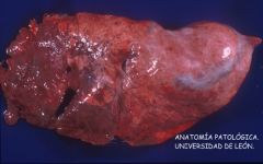
when is abortion seen with BHV-1? what are the lesions and the diagnosis? |
last trimester in unvaccinated animals multifocal disseminated necrosis in liver and lungs with IN IB in fetus IPX and PCR |
|
what virus is responsible for this condition? how is it transmitted naturally? when are cattle seropositive? what family and type is this virus? |
bovine leukemia virus transmitted via blood inoculation: in infected leukocytes (Flies needles dehorning) infected cattle are seropositive retroviridae, deltaretrovirus |
|
what is pictured here? what virus causes this? what cells are transformed and is there viremia? |
persistent lymphocytosis in BLV B cells infected and transformed, no free virus transmitted in transfered BLV infected lymphocytes |
|
where are typical sites of BLV lymphoma? what are the clinical signs? |
HALS: heart: hydropericarium, hydrothorax, and edema of brisket abomasum: diarrhea and melena lymphnodes: protruding eyeballs spinal cord: posterior paralysis |
|
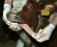
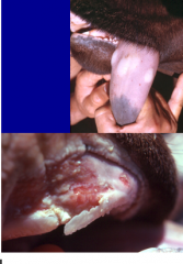
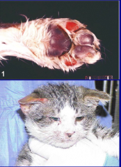
what is this calf with BVD mucosal disease? what are the clinical signs? |
mucosal disease due to PI mucopurulent nasal discharge and crusty reddened nose, erosions in its mouth |
|
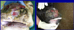
what is this disease present and what virus is it? who does it occur in and how would you diagnose? |
mucosal disease of BVD PI animal profuse watery diarrhea, anorexia, mucopurulent nasal discharge, erosive/ulcerative stomatitis 100% fatality rate IHC on ear notch |
|
what is seen here? what diseases cause this? what disease will not cause these? |
erosions on the tongue and esophagus MCF and BVD mucosal disease, BHV-1 in young calves and rinderpest virus make similar lesions BTV will not cause these lesions |
|
|
how does a calf become PI with BVDV? what is the primary transmission of BVDV? what is the viral family and type? |
infected in utero by 45-125d transmission primarily via PI animals excreting NCP BVDV in all secretions flaviviridae and pestivirus |
|
|
what abnormality can occur in BVDV infected calves? |
cerebellar hypoplasia, calves are ataxic with base wide stance |
|
what test is this for BVDV? what sample is it performed under? who does it detect virus in? |
Immunohistochemistry on ear notch sample, BVD virus antigen is reddish/brown in infected cells of hair follicle transiently viremic animal will be negative via ear notch |
|
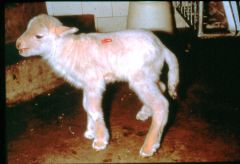
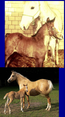
what is the name of this disease? what are the signs? what virus is it related to? |
hairy shaker- border disease hair rather than wool, trembles and ataxic, PI with virus closely related to BVDV |
|
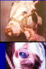
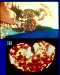
what disease is this? what viruses cause this disease? what is this lesion? what are the reservoirs? |
bovine malignant catarrhal fever: alcelaphine herpesvirus 1 and ovine herpesvirus 2 corneal opacity, starts at the limbus and progresses towards the center- stromal keratitis wildebeest and sheep carry the virus |
|
what disease is this and what viruses cause this disease? what are the family and type of virus? who does it infect? |
malignant catarrhal fever alcelaphine herpesvirus 1 and ovine herpesvirus 2 herpesviridae and gamma herpesvirus disease seen in cattle and deer |
|
in what disease do you see swollen prescapular lymph nodes and laminitis? |
malignant catarrhal fever |
|
what is the pathognomonic lesion (pictured here) with MCF? where does the virus replicate? |
necrotic vasculitis- pathognomonic lesion in cattle virus replicates in vascular endothelial cells, CD8 cells kill these viral infected cells lymphoproliferation within various tissues with widespread perivascular lymphoplasmacytic infiltration |
|
who else does MCF infect and cause high mortality in? what clinical signs are seen? what else causes high mortality in this species? |
infects and causes high mortality in deer MCF and EHD mucopurulent nasal and ocular discharge |
|
what virus causes this? who else does this virus infect? what is the virus family and type? |
bluetongue virus wild and domestic ruminants (rarely clinical disease in cattle) reoviridae and orbivirus |
|
what insect is this? what virus of sheep and deer does it transmit? what is the extrinsic incubation? when does disease occur? |
cullicoides transmits BTV extrinsic incubation: 8-10d fall and summer |
|
what are the clinical signs of BTV? |
sheep: nasal discharge and excessive salivation, mucopurulent, tongue and gums are swollen and purple, catarrhal stomatitis, lameness |
|
what disease is this? what condition? |
bluetongue, erosive lesions |
|
what lesions are these and what disease are they pathognomonic for in sheep? what about deer? |
hemorrhage at base of pulmonary arty in sheep patho for BT in deer can be BT or Epizootic hemorrhagic disease |
|
what are these conditions and what virus causes these issue? |
fetus or newborns infected with BTV arthrogryposis hydranencephaly |
|
what is this condition and what causes it? what is the difference from hydrocephalus? what does this condition cause in cattle and why does it happen? |
fetal infection BTV hydranencephaly: accumulation of fluid within white matter hydrocephalus: fluid accumulation in ventricles causes dumb calf- BTV attenuated vaccine virus |
|
what is this condition called? |
arthrogryposis |
|
what are the clinical signs of BTV in cattle? |
usually sub clinical or inapparent, serve as amplifiers characterized by lameness, erosions in nostrils and by peeling of the skin on nasal septum |
|
what disease is this that's seen in WTD? what is it transmitted by? when is disease observed? what is seen? what type is it? |
epizootic hemorrhage disease cullicoides, mortality high in deer and observed in late summer/fall extensive hemorrhages in the muscles orbivirus |
|
what form of EHD is this? what did this deer die as a result of? |
pulmonary form: deer die as a result of acute lung edema, frothing at mouth/nostril, interlobar fluid accumulation |
|
what disease is this? |
epizootic hemorrhagic disease |
|
|
what are the three viruses that can infect deer and cause a high mortality? what is one disease associated with? |
epizootic hemorrhagic disease, bluetongue virus and malignant catarrhal fever MCF acquired from sheep |
|
what vesicular disease is this? |
vesicular stomatitis |
|
what are the two vesicular diseases that infect cattle? how are these transmitted? what samples do you collect? what family/type are these viruses? |
vesicular stomatitis and F&M disease VSV: vesiculovirus, rhabdoviridae, sand flies and black flies FMD: aphtovirus, picornaviridae, saliva, milk and other body fluids vesicular fluid and overlying epithelium |
|

what two vesicular diseases cause these lesions? what multisystemic virus can also cause this? |
vesicular stomatitis virus and foot and mouth disease also caused by bluetongue virus |
|

what are the four vesicular diseases that affect pigs? how are each of these transmitted and what type of virus is each one? |
VSV: vesiculovirus in rhabdoviridae, black flies and sand flies vesicular exanthema of swine: calicivirus, undercooked sea lion meat FMD: aphtovirus in picornaviridae, saliva, milk, other body fluids swine vesicular disease: enterovirus type 9 and picornaviridae, body fluids |
|
what virus is this? what is the family/genus? |
vesicular stomatitis vesiculovirus in rhabdoviridae |
|
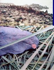
what virus is this? what type of virus is it and what disease in swine is it related to? |
san miguel sea lion virus calicivirus related to vesicular exanthema of swine |
|
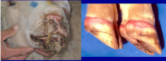
what is the clinical signs of vesicular disease in each species? |
fever anorexia and salivation cattle and horse: vesicles on dorsum of tongue, dental pads, buccal mucosa, teats, coronary bands (lameness) swine: vesicles on feet and snout causing lameness and sometimes sloughing the hoof capsule hard in sheep (FMD) because vesicles dehydrate quickly, don't see lameness |
|
what do vesicles rupture to form? |
erosions which will heal without scarring |
|
what are the viruses that can cause ITB? what 3 are the most important? what bacterial pathogen is associated with ITB? |
canine parainfluenza, canine influenza, canine adenovirus 2, CAV-1, CDV, CHV-1, Reovirus 1,2,&3 complexed with infection of bordetella bronchiseptica and mycoplasma |
|
what respiratory illness is seen in this dog? what is the virus that causes it if this is an adenovirus? (family?) what specific disease does this virus cause? |
infectious tracheobronchitis canine adenovirus type 2 infectious canine laryngotracheitis adenoviridae |
|
what respiratory virus has adapted from horses? |
canine influenza H3N8 |
|
what feline respiratory virus is this? what disease is it? what are the clinical signs? who does it infect and do they recover? |
feline herpesvirus 1: feline viral rhinotracheitis causes severe eye lesions (keratitis, corneal ulcers, descemetocele) oral erosions/ulcers, sneezing, nasal discharge, anorexia, fever infects young kittens and remain latently infected after recovery |
|
what clinical sign is seen here? if accompanied by respiratory disease and nasal discharge, what virus is it? |
conjunctivitis, FHV-1, feline rhinotracheitis can lead to ulcers and blindness |
|
what condition is seen on the left? what disease is this common with? what diagnostic test is on the right? |
descemetocele caused by FHV-1 FA showing FHV-1 replicating in nucleus of corneal cells |
|
if no keratitis or conjunctivitis is seen, what viral infection is this? what are clinical signs? who does it infect and do they clear infection? |
feline calicivirus infection stomatitis, gingivitis with ulcers at border of tongue and hard palate, rarely causes eye infections, sneezing, nasal discharge, anorexia, fever, LAMENESS in all ages of cats in absence of respiratory disease infects younger cats and some remain persistently infected in oropharynx |
|
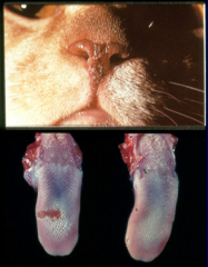
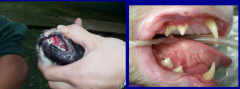
what conditions are seen here and what infection causes it? |
ulcers on nasal septum and oral ulcers (virus on nose from licking) calicivirus |
|
what conditions are seen here and what virus causes it? |
skin sloughing and ulceration of pad-haired skin junction- virulent systemic calicivirus infection systemic disease resulting in facial edema and ulceration of pinna |
|
what are the viruses that cause bovine respiratory disease? which are the most important? which 3 are associated with interstitial pneumonia? which 2 are associated with bovine enzootic pneumonia of young calves? |
respiratory dz: BHV-1. parainfluenza-3 (PI-3), respiratory syncytial virus (BRSV), coronavirus, adenovirus interstitial pneumonia: PI-3, BRSV, and adenovirus bovine enzootic pneumonia: PI-3 and BRSV |
|
what disease is affecting this lung? what viruses cause this? what 2 viruses cause enzootic pneumonia in calves? |
interstitial pneumonia, associated with PI3, BRSV, and bovine adenovirus PI-3 and BRSV cause enzootic pneumonia |
|
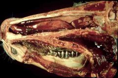
necrosis of nasal epi and turbinates. what is another name for this disease? what virus caused this infection? what is the respiratory form of this disease? what is not seen? |
Red nose bovine herpesvirus type 1: infectious bovine rhinotracheitis no mouth lesions seen |
|
what virus causes this disease (accompanied by interstitial emphysemaa)? |
bovine respiratory syncytial virus |
|
this is an infection with BRSV. what is this condition? what is this virus also responsible for? what would you seen on histopath? |
interstitial emphysema in bovine lungs also causes interstitial pneumonia see large multinucleated syncytial cells in lung tissue |
|
this tissue is from a bovine lung, what do you see? what two viruses cause this? |
giant syncytial cells PI-3 and BRSV |
|
what two viruses cause sheep and goat lungs to have this appearance? what is the family/type of these viruses? what is the incubation period of both and what do they cause? who do they cause disease in? |
Ovine progressive pneumonia virus (visna/maedi virus): lentivirus belongs to retroviridae, chronic progressive pneumonia in adult sheep (incubation period 2-3yrs) ovine pulmonary adenocarinoma: Jaagseikte sheep retrovirus, betaretrovirus in retroviridae, fatal progressive pneumonia in adult sheep (incubation period 2-3yrs) |
|
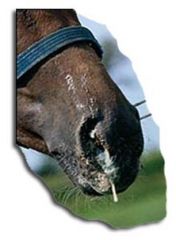
this disease is characterized by the development of bronchial alveolar adenocarinoma. what disease is it? what virus causes it? what is the pathogenesis and the pathognomonic diagnosis? |
ovine pulmonary adenocarcinoma: jaagseikte sheep retrovirus transforms pneumocytes of the lungs which secrete excessive amounts of fluid that fill up the alveolar space on necropsy fluid will pour out of the sheep nostril when the head is down or when animal is held up |
|
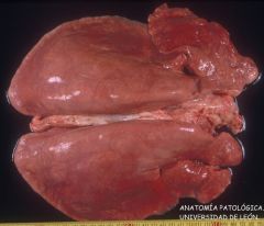
these lungs are heavy with no increase in fluid. what disease is this and what virus causes it? what is the pathogenesis and why are they lungs heavier? |
ovine progessive pneumonia: visna/maedi virus transforms macrophages which infiltrate the lungs and other tissues including the udder and brain infiltration of cells causes the increased weight of lungs |
|

what are the important respiratory viruses of horses? |
Equine herpesvirus type 1, equine herpesvirus type 4, Equine influenza, equine viral arteritis, equine rhinitis A virus, equine adenovirus, equine reovirus, equine herpesvirus 8, hendra virus, african horse sickness |
|

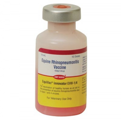
what is the common name of EHV-4? what are the clinical signs and who does it infect? what are the samples for diagnosis? what vaccines are available? |
EHV-4: equine viral rhinopneumonitis rhinopneumonitis seen in fall in foals and yearlings: fever, mucopurulent nasal catarrh, conjunctivitis serious respiratory outbreaks have been seen Deep nasal and pharyngeal swabs with paired sera ??? |
|
what are the important clinical signs of influenza and rhinitis A virus? are vaccines available? what sample would you submit for diagnosis? |
influenza: explosive outbreaks, high morbidity, coughing, sudden onset, depression, dry hacking cough, mucopurulent nasal discharge and conjunctivitis not commonly seen in acute stage, bivalent inactivated vaccine, no racing for 10 days after vaccine Rhinitis A: fever, pharyngitis and pharyngeal lymphadenitis and copious nasal discharge, becomes mucopurulent, coughing, no vaccine deep nasal or pharyngeal swabs and paired serum |
|
what virus causes fatal infection in arab foals? when do they show signs? when do they die? |
Equine adenovirus infection: usually causes subclinical infections combine immunodeficiency foals show respiratory signs first month or two and die by 4-6 months of age |