![]()
![]()
![]()
Use LEFT and RIGHT arrow keys to navigate between flashcards;
Use UP and DOWN arrow keys to flip the card;
H to show hint;
A reads text to speech;
12 Cards in this Set
- Front
- Back
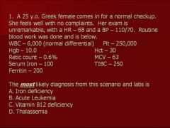
|
D
|
|
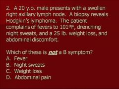
|
D
|
|
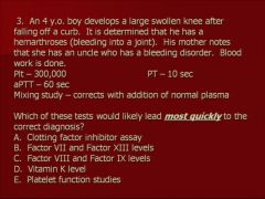
|
C
|
|
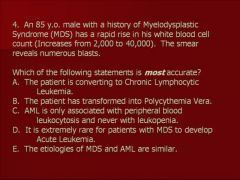
|
E
|
|
|
C
|
|
|
Presentation
- Bone marrow failure • Anemia – fatigue, weakness, shortness of breath • Thrombocytopenia – increased bruising and bleeding • Leukopenia/neutrapenia – infections - Enlarged lymph nodes and/or splenomegaly - Mediastinal mass - Neurologic complaints (headaches, double vision, swallowing difficulty, etc) from leukemia in the central nervous system |
Acute Lymphocytic Leukemia(ALL)
Epidemiology – Incidence 1-3/100,000/year – 3,000 new cases of ALL/year – Peak incidence at 2-3 years – Second peak after 80 years – 10-20% of adult leukemias, but 90% of childhood leukemias Pathology - Acute Leukemia is by definition a cancer of bone marrow progenitors (progenitors are immature marrow cells still capable of division) - Lymphoblasts (immature lymphocytes) proliferate and are unable to mature into normal cells - These blasts eventually dominate the bone marrow, suppressing normal hematopoiesis – therefore decreases red cell, platelet, and neutrophil production - By definition acute leukemia must have 20% leukemic blasts in the bone marrow - Can be T or B cell origin - Often associated with chromosomal (cytogenetic) abnormalities, including the Philadelphia chromosome (9;22 translocation). This particular translocation is associated with a very poor response to conventional treatments |
|
|
- Often found incidentally on routine blood work (patient asymptomatic)
- Can present secondary to bone marrow failure • Anemia – fatigue, weakness, shortness of breath • Thrombocytopenia – increased bruising and bleeding • Neutropenia (lack of neutrophils) – infections - Lymphadenopathy/Splenomegaly - B symptoms (night sweats, weight loss, fever). |
Chronic Lymphocytic Leukemia(CLL)
Epidemiology - Disease of advancing age - Approximately 10,000 new cases per year Pathology - Proliferation of mature lymphocytes in the bone marrow - Majority are of B-cell origin - Usually an indolent process - Does not convert to ALL |
|
|
Presentation
- Lymphadenopathy - B symptoms (night sweats, weight loss, fever). These represent a worse prognosis, and are added to the staging - Can present in extra-nodal sites |
Non-Hodgkin’s Lymphoma(NHL)
Epidemiology - NHL is rapidly increasing in incidence in the U.S., with 50,000 new cases/yr and 20,000 deaths/yr. - Age and any immunodeficiency (especially AIDS) increase risk. Pathology - Malignancy can occur at any stage of lymphocyte development in the lymph node - There are multiple histologic types with differing proliferation rates o Indolent (low-grade)– this grows slowly but is incurable (average survival 8 yrs) – an example is Follicular lymphoma o Aggressive (intermediate) grade – this grows more quickly, but is curable- an example is Diffuse large cell lymphoma- the most common type of NHL, which has a 50-60% 5-year survival o Leukemic equivalent (high-grade) –these grow extremely rapidly and are potentially curable - an example is Burkitt’s lymphoma. - NHL usually starts in the lymph nodes (or other lymphoid tissue) and spreads both via the lymphatics and hematogenously to adjacent nodes, the spleen, liver and/or bone marrow - Stage I- single node region, Stage II- 2 or more nodal groups on the same side of the diaphragm, Stage III- nodes on both sides of the diaphragm involved, Stage IV- any extra-lymphatic involvement (e.g. liver, marrow). |
|
|
Hodgkin’s Lymphoma or Disease (HD)
|
Hodgkin’s Lymphoma or Disease (HD)
Epidemiology - HD is uncommon, 8,000 new cases/yr with 2,000 deaths/yr - Incidence demonstrates a bimodal distribution – peak ages 15-35 and second smaller peak after age 50 Pathology - The characteristic malignant cell is the CD30+ Reed-Sternberg cell, thought to be an antigen presenting cell in the lymph node - HD starts in the nodes and spreads predominantly via the lymphatics to adjacent nodes. - Stage I- single node region, Stage II- 2 or more nodal groups on the same side of the diaphragm, Stage III- nodes on both sides of the diaphragm involved, Stage IV- any extra-lymphatic involvement (e.g. liver, marrow) Presentation - Lymphadenopathy - B symptoms (night sweats, weight loss, fever). These represent a worse prognosis, and are added to the staging |
|
|
Presentation
- Bone pain from lytic lesions or fractures - Weakness and light-headedness from anemia - Infection from hypogammaglobulinemia - Nausea and vomiting, edema from renal failure - Confusion from hypercalcemia |
Plasma Cell Dyscrasias
Multiple Myeloma – most common type of plasma cell dyscrasia Epidemiology - Uncommon, 13,000 new cases/yr, causing 9,000 deaths/yr. - Advancing age, males, African-American race, and radiation exposure associated with an increased risk. Pathology - A malignancy of antibody-producing B-cells. They typically produce a monoclonal antibody that can suppress other antibody formation (hypogammaglobulinemia) - Antibodies are typically IgG and IgA (not IgM) - Myeloma can create lesions in cortical bone leading to hypercalcemia, fractures, and spinal cord compression - Antibody deposition, hypercalcemia, hyperuricemia all can lead renal insufficiency - Myeloma cells typically stay in the marrow but may also produce malignant masses, called plasmactyomas that can be found anywhere in the body - Not a curable disorder |
|
|
Waldenstrom’s Macroglobulinemia
|
Waldenstrom’s Macroglobulinemia –
- Plasma cell dyscrasia associated with increased production of IgM - Does not cause bone destruction or renal failure - Increased amount of IgM can cause hyperviscosity – slow movement of blood that can lead to decreased CNS function from decrease oxygen delivery |
|
|
Rouleaux Formation
|
Rouleaux Formation: In a blood smear, RBC stack up on top of each even in the Good goldilocks area. This is a sign that serum protein levels are high. When does this occur? In plasmacytomas or in multiple myeloma.
|

