Reading...
![]()
Play button
![]()
Play button
![]()
Use LEFT and RIGHT arrow keys to navigate between flashcards;
Use UP and DOWN arrow keys to flip the card;
H to show hint;
A reads text to speech;
41 Cards in this Set
- Front
- Back
|
Animal development
|
birth, maturity and gametogenesis, fertilization, Embryogenesis
|
|
|
Embryogenesis
|
stage between fertilization and birth consists of:
-cleavage:blastula formation -gastrulation-blastula rearranged-gastrula 3 germ layer development -organogenesis |
|
|
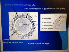
Draw and label mature ovum label
Also what does the corona radiate have? the zona pellucida? vitelline membrane? |
do it
corona> contain follicle cells z.p>matrix of glycoproteins vitelline membrane>protien fibers |
|
|
Monotremes have ___
masupials have _____ Eutherians have ____ |
monotremes=eggs
masupials=pouches eutharians=placentas |
|
|
ovulation occurs as a response to increase of what hormone?
|
Luteneizing hormone
|
|
|
When is the egg fertilizable?
|
24 hours after ovulation
|
|
|
How long can sperm survive in the female reproductive tract?
|
72 hours
|
|
|
Where does fertilization normally occur?
|
the upper 1/3 of the fallopian tube
|
|
|
How many sperm normally reach there?
|
.01 percent
|
|
|
How long does it take sperm to travel up the fallopian tubes?
|
about seven hours
|
|
|
become able to fertilize egg through what?
|
the activation of enzymes in acrosome
|
|
|
what is this activation called? what happens?
|
capacitation
-takes about 7hrs -mobility becomes enhanced and membrane becomes fragile so hydrolytic enzymes in their acrosome can be released. -As swim secreteions from female tract cause mambrane proteins to be removed and cholsesterol that keeps acrosomal rxn. tough depletes. -Sperm gets to egg before capacitation completes> this is to prevent spilling -so must wait to finish |
|
|
the reaction can not happen
|
outside of the female reproductive tract
|
|
|
Place on the egg sperm attaches to
|
the zona pellucida
|
|
|
How does the sperm penetrate corona radiata and attach to the z.p?
|
- penetrates corona radiata assisted by a cell surface hyaluronidase that digests the intercellular cement between granulosa cells causing them to fall away
-after sperm binds to ZP3 gylycoprotein of the zona pellucida which functions as sperm receptor and helps trigger acrosomal rxn. (ivolves breakdown of plasma membrane and release of acrosomal enzymes that digest hole through z.p -[takes hundreds to digest zp and later sperm has better chance of getting in once has been considerably digested. -once clear path single sperm fuses to membrane receptors using two part binding apparatus: -beta: first binds to receptor oocyte membrane -this engages alpha protein that causes insertion into membrane. |
|
|
Preventing polyspermy in humans
|
1. Once sperm enters eggs there are waves of Ca 2+ released by oocytes endoplasmic reticulum into its cytoplasm which activates oocyte to prepare for cell division (also Na+ diffuses in Ca out changes the polarity first fast block)
2. Calcium surges also trigger cortical rxn in which granules located just inside plasma membrane spill enzymes into extracellular space beneath the z.p. These enzymes called Zonal Inhibiting proteins or ZIPS, and they destroy sperm receptors preventing further sperm entry. 3. Spilled materials also bind water, so swells it detaches all sperm bound to receptors (slow block). -Zona pellucida hardens and the secondary oocyte divides which generates the polar body once polar body formed egg and sperm nucleus fuse |
|
|
How many hours after fertilization does the zygote (formed upon fusion of nucleus' cleave)?
|
18 to 36 hours
|
|
|
Location of oocyte, and events as it travels towards the ovary
1.Pre fertilization whats happening 2. Fertilization where and what 3. Cleavage |
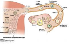
1. Oocyte has been released from ovary and swept by the fimbriae into oviducts
2. occurs in the ampulla of the oviduct (close to ovary), meiosis is completed after the sperm enters oocyte. 3. Begins approximately one day later (18 to 36 hrs) slowest cleavage in animal kingom, during cleavage cilia moves zygote towards uterus. (m |
|
|
How long does zygote float downstream in fallopian tube?
|
moves for about 30 or 40 hours
|
|
|
The first cleavages
|
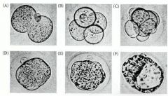
1. one cell divides to produce two cell stage (30 hrs after fert.) happens in tube> divides normal meriodonally
2. about fourty hrs. later two divides into 4> one cell again has meridional division and the other equatorially. (called ROTATIONAL CLEAVAGE) 3. COMPACTION: up to third cleavage and through eight cell stage loose arrangement of cells with space btw. Then at third cleavage Cell adhesion proteins (like E cadherin) are expressed, and blastomeres huddle and compact to form a ball of cells. This arrangement stabilized by tight junctions that form between outside cells of ball, sealing off inside of sphere. Cells in sphere form gap junctions that allow molecules and ions to pass between. 4. Cells of compacted eight cell embryo divide to form 16 cell MORULA> has small group of internal cells surrounded by larger grp of external cells. **descendents of external cells become the trophoblast>produce no embryonic structures but forms the tissue of the chorion, the embryonic portion of the placenta. |
|
|
What do the external cells of the morula become??
|
most of the external cells become the TROPHOBLAST
-doesnt produce embryonic tissue -will form the tissue of the chorion or the embryonic portion of the placenta |
|
|
What will the chorion do? What is it for?
|
=Enables fetus to get oxygen and nourishment from mother
=also secretes hormones that cause mother's uterus to retain fetus and produce regulators of the immune response so that the mother will not reject the embryo |
|
|
What is the crucial outcome of these first divisions? i.e of the creation of the morula, external cells internal cells etc.
|
-the crucial outcome is that generates cells that will stick to uterus.
-thus formation of trophoectoderm cells (cells that become trophoblast cells) is 1st differentiation in mammalian development. |
|
|
After trophoectoderm cells adhere to this lining what happens?
|
will digest path that allows embryo to lodge itself in the uterine wall.
|
|
|
differences between mammals/humans and other animals
|
-cleavage in mammals is slower
-blastomere orientation unique from different type of cleavage specific to mammals known as ROT. CLeavage> where first division is meridional and in the next division one again divides meridionally but the other divides equatorially -this allows the other difference which is that unlike other animals will have odd numbers of cells -unlike almost all other animal genomes, mammalian genome activated during early cleavage and the newly formed nuclei (rather than oocyte cytoplasm) that produce the proteins necessary for cleavage and development. -Also different because mammalians go through COMPACTION: (see previous note card and explain what it is) |
|
|
Okay so earlier cleavages( through 16 cell Morula phase) occurring as embryo moves through oviduct. What is also going on? End when reaches uterus
|
=blastocyst is expanding within zona pellucida (which had been where sperm attached)
=while en route, z.p prevents blastocyst from adhering to oviduct walls (if does happen ectopic pregn.) =when reaches uterus around DAY the 16 cell undifferentiated Morulla 4 must hatch |
|
|
16 cell phase Morula has reached embryo, what now?
|
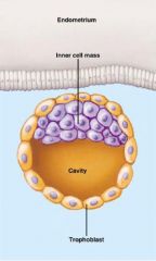
-Inner cells generate the inner cell mass (ICM) which gives rise to embryo and associated yolk sac, allantois, and amnion. By 64 cell stage trophoblast and ICM are separate layers neither contributing cells to other group.
- ICM supports trophoblast by secreting proteins that cause trophoblast to divide. -also a cavity if forming -Also hatching, has happened by day 6: blastyocyst digests small hole through zona pellucida and squeezes through as expands. (sseems trypsin like protease responsible) -CAN NOW MAKE CONTACT WITH UTERUS |
|
|
Attaching to the uterus: with emphasis on
-what makes up the surface of uterine epithelium? -2 phases of attachment? |
-uterine epithelium catches blastocyst on an extracellular matrix containing complex sugars, collagen, laminin, fibronectin, hyaluronic acid, and heparan sulfate receptors.
2 PHASES: 1.period of labile attachment 2. more stable attachment 1..First attachment mediated by L-selectin on trophoblast cells adhering to sulfated polysaccharides on the uterine cells. These sulfated polysaccharides are synthesized in response to estrogen and progesterone secreted by corpeus luteum. 2. After several factors at play to keep tightly bound: trophoblast cells synthesize integrins that bind to uterine collagen, fibronectin, and laminin, and they synthesize heparan sulfate proteoglycan precisely prior to implantation. -once in contact trophoblasts secrete another set of proteases that digest extracellular matrix so can bury in lining. -Implantation about 7 days after fertilization |
|
|
1st 7 days embryo uses food from yolk, after imbeds in wall uses nourishment from mother, how?
what other imp. funct. do trophoblast and chorion perform they secrete _____. what does that do? two things |
-chorion is derived primarily from trophoblasts with suupplementation by mesodermal cells from inner cell mass.
-chorion forms fetal portion of placenta and induces uterine cells to form the maternal portion of placenta called decidua>which becomes righ in blood vessels to provide embryo with oxygen and nutrients -trophoblasts and chorion also secrete HCG or (Human chorionic gonadotropin) that targets corpus luteum to cause it to stop secreting progesterone: which would normall change gluteal phase to menses and embryo would be rejected. we dont want. -HCG also prevents immune attack by mother -supresses FSH and LH so no menses no ovulation |
|
|
Now implantation, perfect development and differentiation within the embro.
origins of early mammalian tissues SET UP for gastrulation |
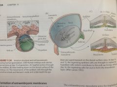
![-first segregation of cells in ICM form 2 layers
-lower layer is hypoblast, and remaining ICM cells become epiblast>together become bilaminar germ disc [day 10 after fertilization]
-hypoblasts delaminate from inner cell mass to line blatocoel cavity whe](https://images.cram.com/images/upload-flashcards/893863/689337_m.jpg)
-first segregation of cells in ICM form 2 layers
-lower layer is hypoblast, and remaining ICM cells become epiblast>together become bilaminar germ disc [day 10 after fertilization] -hypoblasts delaminate from inner cell mass to line blatocoel cavity where they give rise to extramembryonic ectoderm which forms the YOLK SAC. -epiblast cell layer is split by small clefts that eventually coalesce to separate the embryonic epiblast from the other epiblast cells that line the amniotic cavity (amnionic ectoderm) -once amnion lining completed, amniotic cavity fills with fluid-serves as shock absorber for developing embryo and prevents from drying out. (embryonic epiblast thought to contain all cells that will generate actual embryo) |
|
|
Now implantation, perfect development and differentiation within the embro.
origins of early mammalian tissues >>with different pic |
![-first segregation of cells in ICM form 2 layers
-lower layer is hypoblast, and remaining ICM cells become epiblast>together become bilaminar germ disc [day 10 after fertilization]
-hypoblasts delaminate from inner cell mass to line blatocoel cavity whe](https://images.cram.com/images/upload-flashcards/893863/689338_m.jpg)
-first segregation of cells in ICM form 2 layers
-lower layer is hypoblast, and remaining ICM cells become epiblast>together become bilaminar germ disc [day 10 after fertilization] -hypoblasts delaminate from inner cell mass to line blatocoel cavity where they give rise to extramembryonic ectoderm which forms the YOLK SAC. -epiblast cell layer is split by small clefts that eventually coalesce to separate the embryonic epiblast from the other epiblast cells that line the amniotic cavity (amnionic ectoderm) -once amnion lining completed, amniotic cavity fills with fluid-serves as shock absorber for developing embryo and prevents from drying out. (embryonic epiblast thought to contain all cells that will generate actual embryo) |
|
|
Actual gastrulation
|
-gastrulation begins at end of embryo where node forms.
-mammalian mesoderm and ectoderm migrate through primitive streak: migrating cells of the mammalian epiblast lose E-cadherin, detach from neighbors and migrate through streak as individual cells. -cells migrating through node give rise to notochord. -cell migration and specification seem to be coordinated by fibroblast growth factors. -ectodermal precursors are located anterior to fully extended primitive streak -in some instances single cell gives rise to descendants in more than one germ layer or to both embryonic and extraembryonic derivatives. -thus at epiblast stage (discussed previously) lineages have not become separated. -cells migrating into space btw hypoblast and epiblast become coated with hyaluronic acid which they synthesize as they leave primitive streak> keeps them separate as they migrate. -hypoblasts become endoderm (day 14-15 hypoblast replaced with precursors) -migrating cells into space btw hypoblast and embryonic epiblast become mesoderm [made of cells from hypo and epi] (not start till day 16) - |
|
|
While previous is going on extramembryonic cells are doing what?
|
remember, extramembryonic endoderm happens when hypoblast cells delaminate from ICM to line blastocoel cavity which then become the extramembryonic endoderm.
-so while cells from befor migrate, this extramembryonic endoderm is making mammalian tissues that enable fetus to survive in uterus. |
|
|
What is going on with the trophoblasts? Process of forming chorion then placenta.
|
=original trophoblasts constitute a layer called cytotrophoblast whereas the multinucleated cell type forms syncytiotrophoblast.
==cytotrophoblasts initially adheres to the endometrium through a series of adhesion molecules. =Moreover, cytotrophoblasts contain proteolytic enzymes that enable them to enter uterine wall and remodel blood vessels so maternal blood bathes the embryo. These attract secrete paracrine factors that attract maternal blood vessels and gradually displace their vascular tissue such that vessels become lined with trophoblast cells. =syncytiotrophoblast tissue is thought to further progression of embryo into wall by digesting uterine tissue. =shortly thereafter, mesodermal tissue extends outward (extramembryonic mesoderm) =this joins trophoblastic extensions and gives rise to blood vessels that carry nutrients from mom to embryo. =narrow connecting stalk of extraembryonic mesoderm that links the embryo to the trophoblast eventually forms the vessels of the umbilical cord. =fully developed extraembryonic organ, consisting of trophoblast tissue and blood vessel containing mesoderm is chorion and fuses with uterine wall to be placenta. |
|
|
What is the mother part of the placenta again? what is the fetal part?
|
A)maternal>uterine endometrium or decidua
=chorionic villi sit in pockets or sinuses of maternal blood> will have losing of blood vessels that are being broken down, some blood leaks out and that is the initial blood bathing of the fetus. fetal>chorion. -allantois (extra-embryonic) forms small part of tissues in umbilical cord |
|
|
After connection made through placenta
|
=there is rapid exchange
=chorionic villi are highly folded to give high surface area>allows more exchange =willi fill with fetal blood and are surrounded by pools of maternal blood =Os and nutrients diffuse from mother to fetal blood and co2 and wast diffuse from fetus to mother |
|
|
Twins and their placentas: monozygotic twins
|
came from one egg, identical
single embryo whose cells somehow become dissociated from each other -possibly by separationof early blastomeres, or even by separation of ICM into two regions within the same blastocyst. |
|
|
twins: dizygotic
|
two egg fraternal, separate fertilization events.
|
|
|
Ways one embryo can become two.
|
A)can develop into their own blastocysts, and will have two complete and separate chorions indicating that separation occurred before formation of trophoblast tissue at day 5. each will have their own sac
if going to share chorion as in B and C split occurs in the ICP after trophoblast formed. Imp. to note: by day nine embryo has constructed amnion, forms sac that surrounds embryo with amnioc fluid and protects. B)Splitting occurs after trophoblast formation but before amnion formation, resulting in twins having individual amniotic sacs but sharing chorion. before amnion formation but after chorion formation. C)Splitting after amnion formation leads to twins in one amniotic sac and single chorion. DIvision after day nine, at risk of being conjoined. |
|
|
Timing of Placenta formulation: when does it happen?
|
=starts forming third week 12th week finalized, formed ecause need complex system for delivering nutrients and getting rid of waste
=will dominate embryonic nutrition after week 8 |
|
|
various hormones and placenta:
-what does it produce? |
-placenta takes over from chorion bc better connection, however it is not just for getting from the mother but will also produce various hormones.
-produces: estrogen, progesterone, and human chorionic somatomammotropin (placental lactogen) |