![]()
![]()
![]()
Use LEFT and RIGHT arrow keys to navigate between flashcards;
Use UP and DOWN arrow keys to flip the card;
H to show hint;
A reads text to speech;
111 Cards in this Set
- Front
- Back
- 3rd side (hint)
|
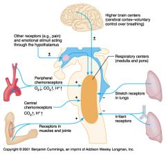
Chemoreptors |
Taste and Smell receptors stimulated by chemicals |
|
|
|
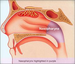
Nasophrynx |
Where the nose and mouth are connected. |
|
|
|
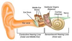
Conduction of deafness |
transmission of nerve waves through the middle ear to the oval window is impaired. |
|
|
|
Nerve Deafness |
transmission of nerve impulses from the cochlea to the auditory complex of the brain is impaired. |
|
|
|
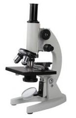
Condensor Lens/Compound Lens |
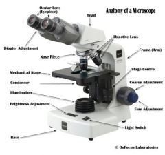
The lens on a microscope that focuses light onto a specimen. |

|
|
|
Base |

The Bottom of the microscope that provides steady support for the weight of the microscope.The light source is part of a base |

|
|
|
Microscope Stand/Arm |

Supports the observation tube, eyepieces, and objectives. When you pick up your microscope, grasp the stand holding the microscope upright, and support it underneath the base with your free hand. |

|
|
|
Ocular lens |

aka 'eye piece', where you look to observe the specimen. It increases the size of the specimen by 10x. The ocular lenses are not attached to the ocular tube so the microscope must be kept upright at all times. |

|
|
|
Nosepiece |

The revolving part to which three objectives are attached. It must be completely "clicked" into position when the objective is changed. |

|
|
|
Objective Lenceses |

Objective lenses should always be used in order of the lwst to the hghst magnification; each magnifies the specimen by the factor marked on the particular lens. Typically, the scanning objective has a signification of 4x, the low power objective has 10x, and the high power is 45x. The total magnification of any specimen seen through a light microscope is a result of the ocular lens and the objective lens |

|
|
|
Stage |

Supports the slide that is held onto it by stage clips and has a hole so that light can shine up through the specimen. |

|
|
|
Condensor |

The lens that focuses the light source from under the stage. |

|
|
|
Condensor Adjustment Knob |

on the right side of the stage, it raises or lowers the stage. |

|
|
|
Iris Diaphragm Lever |

Opens and closes the iris diaphragm in the condenser, regulating the amount of light that comes through. |
|
|
|
Coarse Adjustment Knob |

The lrgr of two knobs located on each side of the microscope arm, it moves the nosepiece up and down for focusing on the slide. |

|
|
|
Fine Adjustment Knob |

This is the smaller knob attchd to the coarse adjustment knob, this refines the focus of the high power objective. |

|
|
|
Depth of Field |
Range of distance. ex: Near vs Far |
|
|
|
Magnification |
To increase the size of a specimen |
|
|
|
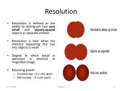
Resolution |
To adjust the microscope to be able to distinguish objects as sperate |
|
|
|
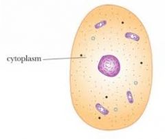
Cytoplasam |
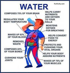
Watery part of the cell. It makes up 60% of the cell. *Water is the most common in our body, for every 100 molecules, 99 of them are water* |
|
|
|
Fun Facts about water |
1) Water is an active molecule. It has a lot of internal Kinetic energy. 2) Water can easily become a gas from a liquid state and evaporate without any problems. 3) Water is POLAR; One end is positive and the other end is negative. This helps water conduct electricity and disloves stuff (it is the best solvent) |
4) Water molecules are so small they can slip through membranes/ 5) Water molecules diffuse away from other molecules which keeps the concentration equal throughout the the cell. |
|
|
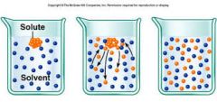
Solutes |
stuff that dissolves in the solute |
|
|
|
Solution |
When the solute dissolves in the solvent it becomes a solution. solute+solvent= solution |

|
|
|
Ions |
ion is a charged atom or molecule. It is charged because the number of electrons do not equal the number of protons in the atom or molecule.
|
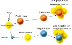
Sometimes solutes dissolve into ions like sodium chloride. Polar molecules don't ionize. But they still have their areas of charge. |
|
|
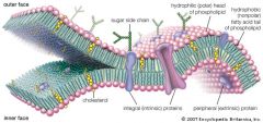
Membrane |
Boundary of cell. They are Selective Permeable (semi-permeable) |
|
|
|
Permeate |
The ability to pass into and out of the cell. |
|
|
|
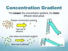
Cocentration Gradient |
The difference of any type of molecule on either side of a solution. *The gradient is what makes the molecules diffuse. |
|
|
|
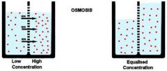
Osmosis |
To reach equilibrium, the cocentration graident follows the flow of WATER; Diffusion of water across a selectively permeable membrane. high to low |
|
|
|
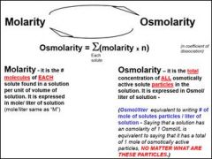
Osmolarity |
Describes the total number of dissolved particles in the solution. *Can be used to determine the flow of water* 1 osmol= 1 mol of solute particles per liter of solution |
|
|
|
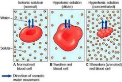
Isotonic Cell |
if the cell has the same osmolarity as the extracelluar enviroment. Water solutes won't move and volume won't change. |

|
|
|
Hypertonic |
If the extracelluar fluid has a higher osmolarity than the cell. Water will flow out of the cell and the cell will shrivel and die. ***CRENATE*** |

|
|
|
Hypotonic |
When the osmolarity of the cell is lower than the cell, more water and fluids will rush into the cell causing it to swell and BUSRT TO DEATH. ***LYSE*** |
|
|
|
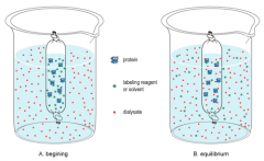
Dialysis |
A process to separate smller molecules from lrgr molecules in a solution. OR Flitering of blood in kidneys |
|
|
|
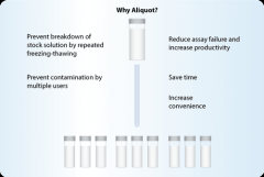
Aliqout |
Sample portion of a solution/ one full squeeze out of a plastic pipette. |
|
|
|
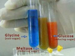
Benedict's Reagent |
Blue, but turns green/orange when it reacts to glucose |
|
|
|
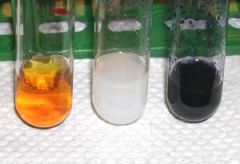
Iodine Reagent |
Dark brown, but turns dark purple when it reacts with Starch |
|
|
|
Acidity |
The concentration of hydrogen ions that are free in the solution (not bound to any molecule) |

|
|
|
Acids |
molecules that release hydrogen ions in a solution. (Water, lemons, etc.) |
|
|
|
Base |
Molecules that accept free hydrogen ions and form charge attractions. (arm and hammer, drano, etc.) |

|
|
|
Fun Fact about Hydrogen Ions |
They're are small and energetic, which helps them asscociate with other molecules easily. They can change other molecules reactivtiy and function. |
|
|
|
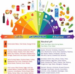
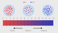
pH Scale |
pH= potency of hydrogen. Used to measure the acidity of something.0-14, 7 is neutral. 0-6=acid 8-14=base *The scale represents a 10 fold increase in ion concentration acidity each #* |

lower the number, the more acidic it is. |
|
|
Equation for acidity |
pH=-log[H+] *Donates h |
|
|
|
Equation for Base 1 |
pOH=-log[oH-] poH=0.3 |
|
|
|
Equation for BAse 2 |
pOH + pH = 14 0.3 +pH =14 |
|
|
|
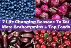
Anthocyanin |
Water souluble pigment that gives vegtables and flowers a pink, purple, or blue colors. pH 1-6 = Red Acid pH 7 = Purple pH 1-6= Green/blue Base pH 11-14= Green Yellow Base |
|
|
|
Buffer |
A substance that can reversibly bind free hydrogen ions. They can resist pH differences--even when bases and acids are added. |
|
|
|
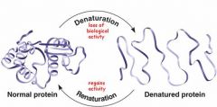
Free hydrogen ions can____ |
effect the folding of protein polypeptide chain. *Temp and ion concentrations can also effect the folding of polypeptide chains* |
|
|
|
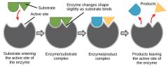
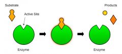
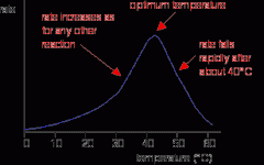
Enzymes |
specailized proteins that catalyze biochemical reactions that are vital to metabolism. ex: turning a substrate into a product. |
|
|
|
Enymes___ |
1) lwr the activation energy for a reaction 2) R not consumed by the reactions they catalyze 3) Do NOT alter the equilibruim of their reactions. |

|
|
|
A major source of _1__ in the body is __2__ |
1) Acid 2) Carbon Dioxide |
|
|
|
Affinity |
used to debscribe the strength of the bond at the active site. |
|
|
|
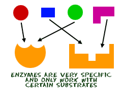
Substrate Specificity |
The 3 dimmensional shape of the active site determines the what ligands can bind to it. This prevents the wrong ligand from binding (most of the time) *regulate enzmye activity* |
|
|
|
Feedback Loops |
When an enzyme is deactivated by the concentration of substate or product. |
|
|
|
Denature |
When large shifts in pH or temperture in the celluar enviroment disable the change caused by the enzyme. |
|
|
|
Rate of an enzyme is determined by __1__ and __2__ |
Concentration of 1) enzyme 2) substrate |
|
|
|
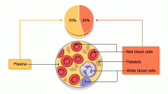
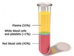
Blood is made up of |
Formed elements: 1) Cells 2) Platelets |
|
|
|
Plasma |
Liquid portion of blood |
|
|
|
Blood Plasma |
Composed of water, protiens ions, and antibodies. |

|
|
|
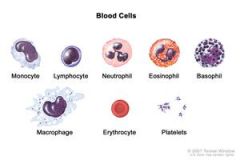
Formed Elements |
The cellular structures of blood. These are: 1) Thrombocytes (platelets) 2) Erythrocytes (red blood cells) 3) Leukocytes (White blood cells) |
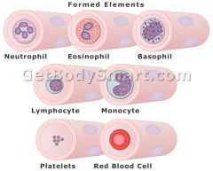
*All formed blood cells are made in the bone marrow* |
|
|
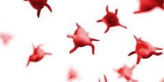
Platelets |
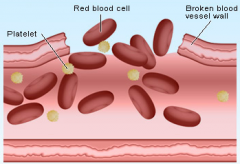
Broken off pieces of lrg cells, they clot injuries (along w/ proteins). |
|
|
|
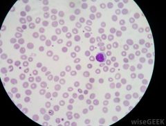
Red Blood Cells |
Most numerous formed elements. They: 1) Carry respiratory gasses on hemoglobin from the lungs to other tissues. |
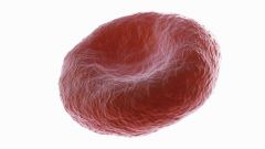
*non-nucleated= cannot replicate* |
|
|
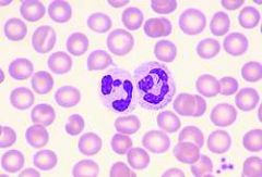
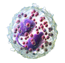
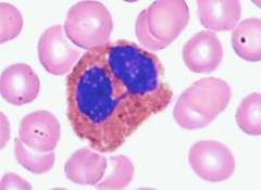
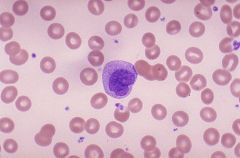
White Blood Cells |
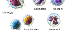
~Part of the bodies immune system~ they fight off infections. They're two groups: 1) Granular (speckled) 2) Agranular (not speckled) |
|
|
|
Neutrophils |

White Blood Cell Granular Most Common Phagocytic (eating) Eats invadors and destroy them |
|
|
|
Eosinophils |
White Blood Cell Granular Rare Parasite hunters |
|
|
|
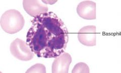
Basophil |
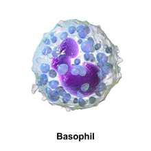
White Blood Cell Granular Rare Allergic Reactions |
|
|
|
Monocytes |
White Blood Cell Agranular The Tony Soprano waste mangement system of the body, they eat everything. Once they leave the circulatory system, they are called macrophages. |
|
|
|
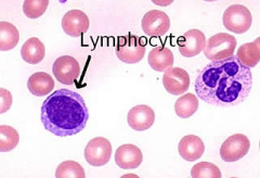
Lymphocyte |
White Blood Cell Agranular Many types and many jobs. |
|
|
|
Naive Lymphocyte |
T-Cells B-Cells *Two of Each* |

|
|
|
Cytotoxic T-Cells |
White Blood Cell Agranular Cytotoxic T-lymphocyte Kill virally infected Cells |
|
|
|
Helper T-Cells |
White Blood Cell Agranular Helper T-lymphocyte Activate and rev up macrophages and B-cells |

|
|
|
Plasma B-Cells |
White Blood Cell Agranular Plasma B-Lymphocytes Responsible for creating antibodies to foreign invaders |

|
|
|
Memory B-Cells |
White Blood Cell
Agranular Memory B-Lymphocytes Remember past pathogens so you don't get sick from them again. |

|
|
|
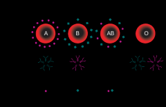
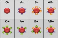
Blood Types |
*Blood Types are based on the protein/antigens of the blood cell* A B AB O |
|
|
|
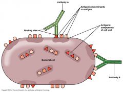
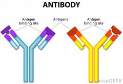
Antigens |
Two Types: A B |
|
|
|
Antibodies
|
They bind to antigens on the blood cell. Two Types: A B |
|
|
|
Aggulation |
When the wrong antibodies bind with the wrong antigens. Usually fatal because it traps the respiratory gasses in the cell. Ex: ANTI-A antibodies BIND to A antigens |
|
|
|
Type A |
Antigen- A antigen Antibodies- B antibodies Can receive from- A,O Can donate to- A, AB |

|
|
|
Type B |
Antigen- B antigen Antibody- A antibodies Can receive from- B, O Can donate to- B, AB |

|
|
|
Type AB |
Antigen- A and B antigens Antibody- No antibodies Can receive from- AB, B, A, O Can donate to- AB *Universal recipient* |

|
|
|
Type 0 |
Antigen- NAKED Antibody- A, B, AB Can receive from- 0 Can donate to- A, AB, O, B |
|
|
|
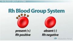
Rh Factor |
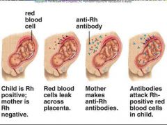
rHesus Factor 50 DEFINED GROUPS However what matters is that pregnant women can have babies that produce different factors that could be negative or positive and can kill mom;Hemolytuc disease of the newborn. |
|
|
|
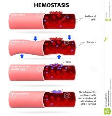
Hemostasis |
Natural form of aggulation; Also called blood coagulation or blood clotting. When you get cut, and your body heals yourself. |
|
|
|
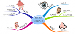
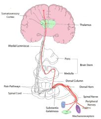
The senses |
Pain Tactile(touch) temperature (hot and cold) Chemical (smell and taste) Vision Hearing Proprioceptic/Kinesthetic-aware of body position Equlibrium-maintain our orientation in space |
|
|
|
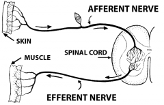
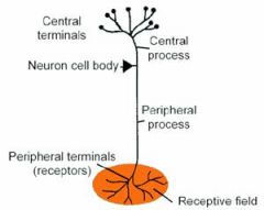
Afferent Nerves |
Help take knowledge from our enviroment to our central nervous system(CNS), which includes the brain or spinal cord. |

|
|
|
3 parts of the Afferent Nervous System: |
1) Sensory Receptors 2) Neural Pathways 3) CNS |
|
|
|
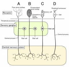
Sensory Receptprs |
Take a stimulus from the environment and convert it into a signal that can be passed on to the CNS. |
|
|
|
Neural Pathway |
carry the signal for the sensory information to the CNS |
|
|
|
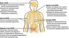
Central Nervous System |
will interpret the sensory information and will make a response if necessary. |
|
|
|
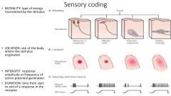
Modality |
Type of stimulus ex: light waves, photons |

|
|
|
Intensity |
strength of stimulus ex: how bright the light wave |

|
|
|
Location |
where the stimulus is occurring ex: in the eye |
|
|
|
Duration |
refers to how long the stimulus lasts ex: brief flash of light |

|
|
|
Sensory Unit |
One sensory afferent neurin and sensory receptor endings |
|
|
|
Receptive Field |
Area of the body that stimulates one sensory unit |

|
|
|
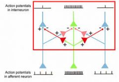
Lateral Inhibition |
most strongly activated signal pathway originating from the center of a stimulus area inhibits the less excited sensory pathways from adjacent sensory receptors by means of lateral inhibitory connections within sensory pathways reducing their response to the stimulus. |
|
|
|
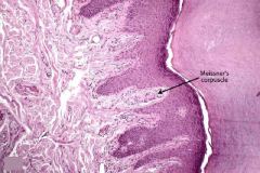
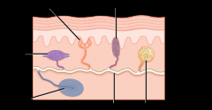
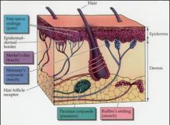
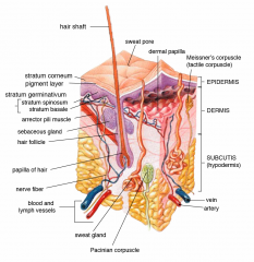
Meissner's Corpuscles |
Sensitive to soft touch. Tactile, light touch |
|
|
|
Merkel's Discs |
Located on the surface layers of our body. Tactile, touch |

|
|
|
Pacinian Corpuscle |
sensitive to deep pressure. They are more plentiful in the palm of our hands, soles of our feet, and in our joints. Deep pressure |
|
|
|
Free Nerve Ending |
pain |
|
|
|
Lamellated (Pacinian) Corpuscle |
corpuscle, deep pressure |
|
|
|
Ruffini Corpuscle |
warmth |

|
|
|
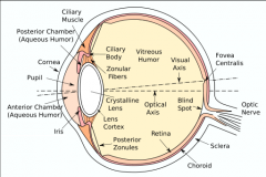
The eye |
a) fovea b) pupil c) lens d) optic discs *blood vessels cross the retina but the brain tells it to ignore it* |
|
|
|
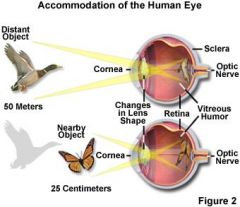
Accomendation |
The rounding of lens, it bends light rays to focus the image on the retina |
|
|
|
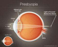
Presbyopia |
when someone ages and loses the elasticity in their eyes. (reducing near vision) |
|
|
|
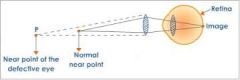
Near Point |
the closet an object can be focused on when the object |
|
|
|
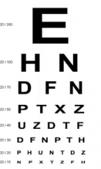
Visual Acuity |
Your measure of vision ability |
|
|
|
Emmetropia |
Good vision 20/20. Light converges on the lens to the back of the eye |

|
|
|
Myopia |
Nearsightedness, 20/50. The eyeball is too long and the light converges on the middle of the eye (in front of the retina) |

|
|
|
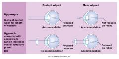
Hyperopia |
Farsightedness, 20/10. Eyeball is too short, so the light hits the back of the retina and doesn't converge at all. |
|
|
|
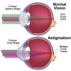
Astigmastism |
Vision is blurry because of an abnormal curvature of the lens. |
|
|
|
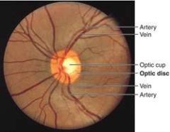
Optic Disc |
No photorecptors (rods or cones) in this area of the eye. Blood vessels and optic nerve converge. This creates a blind spot in your vision. |

|