Reading...
![]()
Play button
![]()
Play button
![]()
Use LEFT and RIGHT arrow keys to navigate between flashcards;
Use UP and DOWN arrow keys to flip the card;
H to show hint;
A reads text to speech;
113 Cards in this Set
- Front
- Back
|
What type of signals are transmitted by the anterolateral-system?
|
-Pain
-Thermal (warm/cold) -Touch (crude) -Pressure (crude) -Tickle and itch -Sexual sensations *slow |
|
|
What time of signals are transmitted by the dorsal column-medial lemniscal system?
|
-Touch (fine)
-Vibration -Skin movements -Joint proprioception -Pressure (fine) *fast |
|
|
C1-C3
|
- Sternocleidomastoid
- Upper traps - 1/3 of diaphram - Neck movement - Shoulder Shrug - Weak Breathing |
|
|
C4
|
- 2/3 Diaphragm
- Levator scapulae - Abdominal breathing |
|
|
C5
|
- Rhomboids
- Serratus Anterior - Deltoid - Rotator cudd - Pectoralis major - Biceps brachii - Brachioradialis - Movement of arm (humerus) at shoulder and shoulder movement - Bend Elbow (flexion of elbow) |
|
|
C6-C7
|
- Pectoralis major
- Latissumus dorsi - Extensor carpi radialis and ulnaris - Triceps - Bend wrist (extension/flexion) - Straighten the elbow (extension) |
|
|
C8
|
- Intrinsic hand muscles
- Bend fingers (Finger extension/flexion) - Fine motor control of hand |
|
|
T1
|
- Intrinsic hand muscles
- Spreads fingers apart and brings back together (abduction and adduction) -Fine motor control of hand |
|
|
T1-T12
|
- Some chest wall
- Abdominal muscles - Balance - Trunk stability and movement |
|
|
L1-L5
|
-Hip flexors
- Hip adductors - Some quadriceps (L3) - Tibialis Anterior (L4) - Toe musculature (L5) - Bends hip, lifts knee (Hip flexion) - Lateral leg raise (Hip abduction/adduction) - Straightens leg (L3, Knee extension) - Pulls foot up (L4, Dorsiflexion) - Wiggling toes (L5) |
|
|
S1-S5
|
- Gastroc/soleus (S1)
- Bladder, bowl, sex organs - Anal and Pelvic muscles - Point foot down (S1, plantarflexion) - Bladder, pelvic floor, external anal sphincter, external urethral sphincter |
|
|
primary structural differences between the skull of a newborn and adult
ossification model(s) during development of skull |
calvaria develops via intramembranous ossification, most bones of cranial base develop via endochondral ossification
|
|
|
primary structural differences between the skull of a newborn and adult
relative size of skull (compared to body) |
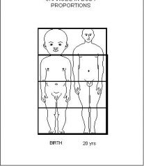
• head height of newborn is ~ 1/4 of entire body; adult head height is ~ 1/8 of body height
• in other words, babies have huge heads relative to the rest of their body! |
|
|
primary structural differences between the skull of a newborn and adult
relative proportions of skull |
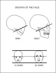
• frontal and parietal eminences are much more notable in newborn skull
• mastoid process is absent at birth; develops during year 1 as sternocleidomastoid strengthens with head movement (Wolff’s law!) • newborn facial skeleton is small compared to calvaria (1/8 of cranium compared to adult facial skeleton = 1/3 of cranium) |
|
|
primary structural differences between the skull of a newborn and adult
sutures |
• frontal suture: present in newborn, typically fused by 8th year (remains in ~8% of adults, called metopic suture)
• mandibular symphysis: fused by end of 2nd year |
|
|
primary structural differences between the skull of a newborn and adult
fontanelles |
• anterior fontanelle: largest, diamond shaped, bordered by frontal and parietal bones, at junction of sagittal, coronal, and frontal sutures (bregma); useful for monitoring growth of calvaria, hydration status, and intracranial pressure
• posterior fontanelle: triangular shape, bordered by parietal and occipital bones, at junction of lambdoidal and sagittal sutures (lambda) • sphenoidal and mastoid fontanelle: lateral aspect of skull, deep to temporalis muscle, small relative to midline fontanelles, and less clinically relevant |
|
|
Which nerves pass through the following areas of the neurocranium (brain case)? Please include the name & number assigned to the cranial nerve.
Supraorbital foramen |
Supraorbital n. (from ophthalmic branch of trigeminal CN V1)
|
|
|
Which nerves pass through the following areas of the neurocranium (brain case)? Please include the name & number assigned to the cranial nerve.
Foramen magnum |
Accessory CN XI (+ spinal cord)
|
|
|
Which nerves pass through the following areas of the neurocranium (brain case)? Please include the name & number assigned to the cranial nerve.
Hypoglossal canal |
Hypoglossal CN XII
|
|
|
Which nerves pass through the following areas of the neurocranium (brain case)? Please include the name & number assigned to the cranial nerve.
Superior orbital fissure |
Ophthalmic CN V1 (from trigeminal), trochlear CNIV, abducent CN VI, oculomotor CNIII.
|
|
|
Which nerves pass through the following areas of the neurocranium (brain case)? Please include the name & number assigned to the cranial nerve.
Foramen ovale |
Mandibular CN V3(from trigeminal)
|
|
|
Which nerves pass through the following areas of the neurocranium (brain case)? Please include the name & number assigned to the cranial nerve.
Foramen lacerum |
no cranial nerves
|
|
|
Which nerves pass through the following areas of the neurocranium (brain case)? Please include the name & number assigned to the cranial nerve.
Foramen rotundum |
Maxillary CN V2 (from trigeminal)
|
|
|
Which nerves pass through the following areas of the neurocranium (brain case)? Please include the name & number assigned to the cranial nerve.
Foramen spinosum |
Meningeal branch (from mandibular CN V3)
|
|
|
Which nerves pass through the following areas of the neurocranium (brain case)? Please include the name & number assigned to the cranial nerve.
Stylomastoid foramen |
Facial CN VII
|
|
|
anterior communicating
|
Connects
anterior cerebral arteries |
|
|
Which cranial bone permits the oculomotor, trochlear, abducent and ophthalmic branch of the trigeminal nerve to exit the cranium?
A. Maxilla B. Temporal C. Ethmoid D. Zygomatic E. None of the above |
E. None of the above
|
|
|
Which of the following muscles have fibers oriented in the superolateral direction (fibers travel up, as they move laterally
“up and out”)? A. Buccinator B. Zygomaticus major C. Corrugator supercilii D. All of the above E. BothBandC |
E. Zygomaticus major and Corrugator supercillii
|
|
|
Which of the following bones are associated with the nasal cavity (ie: make up the walls or septum)?
a) Palatine b) Vomer c) Ethmoid d) All of the above e) BothBandC |
Answer: d) All of the above
|
|
|
palatine
|
- posterior portion of hard palate
- forms wall of nasal cavity - forms part of the floor and lateral wall of the nasal cavity - It also forms a small portion of the floor of the orbit |
|
|
Which of the following bones are paired in an adult?
a) Palatine b) Sphenoid c) Mandible d) None of the above e) BothAandB |
Answer: a) Palatine
|
|
|
paranasal sinuses can be found in which bones?
|
• Cavity lined with mucosa
• Helps to warm and humidify inspired air • Reduce weight of skull • Enhance voice resonance • Born with ethmoid sinuses only |
|
|
hyoid bone
|
• What is unique about the hyoid bone?
• Only bone that does not articulate with any other bone! |
|
|
Most common development defect of the skull?
|
cleft palate
|
|
|
In Guatemala, there are a great number of cleft lips and cleft palates. At what stage of development does this defect occur and what fails to occur?
|
At 9 to 10 weeks in-utero the skin fuses between the nasal septum and the palatine arches. Lack of fusion of this overlying tissue results in clefting – either
cleft lip, unilateral cleft palate and/or lip, or bilateral cleft palate and/or lip |
|
|
Orbicularis oris:
Location/Direction? Action? |
Location/Direction:
Circular fibers around mouth and within lips. Action: Close mouth and purse lips. |
|
|
Mentalis:
|
Location/Direction?
Skin of chin to mandible with vertical fibers. Action? Raise the skin of the chin superiorly; expression of doubt or pouting |
|
|
Buccinator:
|
Location/Direction?
Horizontal fibers; rectangular muscle from alveolar process on maxilla and mandible laterally to the obicularis oris muscle medially. Action? Smiling, chewing (pressing cheeks against teeth). |
|
|
Depressor Anguli Oris:
|
Location/Direction?
Vertical fibers from angle of mouth to inferior border of the mandible. Action? Depresses the angle of the mouth. |
|
|
Zygomaticus major:
|
Location/Direction?
Zygomatic bone to the angle of the mouth. Action? Draws angle of the mouth superolaterally during smiling and laughing. |
|
|
Platysma:
|
Location/Direction?
Broad and thin muscle in the neck, from the fascia of the pectoralis and deltoid muscles to the muscles on the inferior border of mandible. Action? Tenses skin of neck, draws the corners of the mouth down to grimace. |
|
|
Orbicularis oculi:
|
Location/Direction?
Circular muscle surrounding the orbit. Orbital and palpebral (eye lid) components. Action? Palpebral part: close eye lids gently (blinking); Orbital part: strongly closes lids (squinting). |
|
|
Corrugator Supercilii:
|
Location/Direction?
Medial part of the orbital orbicularis oculi to the skin of the eyebrows. Fibers are superiolateral. Action? Draws the medial portion of the eyebrow downward, demonstrating concern. |
|
|
Nasalis:
|
Location/Direction?
Fibers travel from maxilla and nasal cartilage to the aponeurosis of the nose. Action? Part of the muscle widens nostrils, and another part compresses. |
|
|
Frontalis (or frontal portion of
occipitofrontalis or epicranius): |
Location/Direction?
Vertically aligned fibers from the skin of the eyebrows to the epicranial aponeurosis. Action? Elevates the eyebrows, demonstrating surprise. |
|
|
Temporomandibular Joint (TMJ): type of joint? movement? is it stable?
|
• Modified hinge-‐synovial type joint
• Movements? • Flexion/Extension “Elevation/depression” • Protrusion/Retrusion • Lateral translation • Pivot/rotation • Mobile or Stabile? • Very mobile, not too stable…is it? • posterior thickening of joint capsule • anterior dislocation is more common. |
|
|
Which muscle of mastication is NOT
involved with elevation of the mandible? A. Masseter B. Temporalis C. Lateral pterygoid D. Medial pterygoid E. All of the above contribute to elevation of the mandible |
C. Lateral pterygoid
|
|
|
Temporalis:
|
Location/Direction?
Vertically aligned fibers fanning out from the coronoid process of the mandible to the temporal fossa of the parietal bone. Action? Elevates the mandible (closes the jaw), and retracts the mandible. |
|
|
Masseter:
|
Location/Direction?
Superiomedial fibers from lateral surface of ramus of the mandible to zygomatic arch. Action? Elevates (closes jaw) and protrudes mandible. |
|
|
Parietal Bone
|
- Coronal suture
- Squamosal suture - What suture separates the two parietal bones? - Sagittal suture |
|
|
Stylomastoid foramen
|
Facial CN VII
|
|
|
Cribiform plates
|
Olfactory (CN I) axons
|
|
|
Mental foramen
|
Mental n. (from inferior alveolar n., branch from CN V3)
|
|
|
Mandibular foramen
|
Inferior alveolar n. (from mandibular CN V3)
|
|
|
Jugular foramen
|
Accessory CN XI, vagus CN X, glossopharyngeal CN IX
|
|
|
Infraorbital foramen
|
Infraorbital n. (from maxiallary CN V2)
|
|
|
Cranial nerves that originate in the cerebrum
|
-olfactory
-optic |
|
|
Cranial nerves that originate in the midbrain
|
occulomotor, trochlear
|
|
|
cranial nerves originating on pons
|
trigeminal, abducent, facial, vestibulocochlear
|
|
|
cranial nerves originating on the medulla
|
glossopharyngeal, vagus, accessory, hypoglossal, vestibulocochlear (both portions)
|
|
|
Which cranial nerves are considered special sensory?
|
Olfactory CN I, Optic CN II, and Vestibulocochlear CN VIIII
|
|
|
Do any cranial nerves contain sympathetic axons?
|
No
|
|
|
What is the basic function of the olfactory nerve?
|
smell
|
|
|
What is the basic function of the optic nerve?
|
vision
|
|
|
What is the basic function of the occulomotor nerve?
|
eye movement, pupil response to light, raising the eyelid
|
|
|
What is the basic function of the trigeminal?
|
facial sensation, chewing (muscles of mastication
|
|
|
What is the basic function of the facial nerve?
|
muscles of facial expression, taste (anterior 2/3 of tongue), saliva and tear production
|
|
|
What is the basic function of the vestibulocochlear?
|
hearing and balance
|
|
|
What is the basic function of the glossopharyngeal
|
swallowing, gag reflex, speech, taste (posterior 1/3 of tongue), saliva production
|
|
|
What is the basic function of the vagus nerve?
|
muscle of internal organes and swallowing, gag reflex, speech
|
|
|
What is the basic function of the accessory nerve?
|
turn neck and shrug shoulders (sternocleidomastoid and trapezius)
|
|
|
What is the basic function of the hypoglossal nerve?
|
tongue movement
|
|
|
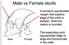
Sex Identification of the Skull
|
- chin (square or rounded)
- supraorbital margin (sharp or dull) - supercillary arches/supraorbital ridge (prominent or not?) - external occipital protuberance (prominent or not?) - mastoid process - angle of mandible - zygomatic bone |
|
|
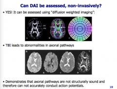
What gives structural support to an axon?
|
Axon is not simply a hollow tube - neurofilaments and microtubles act as structural support and transporters
- TBI can result in structural damage to axons (forms bulb) |
|
|
Chronic Traumatic Encephalopathy (CTE)
Anatomical changes |
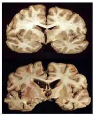
Atrophy: Overall decreased brain mass
- medial temporal lobes - frontal loves - corpus callosum - mammilary bodies - thalamus - hypothalamic floor - hippocampus - amygdala - substantia nigra Increased: - lateral ventricles - third ventricles - cavum septum pellucidum - septal fenestrations |
|
|
Can DAI be assessed, non-invasively?
|
|
|
|
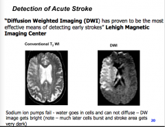
Detection of Acute Stroke
|
|
|
|
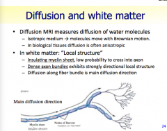
Diffusion and white matter
|
|
|
|
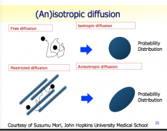
Isotropic diffusion
|
|
|
|
Midbrain sits beteen which two structures? Connects which ventricles?
|
- diencephalon and pons
- 3rd and 4th |
|
|
Midbrain structures?
|
- cerebral peduncles
-->ascending and descending neural tracts -->convey impulses: cerebral cortex to the spinal cord and reverse - corpora quadrigemina -->four rounded eminences -->dorsal portion of the midbrain -->posterior to the cerebral aqueduct |
|
|
Corpora quadrigemina
|
superior colliculi
- reflex center for movement of the eyeballs and head in response to visual and other stimuli - coordinate movements for visual traching inferior colliculi - reflex centers for movements of the head and trunk in response to auditory stimuli - e.g. --turning your head to the source of a sudden sound - What does the relatively large superior colliculi on the sheep brain suggest? |
|
|
Primary function of the Pons? Structures?
|
- relay station: cerebrum to cerebellum (medulla to thalamus)
- latin for bridge - nuclei involved with many functions -->sleep, breathing, hearing and equilibrium middle cerebellar peduncles - white matter tracts connecting the pons with the cerebellum |
|
|
Medulla Oblongata
|
pyramids
- ridges on the ventral surface containing tracts from the cerebral cortex olives - nuclei which relay information from the spinal cord, cerebral cortex diencephalon and brain stem to the cerebellum - autonomic reflex centers - cardiovascular center - cardiac center adjusts force and rate of heart contraction - vasomotor center adjusts blood vessel diameter for blood pressure regulation - respiratory centers - generate respiratory rhythm - control rate and depth of breathing, with pontine centers - ADDITIONAL CENTERS regulate - vomit - hiccup - swallow - cough - sneeze |
|
|
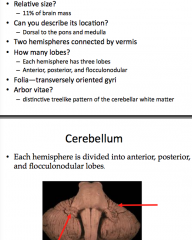
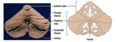
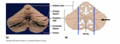
Anatomy of the Cerebellum
|
|
|
|
The outer cortex of gray matter present in the cerebellum is composed primary of: a) myelinated neuron fibers b) connective tissue c) simple cuboidal epithelium d) neuron cell bodies
|
d) neuron cell bodies
|
|
|
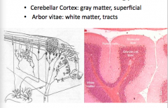
Cerebellum: histology
|
|
|
|
What structures do the superior, middle and inferior cerebellar peduncles connect with the cerebellum?
|
superior
- midbrain, diencephalon, cerebrum middle - pons inferior - medulla oblongata and spinal cord -->fibers in the cerebellum are ipsilateral |
|
|
Function of the cerebellum
|
subconcious, timing and pattern of muscle action "coordination"
|
|
|
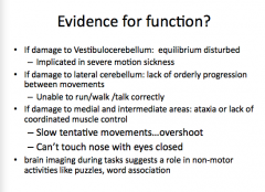
Vestibulocerebellum: flocculonodular lobe
|
- receives input from the equilibrium appatatus and visual pathways
- control posture to maintain balance |
|
|
Medial and intermediate parts of each cerebellar hemisphere function
|
- contain overlapping sensory and motor maps
- one in anterior and two in posterior - each map receives ipsilateral information - ascending sensory (proprioceptive) info from muscles (stretch and joints/tendon) - motor information from descending corticospinal axons; relayed from brainstem and nuclei - congruent map function: position of muscles and the commands going to them (sensory and motor) - overlap of sensory and motor maps acts as a correction device - keep from over or undershooting ie touch nose |
|
|
Lateral parts of hemispheres
|
plan the sequence and timing of complex movements
- receives information from both motor association (premotor, intent to perform action) and somatocensory association areas of the cerebral cortex |
|
|
Evidence for function of cerebellum?
|
|
|
|
The terminal end of the spinal cord is called the __________?
|
conus medullaris
NOT filum terminale |
|
|
Protection of spinal cord
|
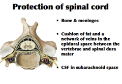
|
|
|
Cerebral Spinal Fluid
|
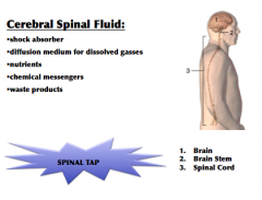
|
|
|
Epidural and lumbar puncture
|

|
|
|
How many pairs of spinal nerves?
|
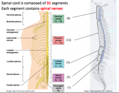
|
|
|
Gray Matter
|
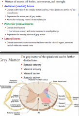
-neuron cell bodies, interneurons, neuroglia
*contain autonomic neurons that innervate the visceral organs |
|
|
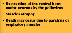
Poliomyelitis
|
|
|
|
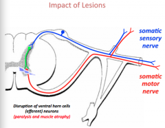
Impact of Lesions
|
|
|
|
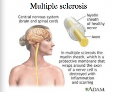
Multiple sclerosis
|
|
|
|
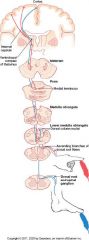
White matter
|
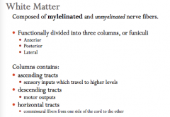
The names of the tracts indicate:
- the white column column, or funiculi, in which the tract travels - where the cell bodies of tract terminate - the direction of the impulse conduction within the tract - e.g. - anterior spinothalmic tract - anterior- anterior funiculi; spino - originates in the spinal cord; thalmic- terminates in the thalamus |
|
|
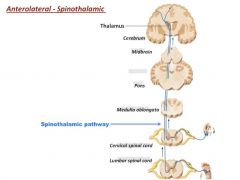
Functions of the anterolateral system
|
to relay information related to thermal (warm/cold) and crude touch or pressure to higher brain centers
|
|
|
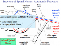
Structure of Spinal Nerves: Autonomic Pathways
|
|
|
|
Autonomic Nervous System (ANS)
|
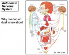
ANS consits of motor neurons that:
- innervate smooth and cardiac muscle and glands - make adjustments to ensure optimal support for body activities - operate via subconcious control - have viscera as most of their effectors |
|
|
Divisions of the ANS
|
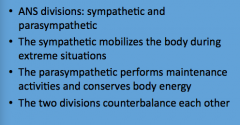
|
|
|
Anatomy of ANS
|
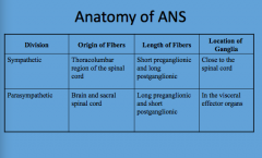
|
|
|
Parasympathetic Division Outflow
|
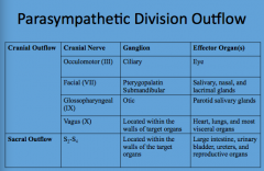
|
|
|
Parasympathetic Division
|
|
|
|
Sympathetic Division
|
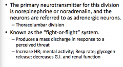
|
|
|
Sympathetic Pathway
|
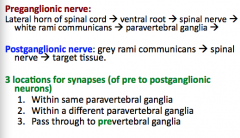
|
|
|
Male vs Female skulls
|
|