Reading...
![]()
Play button
![]()
Play button
![]()
Use LEFT and RIGHT arrow keys to navigate between flashcards;
Use UP and DOWN arrow keys to flip the card;
H to show hint;
A reads text to speech;
147 Cards in this Set
- Front
- Back
|
Which plane divides the body into top and bottom? Right and left? Front and back?
|
- Transverse/horizontal/axial
- Sagittal (median/mid-sagittal) - Frontal/coronal |
|
|
If you have torn both your right and left anterior cruciate ligaments, it is a _______ injury.
a) unilateral b. bilateral |
b. bilateral
|
|
|
An "extra bone besides the navicular bone is called...
|
An accessory bone
|
|
|
What is a sesamoid bone? Function?
|
Bone found witin a tendon
Functions in protection and mechanical advantage |
|
|
Bone formed directly from embryonic connective tissue is known as ______.
|
Mesenchyme
|
|
|
Is long bone growth endochondral or intramembranous ossification?
|
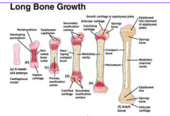
Endochondral
|
|
|
Functions of spongy bone
|
- Lightens bone
- Resists forces from many angles - 3-D branching lattice, aligned with the direction of mechanical stress - High surface area - High metabolic activity (8x that of cortical bone) - creates cavities to contain bone marrow - Ends of long bones - Flat bones |
|
|
Location of Hematopoietic Tissue aka Red Marrow
|
- Red marrow cavities of adults
- Trabecular cavities of the heads of the femur and humerus - Trabecular cavities of flat bones - Red marrow of newborn infants - Medullary cavities and all spaces in spongy bone |
|
|
What are the typical symptoms of a fractured bone? What produces the painful symptoms?
|
- Pain, nausea, swelling, deformation, discoloration
- Nerve endings in the periosteum |
|
|
What do Vessel and Nerves Innervate through?
|
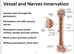
Periosteum
|
|
|
Periosteum function and description
|
(2 layers)
- Outer fibrous layer - Inner osteogenic layer - Osteoblasts (bone-forming cells) - Osteoclasts (bone-destroying cells) - Osteogenic cells (stem cells) - Nerve fibers, nutrient blood vessels, and lymphatic vessels enter the bone via nutrient foramina - Secured to underlying bone by Sharpey's fibers |
|
|
Endosteum
|
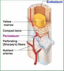
- Delicate membrane on internal surfaces of bone
- Also contains osteoblasts and osteoclasts |
|
|
Which of the following are examples of a cartilaginous joint?
a. The epiphyseal plate in a long bone b. Intervertebral discs in the vertebral column c. The sutures between the bones of the skull d. All of the above e. Only A and B |
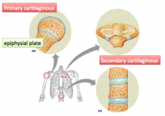
e. Only A and B
|
|
|
What type of cartilage forms cartilaginous joints?
|
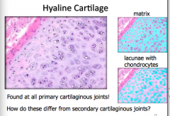
Hyaline Cartilage
|
|
|
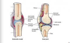
What are the general features of a synovial joint?
|
|
|
|
Which of the following accurately describes cardiac muscle?
a. multiple nuclei & striated b. striated, branching fibers with intercalated discs c. found in walls of blood vessels d. all of the above e. B and C only |
b. striated, branching fibers with intercalated discs
|
|
|
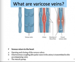
What are varicose veins?
|
|
|
|
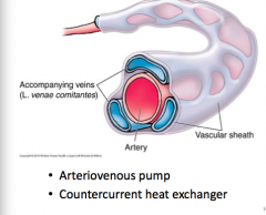
How is blood propelled?
|
|
|
|
What are the functions of capillaries?
|
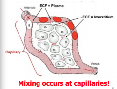
- Gas exchange
- Lungs - Tissue (brain, heart, skeletal muscle, etc..) - Deliver nutrients - Remove waste - Uptake from small intestine - Filtration at kidney |
|
|
What type of nerve fiber is typically found in the ventral root of the spinal cord?
a. Motor b. Sensory c. Both motor and sensory d. None of the above |
a. Motor
|
|
|
How are motor nerves related to muscle?
|
Then send a message from the brain signaling the muscle to contract
|
|
|
If there is damage to a motor nerve, what affect will it have on the muscle is serves?
|
The muscle will not receive the signal to contact; person is weaker doing movements involving that muscle
|
|
|
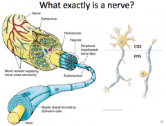
What exactly is a nerve?
|
|
|
|
What are the 4 primary tissue types?
|
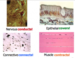
Nervous
Epithelial Connective Muscle |
|
|
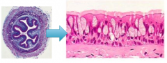
What is the general pattern of tissue arrangement of a tube shaped organ?
|
epithelium-->connective-->muscle-->connective
|
|
|
What structural characteristic of epithelia allow for unique functions?
|
- Cellularity: just cells, no extracellular stuff
- Specialized contacts: tight junctions, desmosomes - Polarity: apical and basal - Supported by connective tissue - Avascular, but innervated - Regeneration via cell division |
|
|
Maintaining the Integrity of the Epithelium
|
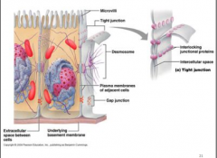
Intercellular connections
- Tight junctions: prevent molecules from passing through intracellular space - Desmosomes: bind cells and forms internal scaffolding, providing structural integrity |
|
|
Epithelia is avascular
|
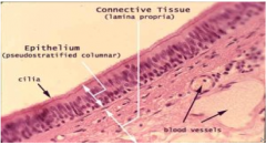
- No space for blood vessels
- blood vessels are loacted in the underlying connective tissue |
|
|
Polarity of epithelia
|
- Two sides with different properties
- Apical (or lumenal) side faces out (free) - Basal side supported by (anchored to) connective tissue via basement membrane |
|
|
Simple squamous epithelium
|
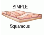
- thin, permeable
- located where rapid diffusion is required where can we find it? - kidney (glomeruli), lung (alveoli), capillaries - endothelium: inside of hollow tubes to reduce friction - mesothelium: lining the ventral body cavity and its organs |
|
|
This type of epithelial tissue is rare in the human body, but can be found in the male urethra, and functions in protection and secretion:
a) simple cuboidal epithelium b) simple columnar epithelium c) stratified columnar epithelium d) stratified squamous epithelium e) pseudostratified columnar epithelium |
c) stratified columnar epitheluim
|
|
|
Stratified Squamous Epitheluim
|
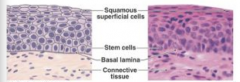
- regeneration from below
- major role is protection located: - mouth - esophagus - vagina |
|
|
Pseudostratified Columnar Epithelium
|
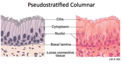
- varies in heights
- single layer - nuclei at different heights - secretes mucus Function: - nasal cavity - trachea - bronchi |
|
|
Endocrine vs exocrine secretion
|
exocrine: releases into a lumen; can be single cells or a multicellular gland
endocrine: ductless, secretes into blood |
|
|
4 classes of connective tissue
|
PROPER
- Loose - Dense BONE - compact - spongy CARTILAGE - hyaline - elastic - fibrocartilage BLOOD |
|
|
Major Functions of Connective Tissue
|
- connects/binds tissue together
- support - protection - defense - repair - insulation - cushions - storage - transportation |
|
|
Origin of connective tissue
|
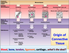
Mesenchyme
|
|
|
Ligaments are composed of very strong connective tissue, which consists of densely packed, parallel connective tissue fibers. When a ligament tears, it is difficult to repair both physiologically and surgically. WHY?
|
this type of tissue has relatively FEW CELLS and vascular supply is POOR
|
|
|
Characteristics of Connective Tissue
|
- mesenchyme as their common tissue of origin
- varying degrees of vascularity why is this relevant? - cells separated by nonliving extracellular matrix (ground substance and fibers) |
|
|
How do the basic components make diverse connective tissue? What if the tissues has more elastin than collagen? What if the tissue has more collagen than elastin? What if more gel component relative to fibrous proteins?
|
- To produce connective tissue with different properties, the basic components are present in different amounts
- Change the type of collagen present - Type I prevalent in tendons, ligaments and bones - Type II prevalent in cartilage - Type III in walls of hollow structures like blood vessels, intestine, uterus: reticular fibers....blend into type Iv - Type IV in basement membrane of epithelia (basal lamina + reticular lamina) |
|
|
Which type of dense connective tissue type often underlies transitional epithelia?
|
elastic
|
|
|
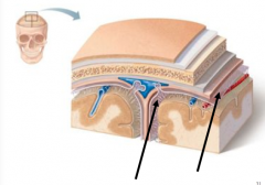
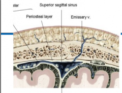
What are the two layers of the dura mater?
|
periosteal layer
meningeal layer |
|
|
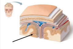
Falx cerebri
Function? Where is this structure found? |
- limit excessive movement of the brain
- longitudinal fissure between cerebral hemispheres - attaches to crista galli of ethmoid bone |
|
|
Tentorium cerebelli
Where is this structure found? |
transverse fissure between cerebrum and cerebellum
|
|
|
Arachnoid mater
|
- delicate, transparent middle layer, loosely covers the brain
- the subarachnoid space lies between the arachnoid and pia mater |
|
|
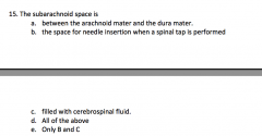
What is contained within the subarachnoid space?
|
cerebral spinal fluid (CSF)
|
|
|
Pia mater
|
- thin, connective tissue membrane
- adheres closely to the surface of the brain - this layer dips into the sulci and fissures |
|
|
In a typical, healthy individual, where do yo expect to find fluid filled space in the brain (as opposed to "potential spaces")?
|
Ventricles and subarachnoid space
|
|
|
Subdural space
|
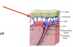
- very low volume
- considered a potential space - "cling" wrap and oil - similar to pleural linings and cavity (lung) |
|
|
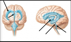
Where are the ventricles of the brain and how to they connect to each other?
|
|
|
|
Cerebral Spinal Fluid: Function?
|
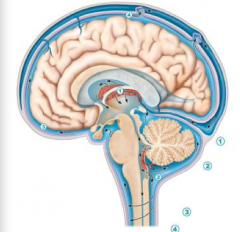
- forms liquid cushion around the brain and spinal cord
- serves as a shock absorber - circulates nutrients filtered from the blood - you can follow a drop of CSF from production to venous blood |
|
|
Where does CSF leave the ventricles to enter the subarachnoid space?
|
- via 2 lateral and 1 median aperture, in the 4th ventricle
- absorbed into superior sagittal sinus via the arachnoid villa |
|
|
What is the choroid plexus made up of? What volume of CSF is produced in a day?
|
- Clusters of capillaries in the ventricles
- 400-500mL (~16 oz) |
|
|
What structure is sensitive to pain, and can produce a headache?
|
When CSF is somewhat depleted the brain sags slightly, pulling on the dura mater. This is thought to cause pain and headache (just one possible cause)
|
|
|
Name the regions of the brain
|
1. Cerebral hemispheres (or cerebrum)
2. Diencephalon: epithalamus, thalamus, and hypothalamus 3. Brain stem: midbrain, pons, and medulla 4. Cerebellum |
|
|
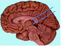
The cerebrum
|
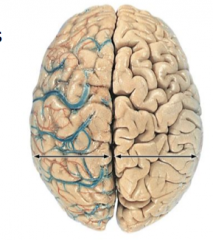
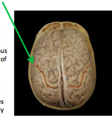
Longitudinal fissure
- separates the cerebral hemispheres L vs. R Hemispheres - Contralateral body control/sense - Have very different functions |
|
|
Gyri (gyrus)
|
elevated ridges
|
|
|
Sulci (sulcus)
|
shallow grooves
|
|
|
Lateral sulcus
|
separates frontal/parietal lobe from the temporal lobe
|
|
|
Central sulcus
|
separates frontal lobe from the parietal lobe
|
|
|
Precentral gyrus
|
primary motor area
responsible for conscious (voluntary) movement of skeletal muscle |
|
|
Postcentral gyrus
|
primary sensory area
receives impulses from the body's sensory receptors |
|
|
Gray matter
|
- 2-4 mm thick
- Neuron cell bodies and unmyelinated fibers |
|
|
White matter
|
- allows for communication over longer distance
- myelinated fiber bundles |
|
|
Lobes of Cerebrum
|
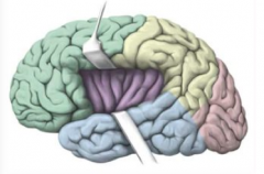
- Frontal
- Parietal - Occipital - Temporal - Insular |
|
|
Frontal
|
- Associated with general intellect
- Voluntary motor control - Emotional behavior - Speech output |
|
|
Parietal
|
- Associated with general sensory input and its interpretation
|
|
|
Occipital
|
- Associated with visual input and its interpretation
|
|
|
Temporal
|
- Associated with auditory, olfactory and gustatory (taste) sensory input and interpretation
- Memory - Speech interpretation (perception and recognition) |
|
|
Insula
|
- Gustatory
- Olfactory - Integrates information from visceral receptors - Integrates autonomic information |
|
|
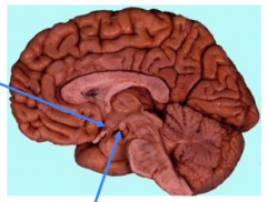
Diencephalon
|

- Located between the cerebral hemispheres, it forms the central core of the brain
- Composed of the thalamus, hypothalamus and epithalamus - Together these surround the third ventricle of the brain |
|
|
Thalamus
|
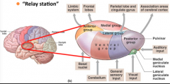
- Gateway to the cerebral cortex
- Sorting out and interpretation of AFFERENT impulses from all over the body - "relay station" |
|
|
Interthalamic adhesion
|
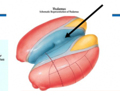
aka intermediate mass
|
|
|
Hypothalamus
|
- Homeostatic control center
Regulates - Emotion - Temperature - Appetite - Thirst - Sleep patterns - Endocrine system - releases dopamine, growth hormone-releasing hormone, gonadotropin-releasing hormone, etc...many of which stimulate pituitary |
|
|
Mammillary Bodies
|
- Part of limbic system (functional brain system)
- Collection of nuclei related to the - emotional response to odors - olfactory reflexes - memory - example: skunks smell bad |
|
|
Epithalamus
|
- Roof of the third ventricle: choroid plexus and pineal body
|
|
|
Pineal body
|
- Synthesizes a hormone involved in sleep-wake cycles: melatonin
- Related to onset of puberty |
|
|
Measurement of TBI Severity
|
- Length of consciousness (LOC)
- Length of post-traumatic amnesia (PTA) - Post-injury period of confusion with deficits in retaining new information and processing new memories; PTA ends when continuous (or near-continuous) memory resumes - Glasgow Coma Scale (GCS) - Focal Neurologic Deficits |
|
|
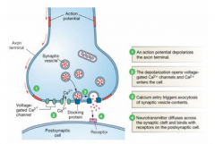
Cell-to-Cell Communication Occurs Via Synapses
|
|
|
|
Chronic Traumatic Encephalopathy (CTE)
|
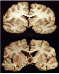
Atrophy: Overall decreased brain mass
- Medial temporal lobes - Frontal lobes - Corpus callosum - Mammillary bodies - Thalamus - Hypothalamic floor - Hippocampus - Amygdala - Substantia nigra Increased: - Lateral ventricles - Third ventricles - Cavum septum pellucidum - Septal fenestrations |
|
|
Cerebral metabolic and energetic effects of concussion
|
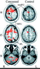
- CT scan showing small hemorrhages and cerebral edema
- Despite the lack of clear anatomical or structural damage following mTBI there is clear evidence of brain activation alterations - Damage to axons TBI can result in structural damage to axons |
|
|
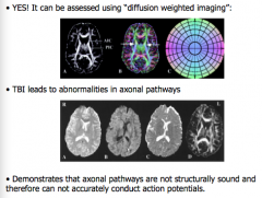
Can diffuse axonal injury (DAI) be assessed, non-invasively?
|
|
|
|
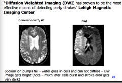
Detection of Acute Stroke
|
|
|
|
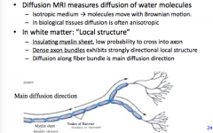
Diffusion and white matter
|
|
|
|
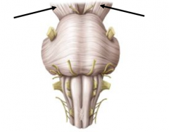
What structures make up the brain stem?
|
- midbrain
- pons - medulla oblongata |
|
|
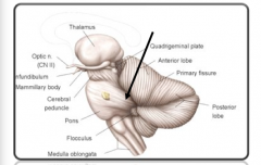
Midbrain
|
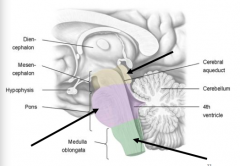
- sits between the diencephalon and pons
- contains the cerebral aqueduct which connects 3rd and 4th ventricle Structures: - cerebral peduncles - corpora quadrigemina |
|
|
Cerebral peduncles
|
- ascending and descending neural tracts
- CONVEY IMPULSES: cerebral cortex to the spinal cord and reverse |
|
|
Corpora Quadrigemina
|
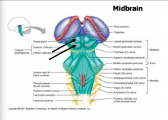
- four rounded eminences
- dorsal portion of the midbrain - posterior to the cerebral aqueduct |
|
|
Superior colliculi
|
- reflex center for movements of the eyeballs and head in response to visual and other stimuli
- coordinate movements for visual tracking |
|
|
Inferior colliculi
|
- reflex centers for movements of the head and trunk in response to auditory stimuli
- e.g. -- turning your head to the source of a sudden sound |
|
|
Superior cerebellar peduncle
|
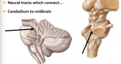
- neural tracts which connect cerebellum to midbrain
|
|
|
Pons
|
Function:
- relay station: cerebrum to cerebellum (medulla to thalamus) - latin for "bridge" - + nuclei involved with many functions - sleep, breathing, hearing, equilibrium Structures: - middle cerebellar peduncles - white matter tracts connecting the pons with the cerebellum |
|
|
Medulla Oblongata: located between what structures?
|

spinal cord and the pons
the medulla is a continuation of the spinal cord which forms the inferior part of the brain stem |
|
|
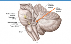
Structures of the Medulla Oblongata
|
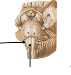
pyramids: ridges on the ventral surface containing tracts from the cerebral cortex
olives: nuclei which relay information from the spinal cord, cerebral cortex, diencephalon, and brain stem to the cerebellum inferior cerebellar peduncles: fiber tracts that connect medulla to cerebellum |
|
|
Inferior cerebellar peduncles
|
|
|
|
medulla oblongata: general functions
|
autonomic reflex centers
cardiovascular center: - cardiac center adjusts force and rate of heart contraction - vasomotor center adjusts blood vessel diameter for blood pressure regulation respiratory centers: - generate respiratory rhythm - control rate and depth of breathing, with pontine centers additional centers regulate - vomit - hiccup - swallow - cough - sneeze |
|
|
What type of joint are intervertebral discs?
|
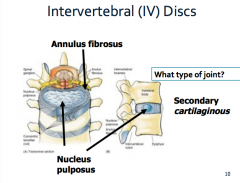
Secondary Cartilaginous
|
|
|
e. B and C
|
|
|
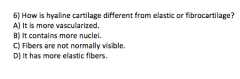
C) Fibers are not normally visible
|
|
|
Which direction would your face turn towards when the left semispinalis muscles contracted?
|
to the right
|
|
|
1. The appendicular skeleton consists of the following bone(s):
ribs vertebrae femur sacrum skull |
Femur
|
|
|
In which of the following regions can a long bone be found?
leg cranium tarsus carpus face |
Leg
|
|
|
Which of the following statements are TRUE?
1. "spongy bone has a lower metabolic activity, relative to cortical bone" 2. compact bone provides strength for weight bearing 3. "compact bone is lighter, relative to spongy bone" 4. "compact bone has more surface area, compared to spongy bone" compact bone is thin in areas of high mechanical stress |
compact bone provides strength for weight bearing
|
|
|
Which of the following is an example of a secondary cartilaginous joint?
epiphyseal plate coronal suture (a cranial suture) the knee interosseous membrane of forearm (syndesmosis) gomphosis None of these are an example of a secondary cartilaginous joint |
None of these are an example of a secondary cartilaginous joint
|
|
|
Functions of the skeleton include:
protection support calcium storage blood cell production all of the above |
all of the above
|
|
|
The left knee is _________ to the right ankle
ispilateral inferior distal contralateral posterior |
contralateral
|
|
|
A cut through which plane, assuming anatomical position, would produce two approximately equal left and right halves?
coronal frontal axial sagittal transverse |
sagittal
|
|
|
Cardiac muscle can be distinguished from both skeletal and smooth muscle by the appearance of:
striations multiple nuclei intercalated discs voluntary contractions lack of striations |
intercalated discs
|
|
|
Small accessory bones that develop between the flat bones of the skull are specifically called:
sutural bones sesamoid bones flat bones irregular bones short bones |
sutural bones
|
|
|
True or False: Since the arteries and veins make up a circuit, about half the total blood volume is contained within the venous system
|
False
|
|
|
True or False: Veins do NOT contain smooth muscle within their walls
|
False
|
|
|
True or False: Veins are more numerous than arteries.
|
True
|
|
|
True or False: Arterioles have a small or narrow lumen, relative to the thickness of the muscular wall.
|
True
|
|
|
True or False: Capillaries provide a site for exchange (of nutrients), while all other blood vessel types provide for blood transport only.
|
True
|
|
|
1. Connective tissue:
supports all epithelial tissue contracts lines internal cavities transmits electrical signals has all of the above attributes |
supports all epithelial tissue
|
|
|
The fiber type that gives connective tissue great tensile strength is:
elastic collagen reticular muscle hemp |
collagen
|
|
|
The fiber type that supports soft organs such as the liver and spleen:
elastic fibers collagen fibers reticular fibers muscle fibers areolar fibers |
reticular fibers
|
|
|
The type of connective tissue that surrounds and cushions organs; significant role in tissue repair and inflammation
Fibrocartilage Irregular Areolar Reticular Regular |
Areolar
|
|
|
How is hyaline cartilage different from elastic or fibrocartilage?
it is more vascularized in contains more nuclei fibers are not normally visible in microscope slide it has more elastic fibers it is less common in the human body |
fibers are not normally visible in microscope slide
|
|
|
What are the characteristics of fibrocartilage?
resists compression and pulling forces Resists compression, reduces friction, absorbs shock Maintain shape of structure while allowing flexibility Strong in many directions but less so in any one direction |
resists compression and pulling forces
|
|
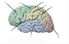
Match each structure on the image below to the correct answer from the list provided. Please note: some answers may be used more than once
A. central sulcus B. frontal lobe C. precentral gyrus of frontal lobe D. postcentral gyrus of parietal lobe E. Parietal Lobe F. Middle gyrus of temporal lobe G. Occipital lobe H. Lateral sulcus |
1 B. frontal lobe
2 C. precentral gyrus of frontal lobe 3 A. central sulcus 4 D. postcentral gyrus of parietal lobe 5 E. Parietal Lobe 6 G. Occipital lobe 7 F. Middle gyrus of temporal lobe 8 H. Lateral sulcus |
|
|
The ______________ separates the two hemispheres of the cerebrum
lateral sulcus longitudinal cerebral fissure falx cerebelli cerebral aqueduct |
longitudinal cerebral fissure
|
|
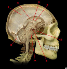
Match each structure on the image below to the correct answer from the list provided. Please note: some answers may be used more than once.
A. Septum pellucidum of cerebral hemisphere B. Corpus callosum of cerebral hemisphere C. Falx cerebelli D. Falx cerebri E. Mammillary body of diencephalon F. Pituitary gland of diencephalon G. Pons of brainstem H. Fornix of cerebral hemisphere |
1 D. Falx cerebri
2 B. Corpus callosum of cerebral hemisphere 3 H. Fornix of cerebral hemisphere 4 A. Septum pellucidum of cerebral hemisphere 5 F. Pituitary gland of diencephalon 6 E. Mammillary body of diencephalon 7 G. Pons of brainstem 8 C. Falx cerebelli |
|
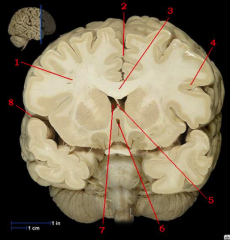
Match each structure on the image below to the correct answer from the list provided. Please note: some answers may be used more than once.
A. fourth ventricle of brainstem B. Lateral ventricle of left cerebral hemisphere C. White matter of left cerebral hemishere D. Lateral sulcus of right cerebral hemisphere E. White matter of right cerebral hemishere F. Fornix of cerebrum G. Lateral ventricle of right cerebral hemisphere H. Septum pellucidum of cerebrum I. Gray matter of left cerebral hemisphere J. Corpus callosum of cerebrum K. Longitudinal cerebral fissure L. Third ventricle of diencephalon |
1 E. White matter of right cerebral hemishere
2 K. Longitudinal cerebral fissure 3 J. Corpus callosum of cerebrum 4 I. Gray matter of left cerebral hemisphere 5 H. Septum pellucidum of cerebrum 6 L. Third ventricle of diencephalon 7 G. Lateral ventricle of right cerebral hemisphere 8 D. Lateral sulcus of right cerebral hemisphere |
|
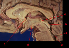
Match each structure on the image below to the correct answer from the list provided. Please note: some answers may be used more than once.
A. Fourth ventricle of brainstem B. Posterior commissure of cerebral hemisphere C. Mammillary body of diencephalon D. Interventricular foramen of diencephalon E. White matter of cerebellum F. Cerebral aqueduct of brainstem |
1 D. Interventricular foramen of diencephalon
2 C. Mammillary body of diencephalon 3 A. Fourth ventricle of brainstem 4 E. White matter of cerebellum 5 F. Cerebral aqueduct of brainstem 6 B. Posterior commissure of cerebral hemisphere |
|
|
A 31 year old man was suspected of having leukemia. It was decided to confirm the diagnosis by performing a bone marrow biopsy. At birth, the marrow of all bones of the body is red and hematopoietic.
True or False |
True
|
|
The fold of dura mater that projects between the cerebral hemispheres and the cerebellar hemispheres is called the _____________ (two words). It extends at an approximate right angle to the falx cerebri. In addition, the transverse sinus (lab 3) is housed within this structure.
|
Tentorium cerebelli
|
|
|
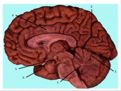
Match each structure on the image below to the correct answer from the list provided. Please note: some answers may be used more than once.
A. Temporal lobe B. Tuber Cinereum of Hypothalamus C. Cingulate (limbic) gyrus D. Primary fissure of the cerebellum E. Parieto-occipital sulcus F. Fourth ventricle |
1 F. Fourth ventricle
2 D. Primary fissure of the cerebellum 3 E. Parieto-occipital sulcus 4 C. Cingulate (limbic) gyrus 5 B. Tuber Cinereum of Hypothalamus 6 A. Temporal lobe |
|
|
Which of the following exclusively describes stratified epithelia?
consists of single cell layer free surface always exposed to inner passageways highly vascular covers surfaces subject to mechanical or chemical stress no exceptions; all of the above are characteristics of epithelia |
covers surfaces subject to mechanical or chemical stress
|
|
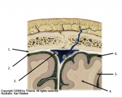
Match each structure on the image below to the correct answer from the list provided. Please note: some answers may be used more than once
A. Dura mater B. Arachnoid mater C. Arachnoid granulations D. White matter E. Pia mater F. Gray matter |
1 A. Dura mater
2 B. Arachnoid mater 3 C. Arachnoid granulations 4 D. White matter 5 F. Gray matter 6 E. Pia mater |
|
|
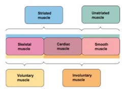
Please match the following with the correct description
A. Striated and Voluntary B. Unstriated and Involuntary C. Striated and Involuntary Cardiac Muscle: Skeletal Muscle: Smooth Muscle: |
Cardiac Muscle: Striated and Involuntary
Skeletal Muscle: Striated and Voluntary Smooth Muscle: Unstriated and Involuntary |
|
|
The flat bones in your skull are formed via ____________ ossification.
Osteochondrol Intramembranous Mesenchymal Endochondral |
Intramembranous Ossification
|
|
|
The long bones in your skull are formed via ____________ ossification.
Osteochondrol Intramembranous Mesenchymal Endochondral |
Endochondral ossification
|
|
|
1. Adults typically have ____ (# of) permanent teeth.
20 24 28 32 36 |
32
|
|
|
Name the different teeth
|
Medial (central) incisor
Lateral incisor Canine First premolar Second premolar First molar Second molar Third molar: wisdom tooth |
|
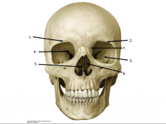
Match each structure on the image below to the correct answer from the list provided. Please note: some answers may be used more than once.
A. suraorbital margin B. greater wing of sphenoid bone C. lesser wing of sphenoid bone D. perpendicular plate E. supraorbital foramen F. maxillary process of frontal bone G. middle nasal concha of ethmoid bone |
1 E. supraorbital foramen
2 A. suraorbital margin 3 B. greater wing of sphenoid bone 4 C. lesser wing of sphenoid bone 5 D. perpendicular plate 6 G. middle nasal concha of ethmoid bone 7 F. maxillary process of frontal bone |
|
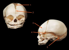
Match each structure on the image below to the correct answer from the list provided. Please note: some answers may be used more than once.
A. frontal suture B. frontal bone C. sphenoidal (anterior lateral) fontanel D. anterior fontanel E. mastoidal (posterior lateral) fontanel |
1 D. anterior fontanel
2 A. frontal suture 3 B. frontal bone 4 C. sphenoidal (anterior lateral) fontanel 5 E. mastoidal (posterior lateral) fontanel |
|
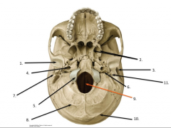
Match each structure on the image below to the correct answer from the list provided. Please note: some answers may be used more than once.
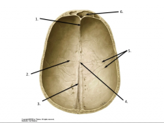
A. carotid canal B. superior sagittal groove C. mandibular fossa D. occipital condyle E. external acoustic meatus F. superior nuchal line G. inferior nuchal line H. mastoid process I. stylomastoid foramen J. pterygoid process K. jugular foramen |
1 C. mandibular fossa
2 J. pterygoid process 3 E. external acoustic meatus 4 A. carotid canal 5 D. occipital condyle 6 K. jugular foramen 7 H. mastoid process 8 G. inferior nuchal line 9 B. superior sagittal groove 10 F. superior nuchal line I. stylomastoid foramen |
|
Match each structure on the image below to the correct answer from the list provided. Please note: some answers may be used more than once.
A. bregma B. frontal crest C. meningeal grooves D. frontal sinus E. coronal suture F. superior sagittal groove |
1 B. frontal crest
2 E. coronal suture 3 F. superior sagittal groove 4 A. bregma |
|
|
The superior orbital fissure is an opening in the _____________.
frontal bone sphenoid bone ethmoid bone temporal bone occipital bone |
sphenoid bone
|
|
|
A 25-year-old man is involved in a motorcycle accident and slams his head into a concrete wall of a bridge. His CT scan reveals that the middle meningeal artery (right side) has ruptured, but the meninges remain intact. Blood leaking from this artery enters which of the following spaces?
subarachnoid subdural epidural subpia cranial dural sinuses |
epidural
|
|
|
As a sports medicine physician working with a young athlete, you would like to confirm your diagnosis of a concussion with brain imaging. Which of the following enables you to view the anatomical or structural damage to the brain following a concussion?
X-ray CT scan MRI ultrasound none of the above can assess the structural damage of a concussion. |
none of the above can assess the structural damage of a concussion.
|
|
|
Membranes, such as the ones lining the ventral body cavity, are organs formed by the combination of which tissues?
epithelial and muscle epithelial and connective connective and muscle muscle and neural connective and neural |
epithelial and connective
|
|
|
A multilayered epithelium with cuboidal basal cells and flat cells at its surface would be classified as ________.
simple cuboidal simple squamous transitional stratified squamous stratified columnar |
stratified squamous
|
|
|
A stroke involving the middle cerebral artery on the left side is likely to cause which of the following symptoms?
Paralysis of left side of face and left upper extremity Paralysis of left lower extremity Complete loss of vision in both eyes Loss of ability to comprehend speech Loss of vision in the right eye only |
Loss of ability to comprehend speech
|
|
|
Why is blood classified as a connective tissue?
blood connect two areas of the body blood provides mechanical support of organs blood stores large quantities of lipids blood develops from mesenchyme blood is NOT classified as a connective tissue |
blood develops from mesenchyme
|
|
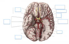
Name the arteries of the brain
|

|