Reading...
![]()
Play button
![]()
Play button
![]()
Use LEFT and RIGHT arrow keys to navigate between flashcards;
Use UP and DOWN arrow keys to flip the card;
H to show hint;
A reads text to speech;
37 Cards in this Set
- Front
- Back
|
When your red cells vary widely in size, you have...
|
...anisocytosis.
|
|
|
MCV stands for...
|
...mean corpuscle volume (RBC cell volume)
|
|
|
RDW stands for.... What is it mathematically? What can cause a high RDW?
|
...Red cell distribution width. Standard deviation of Red cell size.
iron deficiency, bleeding (loss of #), etc. |
|
|
What is microcytosis?
Associates with which measure of red-cells? What can cause it? |
Small red cells
MCV Iron deficiency, thalassemias, sideroblastic anemia |
|
These RBCs have a small....
|
MCV.
|
|
|
What is macrocytosis (re: RBCs) associated with? (6)
|
elevated MCV (~ >100)
Elevated reticulocyte count, B12/folate deficiency (opposite of iron deficienty/thalassemia... here we have a DNA production problem) Thyroid disease, Liver Disease, Chemotherapy, Anti-retrovirals (AZT) |
|
|
Red cells that have too little hemoglobin are said to have....
This is denoted observationally by noting when the area of central pallor is more than ____ the total RBC diameter. What is this measured by? |
... hypochromasia.
1/3 MCH |
|
|
What is MCH?
|
(Hemoglobin / RBC)
|
|
|
Polychromasia refers to red cells that...
|
...have more of a blue-ish tinge.
|
|
|
In general, the blue cells associated with polychromasia are _____ (+/- size) and are probably ________.
|
larger, reticulocytes.
|
|
|
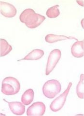
Poikilocytosis refers to red cells that...
|
....vary widely in SHAPE.
|
|
|
What is MCHC? Can you have a high MCHC?
|
amount of Hb / size of red blood cell
no, because cells are already saturated w/ hemoglobin |
|
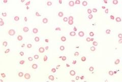
What disorder of red cells does this pictures show?
|
Poikilocytosis
|
|
|
When RBCs look like bulls-eyes, how do we label this disorder?
What diseases is this condition associated with? (4) |
Target cells.
Liver disease, thalassemias, hemoglobin C, after splenectomy |
|
|
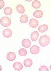
The RBC disorder termed Spherocytes have...
When are these seen? (2) |
...a loss of central pallor.
1. Hereditary spherocytosis 2. autoimmune hemolysis |
|
|
If spherocytes are due to autoimmune hemolysis, are the RBCs larger or smaller? What do we call this?
|
smaller
microspherocytes |
|
|
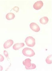
Red cell fragments with sharp edges are called...
They are a hallmark of... |
...schistocytes
Microangiopathic Hemolytic Anemia (MAHA) |
|
|
Microangiopathic Hemolytic Anemia (MAHA) associates with which disorder of RBCs?
|
Schistocytes, cells with jagged edges.
|
|

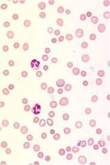
What type of disorder red cells are seen here?
|
Spherocytes
|
|
|
In sickle cell anemia, which two types of disorder red cells can be seen?
|
sickled cells & target cells
|
|
|
thrombocytopenia is....
which RBC disorder is it sometimes associated with? |
.... low platelets.
Schistocytes. |
|
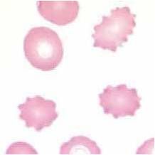
which RBC disorder cells are seen here? Another name for them?
What type of disease are they seen in? |
Echinocytes, Burr Cells.
Renal disease |
|
|
What is the difference between echinocytes and acanthocytes?
|
e: small, reg. projections, renal diz.
a: larger irreg. projections, liver diz. |
|
|
What in liver diz. causes acanthocytes?
|
Elevated BUN.
|
|
Name the cells. They are seen in _____ processes, which are diseases of _______ infiltration.
|
Teardrop cells.
Myelophthisic processes, marrow infiltration. The cells look like this because they have to 'squeeze' out of the marrow. |
|
|
Myelofibrosis
tumor metastatic to marrow Granulomatous diseases Leukemias and lymphomas ....which disordered RBCs can be seen? |
teardrop cells
|
|
|
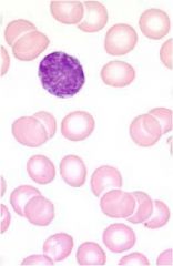
What is the name given to peripheral, small, round purple inclusions within red cells that represent nuclear remnants?
|
Howell-Jolly bodies
|
|

What are these purple inclusion bodies? When are they seen?
|
HJ bodies. Post splenectomy or in the case of splenic hypofunction.
|
|
|
What types of cells can be seen after splenectomy?
|
HJ bodies, target cells, acanthocytes, schistocytes, nucleated red cells.
|
|
|
What are Rouleaux? When are they seen?
|
linear arrangement of red cells typically described as piles of coins on a plate.
In disorders w/ increased levels of IgG such as multiple myeloma or waldenstrom's macroglobulinemia. * also severe hypo-albuminemia. |
|
|
When does red cell agglutination occur? How are these different from Rouleaux?
|
when the cells are coated w/ IgM. Unlike roleaux, the RBC clumps are disorderly and not linear.
|
|
|
What is seen in Fe-Def anemia?
|
Hypochromic, microcytic cells. Increased numbers of platelets can be seen.
|
|
What is most likely the etiology behind the assortment of cells seen here?
|

Fe-def anemia: Hypochromic, microcytic cells w/ elevated platelets.
|
|

Which disorder is likely behind this assortment of cells?
|
Fe-def anemia.
Hypochromic, anisocytosis. Small cells. |
|
|
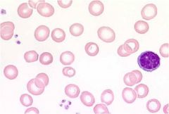
What is seen on a blood smear of someone with megaloblastic anemia?
|
RBCs are macrocytic
Hypersegmented PMNs |
|

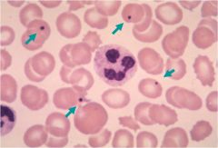
What is most likely the disorder behind this smear? Also, name the abnormalities at the black arrows and those at the green.
|
Autoimmune Hemolytic anemia
(AIHA) Black: polychromasia Green: Microspherocytes |
|

What etiology? What specific abnormalities?
|
Microangiopathic Hemolytic Anemia (MAHA).
Schistocytes, low platelets (none?) |