![]()
![]()
![]()
Use LEFT and RIGHT arrow keys to navigate between flashcards;
Use UP and DOWN arrow keys to flip the card;
H to show hint;
A reads text to speech;
27 Cards in this Set
- Front
- Back
- 3rd side (hint)
|
Connective Tissue |
Most abundant tissue in the body. |
|
|
|
Functions of Connective tissue: |
•Binds structures •Supports body and protects organs •Stores fat •Produces blood cells •Helps repair damage •Protects against infection •Fills spaces |
|
|
|
Matrix between cells may be fluid, as in blood plasma, gel like as in cartilage or solid as in bone. |
•Can usually reproduce. •Typically vascular expect cartilage which is avascular. •May be rigid (ex: bone, cartilage) or flexible (ex: loose connective, adipose (fat), dense connective) |
|
|
|
Major Cells in Connective Tissue: |
A. Fibroblast (most common) Produces fibers by secreting protein into matrix large-star shaped. 1. White fibers: Contains collagen, flexible but only slightly elastic great tensile strength. 2. Yellow fibers: Contains elastin, less strength but will return to its original length. (Elastic) 3. Reticular: Thin collagen fibers that form a supportive network in Connective Tissue. B. Macrophages Carry out phagocytosis and provide important defense against infectious agents. Clear out foreign particles white blood cells. Can move around. C. Mast Cells •Located near blood vessels and contain heparin (prevents clotting) •Enhances inflammation by releasing histamine.
|
|
|
|
8 different types of connective tissue |
Loose Connective (Areolar) Adipose Fibrous (dense) tissue Elastic connective tissue Cartilage Bone Blood Reticular connective. |
|
|
|
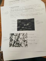
Loose Connective |
Description: •Fibroblast are scattered throughout the tissue and are responsible for producing the fibers. •Mast Cells plasma cells, and macrophage cells are commonly found in the tissue. Composition: Mainly fibroblast cells; contains white and yellow fibers. Mast Cells release histamine. (Causes capillaries to be more permeable during the time of the body is subject to allergies or inflammation. Plasma cells produce anti-bodies to protect the body. Function: •Binds skin to underlying organs and fill in spaces between muscles. •Forms fragile, thin membranes found in the body. |
|
|
|
Loose Connective picture |
|
|
|
|
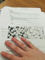
Adipose |
Description: Special cells that store fat globules becomes swollen and pushes nucleus against plasma membrane. Location: •Beneath skin, between muscles, around organs, inside joints, behind eyeballs. •Evenly distributed in infants and children. •In adults, thins and some areas - remains in others. Males: Backs, arms, buttocks, and abdomen. Females: breast, thighs and buttocks. Function: •Protection layer for joints, organs and behind the eyeball. •Heat insulation. •Source of energy. |
|
|
|
Adipose picture. |
|
|
|
|
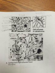
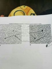
Bone (Osseous tissue) |
Description: •Most rigid type of connective tissue. •Bone cells are called osteocytes. •There are housed in cavities called lacunae and are found in the circular layers of matrix called lamelle. Composition: Matrix is composed of calcium and phosphate salts and collagen protein. Also within the matrix are longitudinal tubed called Haversian (oestonic) canals. Haversian canal house the blood vessels and nerves. Haversian canals and osteocyte are connected by minute tubules canaliculi which ensure transportation between the blood vessels and the bone cells. Function: •Protect internal body parts. •Provide internal support. •Provide a point of attachment. •Forms blood cells. •Store minerals. |
|
|
|
Bone picture |
|
|
|
|
Cartilage (Semi-Rigid Connective Tissue) |
Description: Matrix: Made up of elastic & collagen fibers embedded in gel like substance. Cells: •Chondrocytes located in lacunae (spaces) •Considered to be avascular expect for perichondrium (fibrous tissue on surrounding surface) •Slow repair and cellular reproduction. Functions: Support, frame work and attachment, protects underlying tissues. |
|
|
|
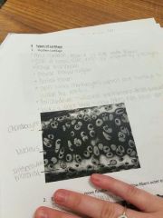
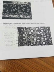
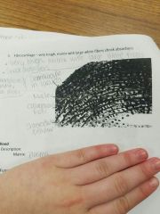
3 types of cartilage |
1. Hyaline Cartilage: •Most common, matrix of fine white fibers. •Ends of bones, nose, rings of respiratory passages. •Provide framework. •Provide flexible support. •Protect tissues. •Cells called Chondrocytes which are housed in the lacunae within the matrix. •Perichondrium - vascular membrane that surrounds the cartilage. •Lacks a direct blood supply (does not heal well) 2. Elastic cartilage - More flexible, matrix of yellow fibers outer ears and larynx. 3. Fibro cartilage Very tough, matrix with large white fibers shock absorbers. Knees (mensicus) pelvic grindle, intervertebral disks. |
|
|
|
Hyaline Cartilage picture |
|
|
|
|
Elastic cartilage picture |
|
|
|
|
Fibro cartilage picture |
|
|
|
|
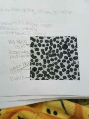
Blood |
Description: Matrix - plasma. Functions: Transportation, blood clotting, anti body production. Cells: Red blood cells (erythocytes) - transport oxygen. White blood cells (lenkeocyte) - immunity. Platelets (thrombocyte) - clotting. |
|
|
|
Blood picture |
|
|
|
|
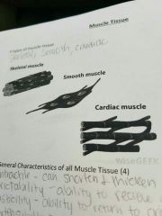
3 types of muscle tissue: |
Skeletal Smooth Cardiac |
|
|
|
General characteristics for Muscle Tissue |
Contractile - can shorter and thicken. Excitability - ability to relieve and respond to stimuli. Elasticity - ability to return to original shape. Extensibility - ability to stretch. Additional information about muscle tissue: Makes up 40% to 50% of total body weight made of elongated cells called muscle fibers. Sacrolemma - cell membrane of fiber. Sacroplasm - cytoplasm of fiber. Function: Motion, maintenance of posture, heat products. |
|
|
|
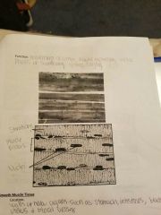
Skeletal Muscle Tissue |
Location: Muscles attached to the bone. Qualities: •Voluntary movement controlled by conscious effort, and nerve impulse. •Fibers are long and tube-like. •Light and dark cross markings called stirations. •Multinucleated many Nuclei. Function: Movement of limbs, facial expression, initial phases of swallowing, talking and singing. |
|
|
|
Skeletal Muscle Tissue Picture |
|
|
|
|
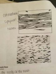
Smooth Muscle Tissue |
Location: Walls of hollow organs such as stomach, intestines, bladder, uterus & blood vessels. Qualities: •Non-stirated and one nucleus. •More then and tapered at the ends when compared to skeletal Muscle. •Involuntary - don't consciously control. Function: •Perstalsis (movement of food through the digestive tract) •Vasoconstriction and vasodilation vessels (controls blood flow) •Emptying of bladder. *** Also called the visceral muscle. |
|
|
|
Smooth Muscle Tissue picture. |
|
|
|
|
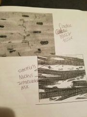
Cardiac Muscle Tissue |
Location: In the walls of the heart. Qualities: •Stirated, involuntary and one nucleus (characteristics of skeletal and smooth muscles) •More Y or V shaped. •Fibers appeared to be branched (bifurcated) Function: Pumping of blood.
Intercalated disk: Special intercellular junctions that give strength and endurance to the tissue (run in the same direction as the stirations) |
|
|
|
Cardiac Muscle Tissue |
|
|