![]()
![]()
![]()
Use LEFT and RIGHT arrow keys to navigate between flashcards;
Use UP and DOWN arrow keys to flip the card;
H to show hint;
A reads text to speech;
22 Cards in this Set
- Front
- Back
|
Nonthrombogenic endothelium covers what structures in the heart? |
Valves and the fibrous cords, chordae tendineae. |
|
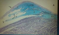
Label the letters and arrow shown in the figure above. |
➡: Atrioventricular valve. C: Connective tissue. Ct: Chordae tendineae. En: endocardium A: atrium V: ventricles M: myocardium |
|
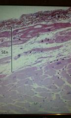
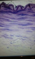
What structure is shown above and label the structures. |
This shows the endocardium of the ventricle of the heart. En: endothelium P: Purkinje fiber SEn: subendocardial layer M: cardiac Muscle |
|
|
What are Purkinje fibers and how are they differentiated from cardiac Muscle? |
They are specialised cardiac Muscle which conducts electrical impulses instead of contact and the are located in the intramuscular septum, where they deliver stimuli by gap junctions. They are stained paler than normal cardiac Muscle due to more glycogen being stored. |
|
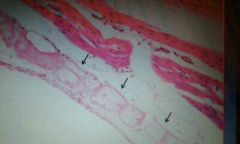
What are the arrows pointing at? |
Purkinje fibers |
|
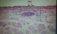
What is the name of the structure above and Label the figure. |
This is the epicardium or the visceral pericardium. Mes: mesothelium CT: Connective tissue N: nerve Ep: epicardium F: fat M: myocardium |
|
|
What are connexons and what are they made out of? |
These are gap junction proteins, also called hemichannels, and use to form gap junctions. They are made up of 6 connexin subunit, and those made from 6 connexin molecules. |
|
|
What are the three main connexons in the heart and where can they be found? |
The main connexons in heart are : Cx43 found in the ventricular myocardium, atheresclerotic plaque, and on astrocytes; Cx40 found in the atrial myocytes; and Cx37 found in induced vascular smooth wall during coronary arteriogenesis. |
|
|
What is the function of Angiopoietin 2? |
It works with VEGF to facilitate cell proliferation and migration of endothelial cells. |
|
Identify the structure shown above. |
An elastic artery |
|
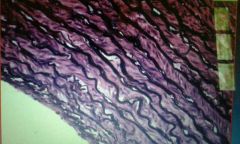
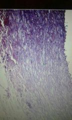
What structure is shown above? |
An elastic artery under a elastic (verhoeff) stain |
|
|
What is a Vasa vasorum? |
These are microvessels that run in the adventitia of large blood vessels. They supply those cells which are to far away from the central lumen. |
|
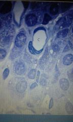
What is the structure shown above and what is its function? Label the diagram. |
This is a Glomus Body. It's a specialized region in the walls of Elastic arteries that act as chemoreceptors. C: capillaries G: Glomus cell S: satellite cell |
|
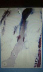
What is the structure above and label the diagram. |
This is a muscular artery. E: endothelium IEL: internal elastic layer SM: smooth muscle V: Vasa vasorum |
|
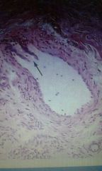
What is the structure above and what is the arrow pointing at? |
This is a small vein and the arrow is pointing at a valve. |
|

What is the structure shown above and label it. |
This is the microvasculature. A: arteriole V: venule C: capillary L: lymph |
|
|
Describe the microvasculature bed. |
Arterioles branch as metarterioles and travel to the post-capillary venules as a channel, called the "thoroughfare channel", lacking smooth muscle. True capillaries branch from the thoroughfare channel and have precapillary sphincters made from smooth muscle which controls the blood that enters the channels. |
|
|
State, describe, and give the locations of the three types of capillaries. |
(i) Continuous capillaries - these have tight junctions between the endothelial cells and a continuous basement membrane. These are found at the beds of skeletal muscles, skin, lungs, and nervous system. (ii) Fenestrated capillaries - these have perforations in the endothelium to allow greater exchange but the basement membrane is still continuous. These are found in the beds of endocrine organs, intestinal wall, kidney, glomeruli, and choroid plexus. (iii) Sinusoids - these have large fenestrations and discontinuity in the endothelium and basement membrane to allow ready exchange of macromolecules and cells. These are found in the beds of the bone marrow, liver, and spleen. |
|
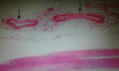
What are the arrows pointing at? |
Continuous capillaries |
|
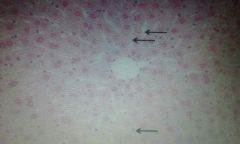
What are the arrows pointing at? |
Sinusoidal capillaries in the liver. |
|
|
What are pericytes? |
These are contractile cells found around the basement membrane of capillaries and post-capillary venules. They help to control blood flow through capillaries, help make up the BBB, they have a role in angiogenesis and skeletal muscle regeneration, and they play a role in certain types of diseases such as neurodegenerative diseases, stroke, and diabetic retinopathy. |
|
What is this structure? |
A lymph vessel |