Reading...
![]()
Play button
![]()
Play button
![]()
Use LEFT and RIGHT arrow keys to navigate between flashcards;
Use UP and DOWN arrow keys to flip the card;
H to show hint;
A reads text to speech;
36 Cards in this Set
- Front
- Back
|
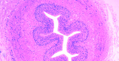
Ureter
The ureter is a muscular tube, composed of an inner longitudinal layer and an outer circular layer of smooth muscle. The lumen of the ureter is covered by transitional epithelium (also called urothelium). Recall from the Laboratory on Epithelia that the transitional epithelium is unique to the conducting passages of the urinary system. Its ability to stretch allows the dilation of the conducting passages when necessary. The ureter connects the kidney and the urinary bladder. |
|
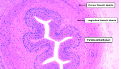
wall structure of _________. Label #'s
|
Wall structure of the ureter:
I. Tunica mucosa - epithelium - lamina propria II. Tunica muscularis* - inner longitudinal - outer circular III. Tunica adventitia so.... 1 – tunica mucosa 2 – tunica muscularis 3 – tunica adventitia 4 – transitional epithelium 5 – lamina propria 6 – umbrella cell |
|
|
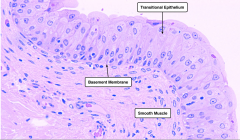
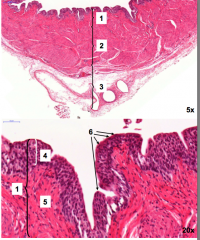
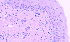
Urinary Bladder
The urinary bladder is lined by transitional epithelium, underneath which are thick layers of smooth muscle interwoven in various directions. This image shows a relaxed bladder where the epithelial cells appear cuboidal. In a distended bladder the epithelial cells are stretched and become more squamous. |
|
|
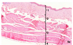
Urinary Bladder,
5x 1 – tunica mucosa 2 – tunica submucosa 3 – tunica muscularis 4 – tunica subserosa + serosa |
|
|
|
|
|
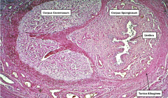
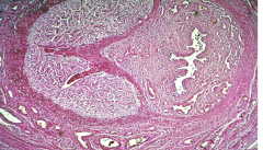
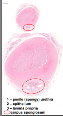
Penis
The penis contains three columns of erectile tissue: two corpus cavernosa and a single corpus spongiosum containing the penile urethra. Note the vast sponge-like arrangement of irregular vascular spaces intercalated between the arteries and veins. |
|
|

penis
|
|
|
penis
|
|
|
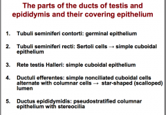
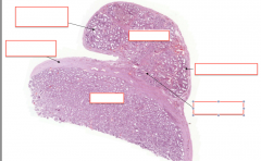
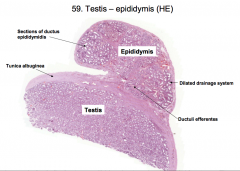
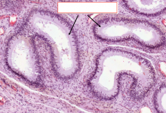
The parts of the ducts of testis and epididymis and their covering epithelium
|
|
|
|
|
|
|
|
|
|
|
|
|
|
|
|
|
|
|
|
|
|
|
|
|
|
|
|
|
|
|
|
|
|
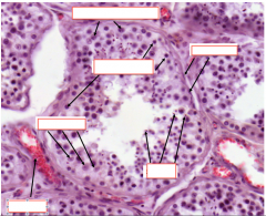
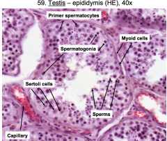
cell types of testis
|
The first two of these are found in the germinal epithelium (layer of the seminiferous tubule in which gamete production and development occurs)
1. Sertoli cells extend from the basement membrane of the germinal epithelium to the lumen of the tubule. • envelope the developing sperm cells • joined together by junctional complexes and form the blood-testis barrier (barrier separates the germinal epithelium into a basal compartment ) containing 1) Sertoli cells and 2) diploid germ cell precursors, and 3) an adluminal compartment , which contains the products of meiosis. 2. Spermatogenic cells •include each of the stages between the spermatogonium and the mature spermatozoan. •least mature cells near the basal layer of the epithelium and the most mature cells near the luminal layer. 3. Leydig (interstitial) cells •located within the loose connective tissue surrounding the seminiferous tubules. •pale due to their high cholesterol content and often contain crystal |
|
|
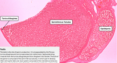
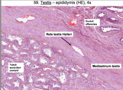
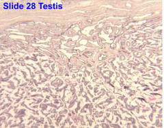
testis
|
Examine the slide w/o the microscope and note the testis, head of epididymis and vas deferens. With low power objective identify the thick dense regular connective tissue capsule surrounding the testis; this is the tunica albuginea . With higher power note that it is covered by mesothelium , the visceral layer of the processus vaginalis. Identify the area of the mediastinum (thickened part of the tunica albuginea) which contains the rete testis (not well preserved in this slide). Note also the muscular vas deferens and the head of the epididymis on this slide. This testis is from an infant so the seminiferous tubules are not well developed.
|
|
|
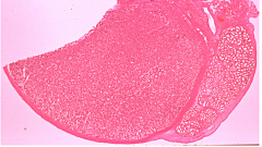
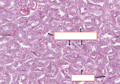
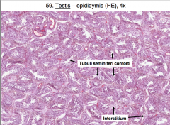
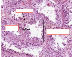
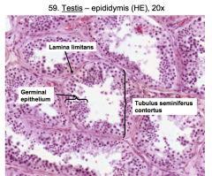
seminiferous tubules and cell types within
|
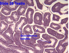
Note the highly coiled structure of the seminiferous tubules seen in cross section. The cell-type population varies from tubule to tubule depending in the stage of the spermatogenic cycle. The endocrine testis consists of the Leydig cells which are found in clusters within the interstitial tissue between seminiferous tubules. The exocrine testis consists of two cell types: the Sertoli cell and the germ cells
1. Spermatogonia (most numerous cell type, adjacent to the basement membrane) 2. Spermatocyte (twice the size of the spermatogonia, in two rows above spermatogonia) 3. Spermatid (small cells adjoining the lumen, can be early or late) |
|
|
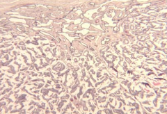
rete testis
|
rete testis in the mediastinum. The rete are communicating channels between the straight tubules and efferent tubules (ductules). They are lined by simple cuboidal epithelium . No smooth muscle is present in this region.
|
|
|
rete testis in the mediastinum. The rete are communicating channels between the straight tubules and efferent tubules (ductules). They are lined by simple cuboidal epithelium . No smooth muscle is present in this region.
|
|

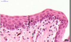
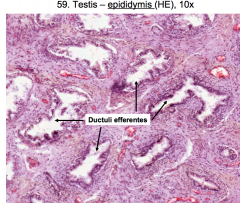
epididymus and efferent ductules
|
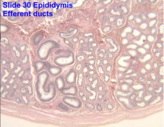
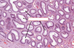
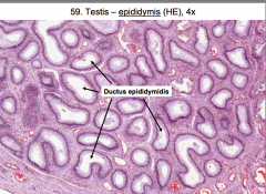
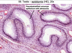
The efferent ductules have epithelia of variable height. The epididymal epithelium (pseudostratified columnar with stereocilia) is regular in height so that the luminal margin is even. Remember, this is a single tortuous duct. It has a thin layer of smooth muscle surrounding it.
|
|
|
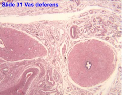
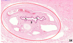
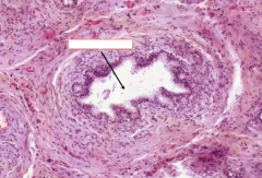
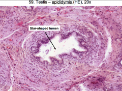
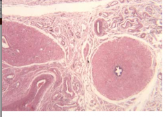
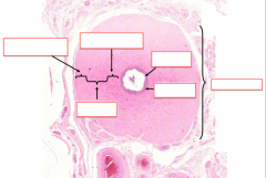
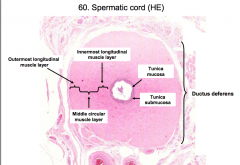
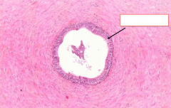
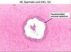
The vas deferens (ductas deferens) has a thick muscular wall and a narrow star-shaped lumen. You should note that the smooth muscle is arranged in 3 layers ; inner and outer longitudinal and middle circular. The lining epithelium is pseudostratified columnar epithelium .
|
|
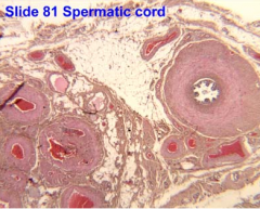
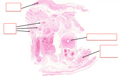
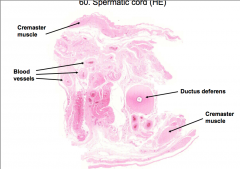
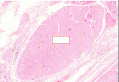
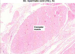
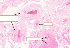
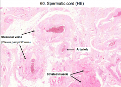
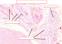
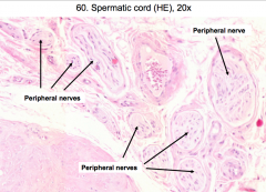
spermatic cord
|

In the spermatic cord, there are bundles of nerve fibers , skeletal muscle , adipose tissue and blood vessels . Note the numerous veins (filled with blood) are atypical in that they resemble arteries and have (2) layers of smooth muscle. This is part of the pampiniform plexus of veins.
|
|
|
SEMINAL VESICLES
|
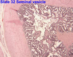
Note the foldings of the mucosa, the epithelium which can be a mixture of pseudostratified columnar, or simple columnar, and the muscularis (an ill-defined inner circular and an outer longitudinal muscle coat). Under low power there are apparently isolated profiles of seminal vesicle epithelium which represent sections through an elongated sac which is folded upon itself
|
|
|
|
|
|
|
|
|
|
|
|
|
|
|
|
|
|
|
|
|
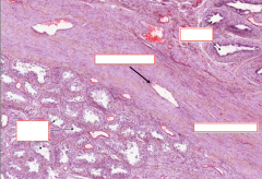
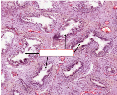
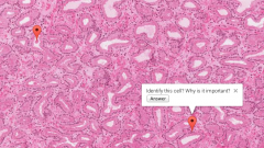
prostate:
Basal cell. Its presence distinguishes benign glands from adenocarcinomas. the other is: Prostatic concretion. Helps to identify the prostate. |
|
|
ID prostate
|
heterogeneous epithelium of the prostate and the abundant stroma consisting of smooth muscle and connective tissue.
ejactulatory duct opens to prostatic urethra. epithet above this is the urothelium, below the opening is a slow conversion to stratified columnar epithet(?) |