Reading...
![]()
Play button
![]()
Play button
![]()
Use LEFT and RIGHT arrow keys to navigate between flashcards;
Use UP and DOWN arrow keys to flip the card;
H to show hint;
A reads text to speech;
64 Cards in this Set
- Front
- Back
|
three histologic fx of hepatitis
|
1. Portal-based portal inflammation
2. Interface hepatitis = piecemeal necrosis 3. Focal necrosis – Foci of lobular inflammation & necrosis of single hepatocytes |
|
|
how diagnose hep A, how determine if immunity
|
IgM HAV for diagnosis
IgG HAV for immunity determination |
|
|
type of virus : dna vs. rna HAV
|
ss RNA, picornovirus
|
|
|
type of virus: HBV
|
partly ds DNA
|
|
|
treatment for HBV
|
α-interferon, lamivudine
|
|
|
how diagnose HBV
|
HBsAg or HBcAg
|
|
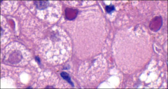
see this, think
|
ground glass hepatocytes: hep b (proliferating ER)
|
|
|
two histo fx for HBV
|
sanded nuclei (pink nuclear inclusions with HBcAg in it
ground glass cytoplasm (contains HBsAg and HBcAg) |
|
|
histol fx for HCV
|
fat, lymphoid aggregates and bile duct damage
|
|
|
if see fat, lymphoid aggregates and bile duct damage, think
|
HCV
|
|
|
ab for AIM hepatitis
|
ANA, ASMA, LKM1, soluble liver antigen or LP (liver
pancreas) antibodies |
|
|
trx for AIM
|
only hepatitis that responds to steroids
|
|
|
histo fx for AIm hepatitis
|
Marked interface inflammation with bridging necrosis (PT to CV)
and plasma cells!!! |
|
|
how detect hep D
|
can do ihc
|
|
|
histologic fx of cholestasis
|
– Portal based damage
– Dilated bile canaliculi with bile plug – pseudoxanthomatous change or feathery degeneration • Bile duct injury and loss • Ductular (cholangiolar) proliferation • Cholestatic Mallory bodies, zone I • Jigsaw cirrhosis (segmental disease) |
|
|
clinical findings for cholestatic dz
|
Elevated Alk phos, bilirubin, GGT
– Pruritis, jaundice, pale stools |
|
|
ihc in cholestasis
|
perivenular hepatocytes are CK7+
|
|
|
two copper stains for cholestasis
|
Rhodanine and victoria blue
|
|
|
clinical associations PBC
|
women, middle age, HLA DR type, AIM associations (scleroderma, Sjogren's)
often AMA positive elevated IgM |
|
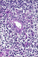
what
|
primary biliary cirrhosis
-Cholestasis • Florid duct lesion – Bile duct injury • Granulomas |
|
|
clinical findings/associations with PSC
|
males, UC, cholangioca,
beading on ERCP |
|
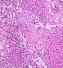
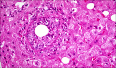
what
|
Periductal fibrosis
– Ductopenia |
|
|
macrovescicular steatosis causes (just read)
|
Alcohol
– Diabetes – Drugs – Obesity – Deficient diet (TPN) |
|
|
reye's
|
fatty liver and encephalopathy
kids, flu-like illness, asa microvesicular/no inflammat |
|
|
findings of steatohepatitis
|
Steatosis
Ballooned hepatocytes Mallory-Denk bodies – Zone 3 (perivenular/pericentral) fibrosis -Inflammed lobules (neutrophils) Megamitochondria |
|
|
what are mallory bodies composed of
|
intermediate filaments, p62 and ubiquitin positive
|
|
|
what are megamitochondria and where found
|
in steatohepatitis, eosinophilic globules
|
|
|
what is amiodarone a/w and what specific histologic findings
|
steatohepatitis
Foamy granular hepatocytes from phospholipids which show lysosome inclusions by EM |
|
|
histologic findings of acetaminophen
|
hepatocellular necrosis with inflammation
|
|
|
see granulomas in liver think
|
infection MAI
cholestasis - PBC |
|
see this, what to think
|
Q fever, ring granuloma, Coxiella burnetii
|
|
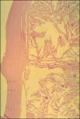
what
|
echinococus, laminated layer (can be gms +)
|
|
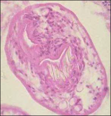
what
|
echinococcus, scolex, gms+ hooklets, birefringent
|
|
|
describe molecular allelic options for alpha 1 antitrypsin deficiency
|
M - normal allele
PiS and PiZ- mutated alleles PIZZ is bad |
|
|
mutations of heretidary hemochromatosis
|
HFE gene mutations
2 most common – C282Y, highest iron overload - H63D |
|
|
sx of hereditary hemochromatosis
|
Cirrhosis, diabetes and skin pigmentation
|
|
|
Hereditary hemochromatosis inheritance pattern
|
autosomal recessive
|
|
|
other HH mutations
|
• 1 HFE
• 2A hemojuvelin- (kids) • 2B hepcidin (kids) • 3 transferrin receptor2 • 4 ferroportin |
|
|
if see iron-free foci in HH, what should you think of
|
HCC
|
|
|
wilson dz mutation
|
Mutated ATP7B gene, a copper transport protein in Golgi with accumulation of copper in liver and other organs
|
|
|
tests for wilson dz
|
↓Cerruloplasm—not sensitive or specific
– ↑Hepatic copper concentration— best test |
|
|
histol fx of wilson's dz and stains
|
Steatosis, hepatitis, glycogenated nuclei, abnormal mitochondria
Mallory hyaline, copper by Rhodanine or Orcein stains |
|
|
see black pigment in liver think
|
dubin-johnson syndrome
|
|
|
mutation of dubin-johnson syndrom
|
cMOAT/MRP2 gene mutation
|
|
|
lab finding in dubin-johnson syndrome
|
isolated conjugated hyperbilirubinemia
|
|
|
histochemical stains to confirm dubin johnson syndrome
|
PASD and Fontana Masson stain
pigment |
|
|
ischemic affects what zone first
|
zone 3 necrosis
|
|
|
gross pathology of ischemic liver
|
nutmeg liver (note can see in tylenol toxicity)
|
|
|
histologic findings in budd-chiari
|
Sinusoidal dilation and hepatocyte atrophy
|
|
|
what is hepatoportal sclerosis
|
weird process by which portal regions are abnormally close to central veins, non cirrhotic
|
|
|
see nodular regenerative hyperplasia on histology, think what clinically
|
portal HTN
recall: vague nodules, no fibrosis |
|
|
hepatic adenoma: clinical associations
|
ocps
glycogen storage dzs |
|
|
treatment for hepatic adennoma
|
surgery to prevent bleeding/rupture
|
|
|
key fx of hepatic adenoma
|
1-2 layers thick, no PT, dilated sinusoids
|
|
|
histologic fx of FNH
|
central stellate scar**
liver divided into nodules by thickwalled arteries in fibrous septa ductular proliferation (CK7+) |
|
|
what is a high grade dysplastic nodule in liver
|
in cirrhotic liver, a nodule >1 cm
|
|
|
background and prognosis in fibrolamellar HCC
|
noncirrhotic background
better prognosis |
|
|
what's inside polygonal cells of fibrolamellar HCC
|
- mito in cytoplasm
- pale body inclusions |
|
|
ddx of central scar in liver
|
fnh, fibrolamellar hcc
|
|
|
ihc in cholangioca
|
Mucicarmine+, CK7+, HepPar1-
|
|
|
infectious a/w cholangioca
|
Clonorchis sinensis, Opisthorchis),
|
|
|
big thing for staging of hcc and cholangioca
|
hcc - size (5cm)
cholangioca: periductal invasion |
|
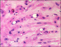
what, ihc
|
hemangioma (dendritic) (intracytoplasmic
lumina/vacuoles w RBCs) CD34, Factor VIII |
|
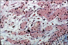
what, ihc
|
epithelioid hemangioma (epithelial cells occlude vessels)
CD34, Factor VIII |