Reading...
![]()
Play button
![]()
Play button
![]()
Use LEFT and RIGHT arrow keys to navigate between flashcards;
Use UP and DOWN arrow keys to flip the card;
H to show hint;
A reads text to speech;
74 Cards in this Set
- Front
- Back
|
What are the lymphoid tissues?
|
- Lymph node
- Spleen (particularly the white pulp) - Mucosa-associated lymphoid tissue (MALT) - Thymus |
|
|
What are the types of structures in the Lymph Node?
|
- Vascular structures
- Lymphatics - Hilum - Capsule - Subscapular sinus - Sinus - Paracortex - Follicles |
|
|
What are the vascular structures in the lymph node?
|
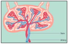
- Primary vein
- Primary artery → gives rise to different tributaries |
|
|
What are the lymphatic structures in the lymph node?
|

- Afferent lymphatics
- Efferent lymphatic |
|
|
What is the function of the Afferent lymphatics?
|

Carry lymph fluid from other portions of body to LN to be filtered
|
|
|
What does lymph fluid contain?
|
- Ags (bacteria, foreign substances, viruses)
- Inflammatory cells (macrophages and dendritic cells) |
|
|
How does the lymph fluid (cells and acellular material) enter the LN?
|

In lymph fluid via afferent lymphatics
|
|
|
What is the function of the Efferent lymphatics?
|
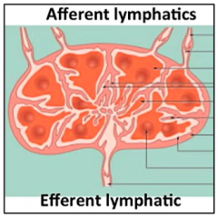
- Where filtered lymph fluid exits the LN
- Carries lymph fluid from LN back through the thoracic duct system |
|
|
What structures are found ini the hilum of the LN? How can you differentiate them?
|
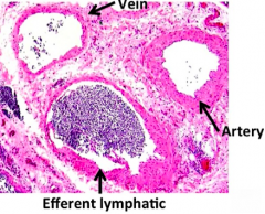
- Vein: thin walled
- Artery: thicker walled - Efferent Lymphatic: relatively thin walled wiht many cells in its lumen |
|
|
What is found in the Efferent Lymphatics?
|
Lots of lymphocytes (vs afferent lymphatic) that are picked up through transport through the LN
|
|
|
Where do afferent lymphatics branch first upon entering the LN?
|
Branches in the subscapular space
|
|
|
What covers the lymph node?
|

Very thin fibrous capsule
|
|
|
What is found under the thin fibrous capsule? What is it made of?
|
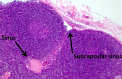
Subscapular Sinus
- Space is created by very thin walled ECs composing this lymphatic sinus |
|
|
What do the subscapular sinuses give rise to?
|
- As they dive into the parenchyma even further they give way to the Cortical Sinuses
- Cortical sinuses give way to medullary sinuses in central LN |
|
|
What do the cortical and medullary sinus contain?
|
- Mostly acellular material (mostly material that needs to be filtered or presented to immune cells)
- Only cells you'd expect to see are APCs (macrophages and sometimes some lymphocytes) |
|
What are the pink areas?
|

Sinuses (cortical and medullary
|
|
|
What are the components of the LN architecture?
|

- Paracortex
- Follicular structures |
|
|
Where is the paracortex of the LN? Structure?
|
- Interfollicular area (between the follicles)
- Generally very vascular - Predominance of T cells |
|
|
What is found in the follicular structures?
|
B cells (where development occurs)
|
|
|
How can you differentiate a benign LN from a malignant LN?
|
- Benign LN: intact architecture
- Malignant LN: disturbed or distorted architecture |
|
|
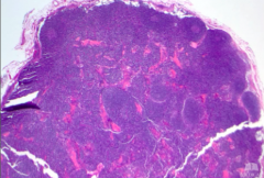
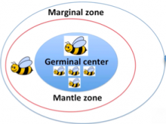
What are the layers of the follicle?
|
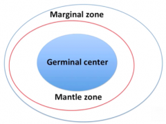
- Germinal center (central)
- Mantle zone (outside germinal center) - Marginal zone (outermost) |
|

What happens in the germinal center of a follicle?
|
- Where significant B cell development occurs
- Frequently where B cells encounter Ag and proliferate in response to antigen |
|

What happens in the mantle zone of a follicle?
|
B cells reside here
|
|

What happens in the marginal zone of a follicle?
|
B cells reside here
|
|
|
What components of the follicle do you see in a normal LN?
|
- Frequently see germinal center and mantle zone
- Less frequently see a marginal zone (more common to see it in the spleen follicle) |
|
|
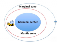
What happens in the first step of B cell development?
|

Naive B cells enter LN
- Never encountered Ag - Exist in mantle zone |
|
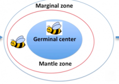
What happens in the second step of B cell development, after naive B cells enter the LN?
|

Upon Ag presentation / exposure:
- B cells migrate into germinal center - Germinal center is where B cell development, somatic hyper-mutation, and proliferation occurs |
|
What happens in the third step of B cell development, after naive B cells are exposed to Ag and enter germinal center?
|
- B cells are ready to proliferate
- B cell proliferation occurs in germinal center (becomes very crowded area of follicle) |
|

What happens in the fourth step of B cell development, after B cells proliferate in germinal center?
|

- B cells migrate out to reside within marginal zone (not easily visualized)
OR - B cells traverse into blood |
|
|
What is the most crowded part of the follicle of a LN?
|
Germinal center (central part)
|
|
|
What is the thin rim around the germinal center of a LN follicle?
|
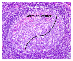
Mantle Layer (very dark, dense small cells)
|
|
|
What can the germinal center be divided into morphologically?
|
Light Zone
Dark Zone |
|
|
What happens in the light zone of the germinal center?
|
- Number of smaller cells ("centrocytes")
- Less proliferative (haven't gone onto gain proliferation capacity because they haven't been completely Ag stimulated) |
|
|
What happens in the dark zone of the germinal center?
|
- Contains large proliferating cells ("centroblasts")
- More dispersed, delicate chromatin (more blast-like) |
|
|
Is it reactive or malignant to have light zone / dark zone polarity in a LN?
|
Normal reactive morphology contains polarized follicle w/ designation into dark and light zones
|
|
|
Where are centrocytes found? What do they look like?
|
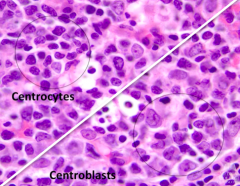
- Light zone
- Smaller cells - Coarser and condensed (purple) chromatin - Irregular nuclei (cells are just being stimulated and just starting to undergo processes of Ag exposure) |
|
|
Where are centroblasts found? What do they look like?
|

- Dark zone
- Open and delicate chromatin - Nuclei not very purple - Characteristic blastoid chromatin pattern (more immature) - Larger cells (2-3x size of smaller centrocytes) - Lots of cytoplasm (these cells are proliferating) |
|
|
What is the term for the macrophages found in reactive LN follicles?
|

Tingible Body Macrophages
|
|
|
When do Tingible Body Macrophages appear?
|

When there's cell turnover or cell proliferation (eg, during reactive process)
|
|
|
What is the term for the dendritic cells found in reactive LN follicles?
|
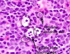
Follicular Dendritic Cells
|
|
|
What is the function of Follicular Dendritic Cells?
|

- Stromal component / meshwork
- Form framework of follicle (and germinal center) |
|
|
What is found in the mantle zone layer?
|
- Naive B lymphocytes that haven't encountered Ag yet
- This is where B cells originally reside before they encounter Ag and migrate into germinal center |
|
|
What is found in the paracortex?
|
- Predominantly houses T cells (helper and cytotoxic)
- High endothelial venules - Dendritic cells and macrophages |
|
|
What are High Endothelial Venules? Where are they found?
|

- Lined by plump, cuboidal endothelial cells
- Lumen within central portion - Found in paracortex |
|
|
What is the function of High Endothelial Venules in the Paracortex?
|
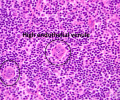
- Important role in migration of lymphocytes to and from the LN
- Allow lymphocytes to ENTER the LN |
|

What does the teal arrow represent?
|

High Endothelial Venule
|
|

What does the small purple arrow represent?
|
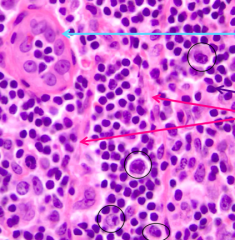
Normal T cell
|
|

What do the red arrows represent?
|

Thin walled vessels
|
|

What does the circled structures represent?
|

Several larger cells w/ big prominent nucleoli
|
|
|
What happens to the lymph node in patients w/ viral syndrome?
|
- LN paracortical area frequently becomes hyperplastic or reactive appearing
- T cells play an important role in mediating viral infections |
|
|
What are the compartments of the spleen?
|

- Red pulp
- White pulp |
|
|
What happens / is found in the Red Pulp of the spleen?
|
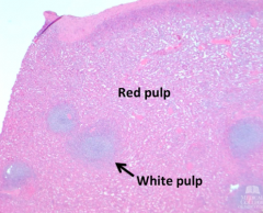
- Filters blood and RBCs
- Majority of the splenic parenchyma |
|
|
What happens / is found in the White Pulp of the spleen?
|
- Immune system: lymphoid tissue component of spleen
- Where immune effect occurs - T cell rich areas and B cell rich areas |
|
|
What is the location rich in T cells in the spleen?
|
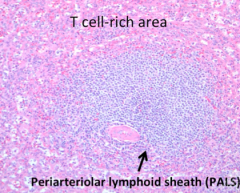
Periarteriolar Lymphoid Sheath (PALS) of White Pulp
|
|
|
What is the location rich in B cells in the spleen?
|
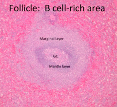
Follicles of White Pulp (histologically mimic LN follicles)
|
|
|
What is found in the Periarteriolar Lymphoid Sheath (PALS) of White Pulp of the spleen?
|

Dominated by helper T cells
|
|
|
What is found in the follicles in the white pulp of the spleen?
|

B cells
|
|

What is in this image?
|

- Arteriole = pink circle in middle
- Lymphocytes = small round blue cells surrounding arteriole (dominated by T cells) - Splenic sinusoids = red pulp in periphery (contains RBCs, macrophages, and CT) |
|

What is in this image?
|
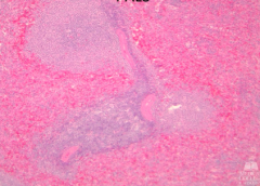
- 3 arterioles (pink areas in center)
- Lymphocytes = pronounced aggregate of small round blue cells surrounding arterioles * This specimen is from a trauma: RBCs are congesting the red pulp (periphery of PALS) - intensely red areas |
|
|
What does a spleen that has undergone trauma look like?
|

RBCs are congesting the red pulp (periphery of PALS) - intensely red areas
|
|
|
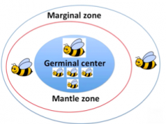
What are the components of a follicle in the spleen?
|

- Germinal Center (GC) - central portion of follicle
- Mantle Layer - zone around GC, dark blue - Marginal Layer - zone peripheral to mantle zone, outermost layer of follicle |
|
|
What is the main difference between follicles in lymph nodes vs spleens?
|

- In spleen the marginal zone is very pronounced
- Unusual to see the marginal zone in a LN unless its very reactive |
|
|
What is found in the germinal center of a follicle?
|
- Centrocytes
- Centroblasts - Macrophages - Dendritic cells |
|

What characterizes the L side vs R side?
|
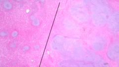
- L: normal spleen
- R: spleen w/ lymphoma |
|
|
What are the features of a spleen with lymphoma?
|

- Big nodules of white pulp (purple, looks like lymphocytes)
- Obliteration of architecture (R side) suggests malignancy - See little red pulp peripherally, but completely lost centrally - Red pulp replaced by big huge nodules of lymphocytes (obliterates normal) |
|
|
What are the sites of Mucosa-Associated Lymphoid Tissue (MALT)?
|
- GI tract (principle location)
- Respiratory tract - Oral mucosa - Ductal mucosa (salivary, breast, etc) |
|
|
Where is there Mucosa-Associated Lymphoid Tissue (MALT) int he GI?
|
Entire tract from stomach to colon
- Pockets of lymphoid tissues that rest right under mucosa |
|
|
Why is it important to have Mucosa-Associated Lymphoid Tissue (MALT) across the entire GI?
|
GI is constantly being bombarded by contents coming through the gut that are foreign to body; very active in what they're encountering (as far as Ags go)
|
|
|
What kind of tissue is important to have in ductal mucosa (eg, salivary or breast)?
|
Mucosa-Associated Lymphoid Tissue (MALT) because you may need an antigenic response
|
|
|
What is the function of the Mucosa-Associated Lymphoid Tissue (MALT)?
|
- Filters luminal (gut, ducts, cavities) contents
- Carries contents to regional LNs via efferent lymphatics |
|
|
Where are Peyer's Patches? What is the organization?
|
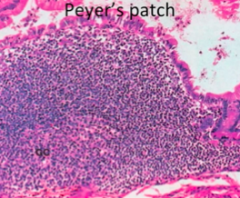
Small Bowel epithelium (bright pink cells)
- Dark blue small lymphocytes are predominantly B cells because it is follicular - Mantle zone surrounds Germinal Center |
|
|
What kind of tissue lines the oral cavity?
|
Stratified squamous epithelium
|
|
|
What is the appearance of Palatine Tonsils?
|
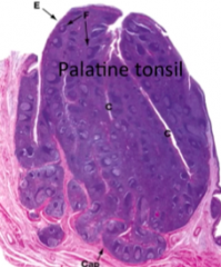
- Look likes a LN at this power
- Contains follicles w/ germinal centres and mantle zones |
|
|
Where is lung Mucosa-Associated Lymphoid Tissue (MALT) located?
|
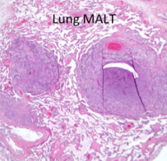
- Alveolar spaces
- Respiratory epithelium - Bronchiole epithelium * MALT is found right under basement membrane of epithelium |