![]()
![]()
![]()
Use LEFT and RIGHT arrow keys to navigate between flashcards;
Use UP and DOWN arrow keys to flip the card;
H to show hint;
A reads text to speech;
50 Cards in this Set
- Front
- Back
|
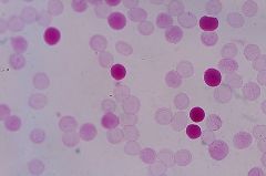
Punch-out vac blasts (L3 burkitt's conversion) |
|
|
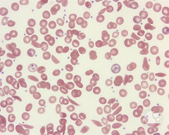
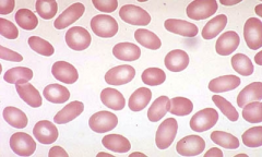
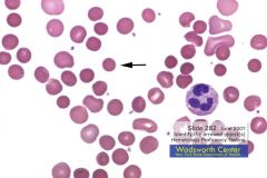
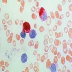
HgB S (sickle cell trait/disease) |
|
|
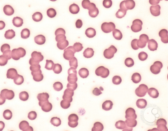
Rouleaux (MM, plasma cell leukemias) |
|
|
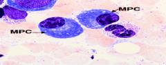
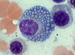
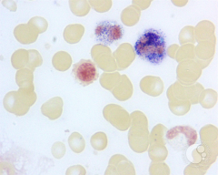
Plasma cell (Ig's are blue) |
|
|
Motte cell (russel bodies Ig's) |
|
|
Flame Cell (IgA, MM, plasma cell leukemia) |
|
|
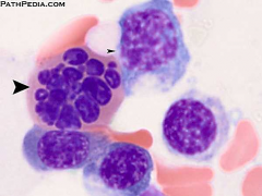
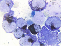
Auer rods |
|
|
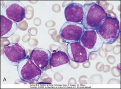
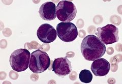
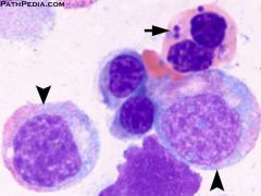
M3 (Promyelo) |
|
|
M3 (Auer rods, Promyelo) |
|
|
M3m (associated with DIC granules activate PLT) |
|
|
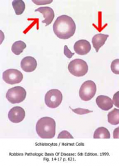
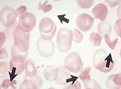
Schistocytes (Hemolytic anemia, DIC) |
|
|
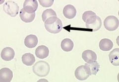
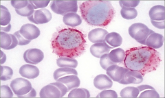
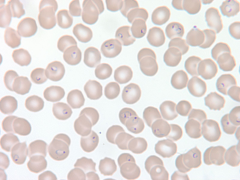
Pappenheimer (Unused iron) |
|
|
Ringed sideroblast (prussian blue) may see in MDS |
|
|
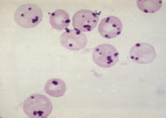
Basophillic stippling (coarse=lead poisoning/sideroblastic anemia) |
|
|
Megaloblastic anemia (look for B12/anti-IF/anti-PC/folate) |
|
|
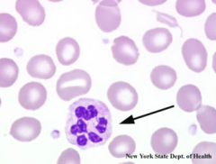
Left shift (CML vs leukomoid) |
|
|
LAP stain (increased in leukomoid, lowered in CML) |
|
|
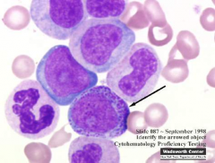
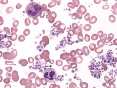
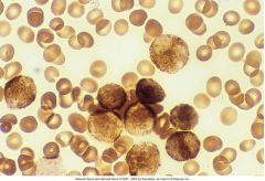
Hairy cell leukemia (TRAP POS) |
|
|
TRAP pos (Hairy cell) acid phosphatase isoenzyme #5 |
|
|
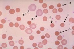
Hypochromic microcytic (Thal, Hgbopathies, sideroblastic anemia, AOI, IDA) |
|
|
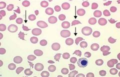
Elliptocytosis (spectrin-ankyrin defect horizontal) |
|
|
M5a (MPO+ SBB+ Spe= Nsp +++ NaF neg) No mature component |
|
|
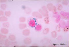
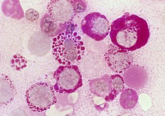
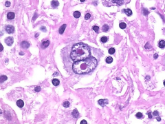
Malaria |
|
|
Malaria (lyses cells to cause problems) |
|
|
Many blasts and NRBC in M6a |
|
|
Granulocytes and nucleated RBC in M6a |
|
|
Nucleated RBC in M6a |
|
|
Pas +, coupled with myelocyte component makes M6a |
|
|
HgB SC |
|
|
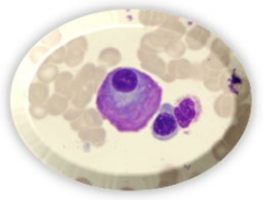
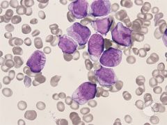
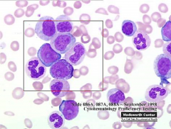
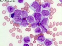
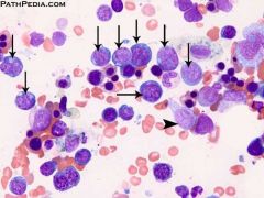
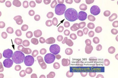
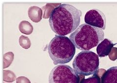
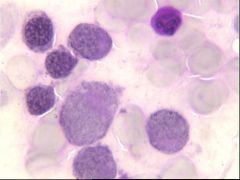
Promyelocytes/Blasts |
|
|
Heinz bodies on Supravital stain (denatured Hgb). Inidcate G6PD def. Alpha thal (H and barts) |
|
|
Smudge cells (ALL/CLL) clear smudging with ALBUMIN |
|
|
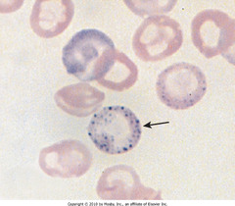
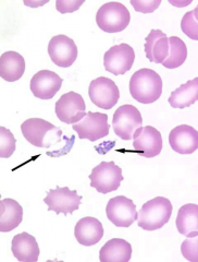
Pelger hewit cells |
|
|
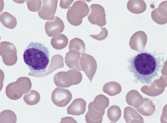
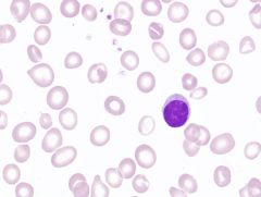
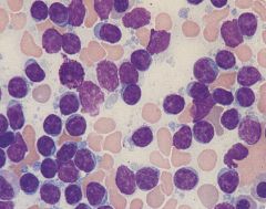
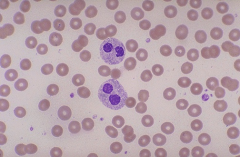
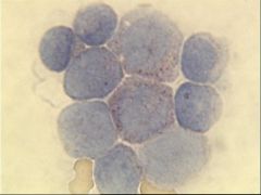
Blasts (LYMPH) |
|
|
PAS + (ALL) |
|
|
Sphereocytes and Retics=hemolysis likely |
|
|
Spherocytes. Confirm with osmotic fragility (increased) |
|
|
Beta thal hemolysis, Alpha-only HgB are unstable and destroy cells. |
|
|
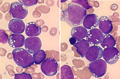
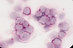
ET or M7 (too many PLT) |
|
|
ET or M7, too many megakaryocytes |
|
|
Kleihauer betke stain=look for FMH. HgB F doesn’t elute in acid. |
|
|
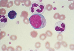
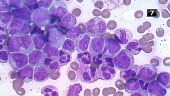
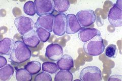
Blasts |
|
|
MPO + |
|
|
SBB + |
|
|
PAS = |
|
|
Spe + |
|
|
Nsp w+ likely M1 |
|
|
M2, increased BASO in blood likely |
|
|
M4 (MPO + SBB+ SE +, NSE +++ NaF w+) SE and NSE indicate myeloid component |
|
|
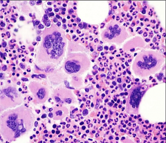
Reed Sternberg cells, Hodgkins lymphoma (see only in lymph) |