![]()
![]()
![]()
Use LEFT and RIGHT arrow keys to navigate between flashcards;
Use UP and DOWN arrow keys to flip the card;
H to show hint;
A reads text to speech;
49 Cards in this Set
- Front
- Back
- 3rd side (hint)
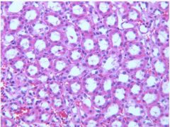
What organ is this and which structures are present? |

Liver - bile duct - hepatic artery - portal vein |
|
|
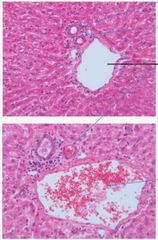
What structure is this? |

|
|
|

What structure is this? |
Portal triad (Portal vein, Hepatic artery, Bile duct) |
|
|

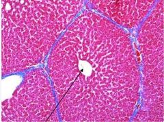
What structure is this? |
Liver lobule |
|
|
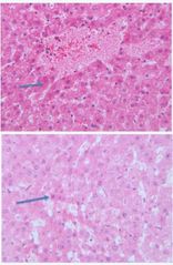
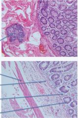
What structure is this and which organ is it present in? |
Kupffer cells Liver |
|
|
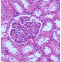
What structure is this and which organ is it located in? |
GlomerulusKidney |
|
|

What structure is this and which organ is it located in? What are the arrows pointing at? |
Proximal convoluted tubules in Kidney
Arrows pointing at brush border |
Tubule |
|
What structure is this and which organ is it located in? |
Distal convoluted tubules |
No brush border, simple cuboidal cells |
|

What organ is this? What are each of the lines pointing to? |

Colon - Taenia coli - Muscularis externa - Submucosa - Mucosa |
|
|
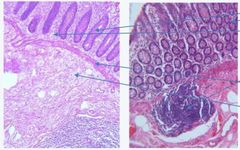
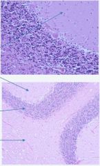
What tissue is this and what are the arrows pointing to? |

Colon Peyer's Patches! |
|
|
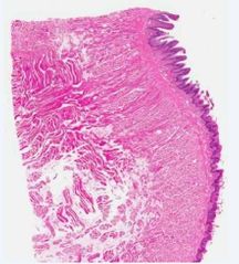
What tissue is this and what are the arrows pointing to? |

Colon |
|
|
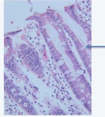
What type of cells is the arrow pointing to? |
Goblet cells |
Located in colon, mucin-secreting epithelial cells |
|

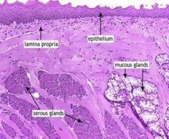
What tissue is this and what are the arrows pointing to? |

Spleen |
|
|

Is this red or white pulp? Which tissue is this found in? |
Red pulp in the spleen |
|
|
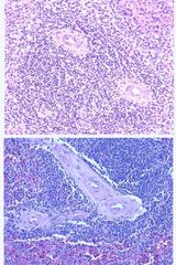
Is this red or white pulp? Which tissue is this found in? |
White pulp in the spleen |
|
|
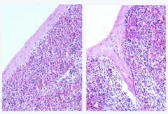
What structures in the spleen are these? |
Capsule & Trabeculae |
|
|

What structure is this and which tissue is it found in? |
Bronchiole
Lung |
|
|

What structure is this and which tissue is it found in? |
Alveoli Lung |
|
|
|
Type of epithilium: bronchi vs bronchioles |
Bronchi - pseudostratified ciliated columnar epithelium
Bronchioles - simple ciliated cuboidal epithelium |
|
|
|
Type of epithilium: type I vs type II alveolar cells |
Type I - flat, squamous cells & cover 90-95% of alveolar surface (gas exchange) Type II - almost cuboidal & secrete pulmonary surfactant |
|
|

What tissue is this? |
Skeletal muscle |
|
|

What is the skeletal muscle structure the arrow is pointing to? |
Intrafusal fibres |
|
|

What tissue is this? |
Cerebral Cortex - Brain |
|
|
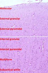
What is this tissue? |
Cerebral cortex |
|
|
What tissue is this and what are the arrows pointing to? |
Brain
Neurons |
|
|

What tissue is this? |
Brain |
|
|
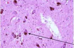
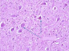
What tissue is this and what is the arrow pointing at? |
Brain Substantia Nigra |
|
|
|
What are Substantia Nigra in the brain (what do they contain and produce)? What disease can their degeneration cause? |
Pigmented neurons containing neuromelanin and produce dopamine. Degeneration is a cause of Parkinson's Disease. |
|
|
What are the layers of the cerebellum? |
First two arrows - molecular layer 3rd arrow - granular layer 4th arrow - white matter (myelinated) |
|
|
|
Which 6 cerebellar cell elements are stained with H&E? |
1 - Golgi cells 2 - Basket cells 3 - Granular cells (excitatory neurons) 4 - Purkinje cells 5 - Purkinje cell dendrites 6 - White matter |
|
|
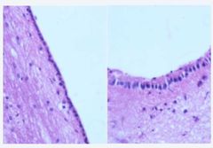
What is this ciliated epithelium between the brain parenchyma and ventricles called? |
Ependyma
- polygonal, cubodial to columnar cells, depending on location in ventricles |
|
|

What are these tufts of capillaries with thin fenestrated endothelial cells, covered by simple cuboidal epithelium called? What do they produce? |
Choroid Plexus
Produces most CSF |
|
|

What is this structure found in the brain called? |
Meninges |
|
|

What are these structures and which tissue are they found in? |
Islets of Langerhans in the Pancreas |
|
|

What are these exocrine ducts found in the Pancreas called? What type of epithelium are they lined with? |
1 - intralobular duct - cuboidal
2 - primary interlobular duct - columnar |
|
|

What is this structure found in the Pancreas called? |
Islet of Langerhans |
|
|

What tissue is this? |
Cardiac muscle |
|
|

What are specialised conducting fibres in cardiac muscle called? |
Purkinje Fibres |
|
|

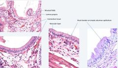
What tissue is this and what are the arrows pointing to? What epithelium lines this tissue? |
Gall bladder 1 - Mucosal folds 2 - Muscular layer Simple columnar epithelium |
|
|
What is this tissue? |
Gall Bladder |
|
|
What is this tissue? What epithelium is the mucosa lined with? |
Tongue Keratinised stratified squamous epithelium |
|
|
What is this tissue? |
Tongue |
|
|

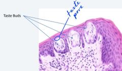
What is this structure found in the tongue called? |
Papillae |
|
|

What are these structures in the tongue called? |
|
|
|

What tissue is this and what are each of the layers called? |
Skin
1 - Epidermis 2 - Dermis 3 - Hypodermis |
|
|
|
What epithilium is the epidermis made up of? |
Keratinised, stratified squamous epithelium |
|
|

What tissue is this? |
Skin |
|
|

What structures are these and which tissue are they found in? |
Hair follicles found in Skin |
|
|

What is this structure found in the skin called? |
Pacinian corpuscle Detects movement and impact, provides tactile information |
A mechanoreceptor found in subcutaneous layer of skin |