Reading...
![]()
Play button
![]()
Play button
![]()
Use LEFT and RIGHT arrow keys to navigate between flashcards;
Use UP and DOWN arrow keys to flip the card;
H to show hint;
A reads text to speech;
58 Cards in this Set
- Front
- Back
|
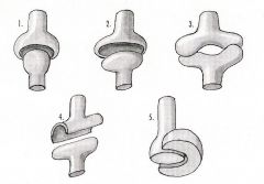
Type of motion for MCP? (3)
|
Flexion - extension
Abduction - adduction Circumduction - rotation |
|
|
Type of motion for PIP?
|
Flexion - extension
|
|
|
ROM for MCP?
|
-15 to 90
RD 10 - 20 UD 20 - 40 |
|
|
Tends toward stiffness of for MCP? PIP?
|
MCP = extension
PIP = flexion |
|
|
When are collateral ligaments tight for MCP? PIP?
|
MCP = flexion (+CAM effect)
PIP = All positions (No CAM effect) |
|
|
Is hyperextension possible for MCP? PIP?
|
MCP = yes
PIP = no |
|
|
Dimensions of Volar plate length for MCP and PIP?
|
MCP = 10mm (flexion), 15mm (extension)
PIP = 9mm |
|
|
Dimensions of Deep transverse MC ligament for MCP?
|
10mm
|
|
|
Dimensions Collateral ligament for MCP?
|
14-18mm
|
|
|
List the volar plate attachments at the MCP (7)
|
Deep Transverse MC ligament
Interosseous fibers Palmar Aponeurosis Accessory collateral ligament Sagittal band 1st dorsal interossei (radial index digit) Abductor digiti quinti (ulnar small digit) |
|
|
Name the volar plate attachments of the PIP (2)
|
P2 base (firmly)
P1 neck (loose) |
|
|
Volar plate strength of the MCP and PIP?
|
MCP = 6-8 kg
PIP = 16 kg |
|
|
Site of Volar plate rupture for MCP and PIP?
|
MCP = proximal
PIP = distal |
|
|
The PIP has significant __1__ stability secondary to __2__
|
1. Lateral
2. Bony anatomy |
|
|
The __1__ provide a strong attachment to the volar plate at the __2__ aspect of the PIP
|
1. Check ligaments
2. proximal |
|
-
|
|
|
|
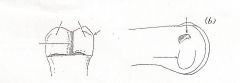
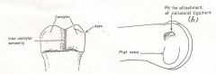
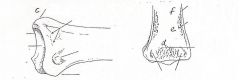
PIP Joint Bony Anatomy - Head of the proximal phalanx:
- 2 condyles separated by the __1__ - Lateral condyle has a __2__ directed apex - There is a lateral pit __3__ to the mid-axis for attachment of the __4__ |
1. intercondylar cavity
2. Transversely 3. dorsal 4. collateral ligament |
|
-
|
-
|
|
|
The Base of the Middle Phalanx has a __1__- shaped ridge that articulates with the __2__ of the __3__
|
1. saddle
2. intercondylar cavity 3. Head of the proximal phalanx |
|
|
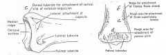
Name the 3 turburcles of the Base of the Middle phalanx and what inserts there
|
Dorsal = Central slip
Lateral = collateral ligament Palmar = C2 pulley and capsule |
|
|
On the Base of the Middle Phalanx there are roughenings for...(3)
|
Palmar plate
FDS tendon A4 fibrous flexor sheath |
|
-
|
-
|
|
|
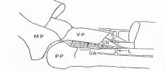
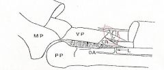
The Volar plate of the PIP attaches to the __1__ and __2__ margin of the proximal phalanx, also referred to as __3__
|
1. Middle phalanx
2. volar-lateral 3. triangular check ligaments |
|
|
The PIP volar plate has a free edge centrally over __1__ allowing for greater __2__ and formation of the __3__
|
1. P1
2. flexion 3. "retrocondylar recess" |
|
|
PIP Capsule: The synovial capsusule is stabilized by connecting tissue on all sides
-Dorsal: __1__ -Volar: __2__ -Lateral: Superficial - __3__ / Deep __4__ |
1. Extensor mechanism (central slip, lateral bands)
2. Volar plate, check ligaments, flexor sheath attachment 3. Transverse ligaments, oblique retinacular ligaments 4. capsule, collateral ligaments, accessory collateral ligament |
|
|
__1__ lie on the slopes of the condyles of the proximal phalanx head. They are dorsal in __2__ and slide volarly in __3__
|
1. Lateral bands
2. extension 3. flexion |
|
|
The Collateral ligament lies __1__ to the apex of joint angulation when in __2__
|
1. Dorsal
2. extension |
|
|
The Check Ligament is a normal confluence of __1__. They are reflected fibers of the __2__ and the lateral margin of the __3__
|
1. fascial structures
2. flexor sheath 3. volar plate |
|
|
Pathologic thickening of the Check Ligaments are called?
|
Checkreign Ligament (swallotail extensions of volar plate along volar proximal phalanx which may thicken producing PIP flexion contracture)
|
|
|
The Checkrein ligament plays a role in ___1___
|
PIP Flexion Contracture
|
|
|
PIP Vascularity
-The Proper Digital Artery gives off the __1__ in the region of C1 just proximal to PIP -The Proximal Transverse Digital Artery passes between __2__ and __3__ in the __4__ toward midline -The Proximal Transverse Digital Artery supplies the __5__ and __6__ |
1. Proximal transverse digital artery
2. bone 3. check ligament 4. "fibro-osseous tunnel" 5. VBS & VLP |
|
|
MCP Joint: Bone Anatomy
-The trapezoidal head is wider __1__, thus stabily in __2__ and mobility in __3__ - CAM effect of collaterals is due to __4__ origin of the collateral ligament __5__ to the axis of rotation |
1. volarly
2. flexion 3. extension 4. eccentric 5. dorsal |
|
|
MCP Joint Bone Anatomy:
-Volar bony prominences on the MC head aid in increasing collateral ligament tension in flexion from __1__ -Radiovolar: less pronounced from __2__ -Ulnovolar: less pronounced from __3__ |
1. 60-90
2. Index to small 3. Small to index |
|
|
MCP Joint Bone Anatomy:
__1__% of thumb MC heads are flat, thus the joint then acts more like a __2__ |
1. 10
2. Hinge |
|
|
MCP Joint: Deep Transverse Metacarpal Ligament (Interpalmar Plate Ligament)
- There are __1__ (#) ligaments - Interossei pass __2__ (volar/dorsal) to the Interpalmar plate ligament -Lumbricals and NV bundle pass __3__ (volar/dorsal) to the Interpalmar Plate Ligament |
1. 3
2. dorsal 3. volar |
|
|
MCP Joint Capsule: The Synovial Capsule is stabilized by connecting tissue on all sides
-Dorsal: __1__ (2) -Volar: __2__ (2) -Lateral: Superficial __3__ (2) / Deep __4__ (3) |
1. Extendor tendon, Sagittal band
2. Volar plate, Deep transverse metacarpal ligament 3. Sagittal bands, Intrinsic muscles 4. Capsule, Collateral Ligament, Accessory Collateral ligament |
|
|
Dorsal Dislocation of MCP:
-Volar incision for reduction is high risk to __1__ -Dorsal incision for reduction: __2__ is felt to be the primary reduction block - it is split __3__ to allow reduction |
1. digital nerves
2. volar plate 3. longitudinally |
|
|
Irreducible dorsal dislocation of the MCP causes skin "dimpling" secondary to?
Pretendinous bands are? |
Pretendinous bands and transverse fibers of palmar fascia connecting to skin
Longitudinal bands of palmar fascia |
|
|
Thumb MCP Joint: Bony Anatomy
- Type of joint? (90% _1_, 10% _2_) - Is there similar joint contact in flexion and extension? __3__ - Sesamoids are located in the __4__. - Radial Sesamoid characteristics __5__ - Ulnar sesamoid characteristics __6__ - ROM for flexion - extension |
1. condyloid
2. hinge 3. yes 4. volar plate 5. Flexor pollicis brevis attachment, larger sesamoid 6. Adductor pollicis brevis attachment, "Gillette's scaphoid" - carved facet. 7. 8 - 53 flexion - extension |
|
|
Thumb MCP Joint: Capsule
- Superficial Layer - Ulnar (2) - Radial (2) - Dorsal -Deep Layer (6) |
Ulnar - Adductor pollicis, Dorsal aponeurosis of adductor pollicis
Radial - APB, FPB Dorsal - EPL, EPB Deep: Ulnar collateral ligament, ulnar accessory collateral ligament, radial collateral ligament, radial accessory collateral ligament, dorsal capsule, FPL |
|
|
Thumb MCP Joint Capsule:
- Superficial and deep layers fuse with the volar plate __1__ (dorsal/palmar) - The adductor aponeurosis stretches from the ulnar margin of the __2__ and __3__ dorsally to the __4__ -The capsule deep layer attaches to within __5__ mm distal to articular surface -The volar plate proximal is __6__, and distal __7__ |
1. palmarly
2. volar plate 3. ulnar sesamoid 4. EPL 5. 1.5 - 2 mm 6. thin/pliable 7. Thick/rigid |
|
|
Thumb UCL injury:
- Diagnosis: - Lack of a firm end-point on stress in extension (__1__) and __2__ degrees flexion (__3__) - __4__ degrees of absolute __5__ deviation on stress - __5*__ degrees difference in radial deviation compared to contralateral thumb - __6__ ligament may tear proximally or distally -What is Stener Lesion? __7__ - What must be injured to have Stener lesion? __8__ - Injury to the collateral and accessory collateral = __9__% chance of Stener |
1. accessory collateral lig
2. 30 degrees 3. collateral ligament 4. 30-40 degrees 5. radial 5*. 15 degrees 6. Accessory collateral ligament 7. interposition of adductor aponeurosis between torn ends of collateral ligament 8. Accessory collateral ligament 9. 89% |
|
|
The CMC Joint of the Thumb = ?
|
Trapezio-Metacarpal joint
|
|
|
Trapezio-Metacarpal Joint: Bony Anatomy
-2 reciprocally opposed __1__ whose axes are __2__ - There is significant inherent BONY instability (no congruence) -> ligamentous restraint and dynamic __3__ stabilization are primary |
1. saddles
2. perpendicular 3. APL *2 reciprocally opposed "Pringle" potato chips |
|
|
Trapezio-Metacarpal Joint Bony Anatomy: Trapezial surface
- __1__ running ridge crossing the greatest diameter - Two parts: __2__ - The groove crosses the ridge perpendicular connecting the 2 parts |
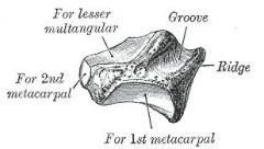
1. Obliquely
2. Palmar - Radial :: Slightly concave Dorsal - Ulnar :: Slightly concave |
|
|
Trapezio-Metacarpal Joint: Metacarpal Base
-__1__ base with a __2__ and __3__ beak -There is a groove corresponding to the obliquely running ridge on trapezial surface |
1. Pentagonal
2. volar 3. ulnar |
|
|
Thumb CMC Joint: LIgamentous Restraint:
3 axes of motion |
Flexion - Extension
Adduction - Abduction Pronation - Supination **Motion is coupled = no independent axis of motion** |
|
|
Name the 5 ligaments of the Thumb CMP joint
|
Anterior oblique
Posterior oblique Dorsoradial Ulnar collateral Intermetacarpal |
|
|
What 3 Thumb CMC ligaments are intracapsular and what 2 are extracapsular?
|
Intracapsular = Anterior oblique, Posterior oblique, Dorsoradial
Extracapsular = Ulnar collateral, Intermetacarpal |
|
|
The Anterior Oblique and Ulnar collateral ligaments of the Thumb MCP are taut in what positions?
|
Extension, abduction, pronation
|
|
|
The Posterior Oblique and Intermetacarpal Ligaments are taut in what position?
|
Abduction, opposition, supination
|
|
|
When is the dorsoradial ligament taut?
|
Supination (extreme)
|
|
|
This ligament is the primary stabilizer of the thumb CMC
|
Anterior oblique
|
|
|
Anterior Oblique Ligament:
- runs from the thumb __1__ ulnarly to the __2__ - located opposite the __3__ - it is taut in __4__, limiting __5__ translation -Degeneration and detachment correlates with extent of __6__ |
1. MC beak
2. trapezium 3. APL dorsal expansion 4. pronation 5. dorsal 6. CMC cartilaginous disease |
|
|
3 types of CMC cartilaginous changes?
|
Chondromalacia - dorsal: no correlation with attrition of AOL
Chondromalacia-Palmar: Positive correlation with attrition of AOL Ebernation: only in palmar joint and always adjacent to AOL |
|
|
This CMC ligament is likely a check-rein to radial subluxation, dislocation
|
Dorsoradial ligament
|
|
|
Radial Artery Course:
- distal forearm: -radial to __1__ -Floor of the snuffbox is between __2__ -Runs distally along the __3__ joint toward the thumb and index __4__ -Travels distally between __5__ -Enters the palm to form the __6__ between the 2 heads of the __7__ and is adjacent to the __8__ nerve |
1. FCR
2. 1st and 3rd extensor compartments (1st = APB, EPB / 3rd = EPL) 3. Scaphotrapezium-Trapezoid joint 4. MC bases 5. 1st dorsal interossei 6. Deep palmar branch 7. adductor 8. deep motor branch of ulnar nerve |
|
|
CMC Joint Reactive Forces:
- Pinch: __1__ - Shear stress: __2__ |
1. 13x
2. 26x |