Reading...
![]()
Play button
![]()
Play button
![]()
Use LEFT and RIGHT arrow keys to navigate between flashcards;
Use UP and DOWN arrow keys to flip the card;
H to show hint;
A reads text to speech;
45 Cards in this Set
- Front
- Back
|
|
|

|
|
|

|
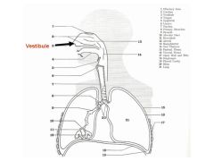
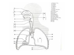
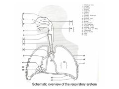
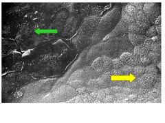
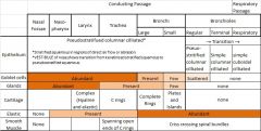
Surface topography of nasal cavity seen at 3
|
|
|
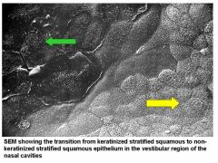
Coming in from outside, would see a fully keratined epithelium at GREEN arrow.
Then becomes nonkeratinzed epithelium at YELLOW arrow |
|
|

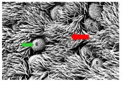
Going further back in more protected regions of respiratory cavity, see respiratory epithelium. The respiratory epithelium is a pseudostratphied columnar epithelium characterized by numerous goblet cells at GREEN arrows dispersed among cilliated cells (RED arrow)
|
|
|
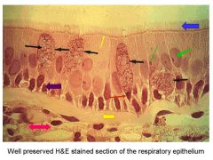
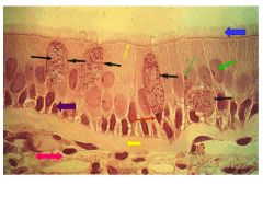
Respiratory epithelium
BLACK: mucin in goblet cells RED ARROW: nucleus of goblet cell large GREEN arrow: nucleus of cilliated cell. Note that it is situated higher than the nucleus of the goblet cell. The nucleoli of the cilliated cell is often obvious, can be seen at small green arrow. BLUE arrow: Numerous cilia can be seen on the free surface of the ciliated cell Subadjacent to the cilia are numerous basal bodies (small YELLOW arrow) most basally located = basal cells --> progenitor cells that can give rise to ciliated or goblet cells 3 layers of cell nuclei at 3 layers in epithelium that lie on same membrane thus, PSEUDOSTRATIFIED COLUMNAR Large YELLOW area: thick basement membrane PINK arrow: lamina propria is at two headed Connective tissue. |
|
|
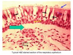
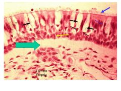
Large BLACK arrows: regions in goblet cells that once contained mucin that was leeched out during processing
small BLACK arrows: goblet cell nuclei YELLOW: basal cell nuclei small BLUE arrow: nucleus of ciliated cell large BLUE arrow: cilia from surface of ciliated cell large GREEN area: thick basement membrane upon which respiratory epithelium rests under ALL that that is connective tissue of lamina propria |
|
|
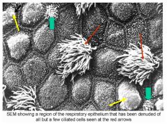
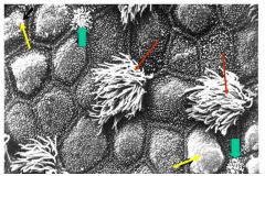
SEM (read slide)
RED arrows: cilliated cells YELLOW: apial portions of goblet cells that are swollen from accumulated mucinas accumulated mucin expands the apical plasmolemma, apical microprojections become encorporated into this membrane. Apical microporjections accumulate along cell borders and reveal hexagonal or pentagonal borders GREEN: brush cells. These can be identified by brush like microvilli on apical surfaces |
|
|
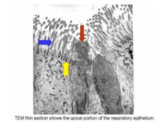
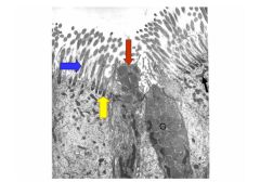
TEM thin section of respiratory epithelium
G = goblet cell. Filled w mucin granules swollen apical portion seen at red BLUE is cilia YELLOW is basal body supporting cilia both ciliated and goblet cells contain numerous mitochondria, which is demarcated by BLACK arrow |
|
|
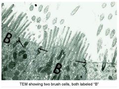
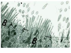
Two brush cells both labeled B.
Note microvillious projections emerging from apical portions of cells apical intracellular juctions can be seen between cells |
|
|

If you were to look into nasal cavities, would look pink/red from blood and underlying vessels
3 shell like structures called conchi because they curve down and look like shells upper/superior, middle, and lower/inferior conchi help create turbulance and facilitate contact of air with respiratory epithelium within conchi are large venous plexi called swell bodies can become engorged with blood and result in stuffy nose if you look at roof of nasal cavity, would look yellowish brown in contrast to pink of respiratory epithlium. Yellowish brown is olfactory region of nasal cavity specialized for smell. Olfactory Epithelium extends from middle roof of nasal cavity to upper surface of superior conchi and upper lateral side of nasal septum |
|
|
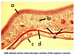
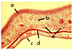
Superior conchae
olfactory epithlium at A thicker than respikratory epithlium (C) glands of bowman = serious glands = B watery secretions from glands of bowman help to bathe surface of olfactory epithelium helping to sense new odors nerves present at e arise from olfactory cells – phila olfactoria |
|

|
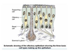
Olfactory epithelium shows 3 basic cell types:
1. supporting / sustentacular cells that contain brownish pigment similar to lipofuscin and gives it yellow/brown color 2. bipolar neurons making up sensory olfactory cells 3. basal cells since all 3 rest on same basement membrane, this epithelium like the respiratory epithelium, is psuedostratphied columnar epitheliumalso note the duct passing through olfactory epithelium and arising from underlying bownam glands |
|
|
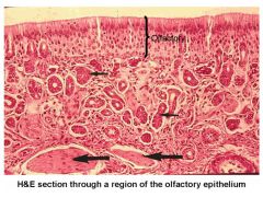
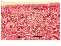
Bracket = olfactory epithelium
SMALL BLACK arrows = glands of bowman LARGER BLACK arrows = bundles of fibers making up fiber olfactoria |
|
|
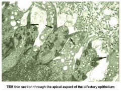
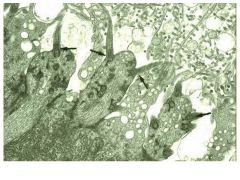
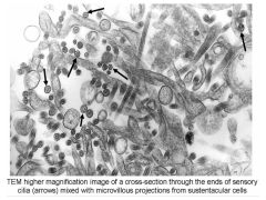
See slide.
Small black arrows indicate sensory cilia emerging from olfactory vesicle 10-15 nonmotile sensory cilia arise from each olfactory cell and extend for 100-200 um parallel to surface of mucosa start with 9+2 arrangement but become thinner and end with 1-2 microtubules in tapered ends |
|
|
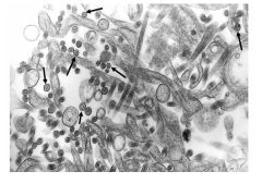
See slide.
Cross sectional images of microtubules in tapered ends. |
|
|

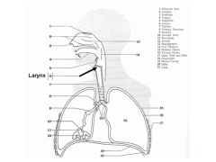
Next talking about larynx.
|
|

|
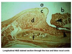
See slide.
A = true vocal cord covered by nonkeratinized stratified squamous epithelium due to the trauma associated with phonation The large vocalious muscle at d makes true vocal cord easy to recognize B = false vocal cord. Has respiratory epithelium and numerous serous mucus glands e = so called ventricle .. protective region, covered by respiratory epithelium thyroid cartilage at f provides support for larynx |
|

|
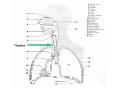
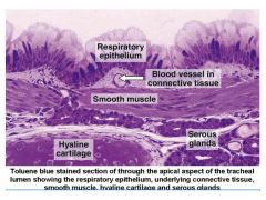
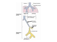
Trachea is about 11 cm long and 2-2.5cm in diameter
starts at larynx and ends when it divides to two 1’ bronchi hilar region where it enters lungs is also where blood and lymphatics enter lungs 16-20 horseshoe rings of hyaline cartilage |
|
|
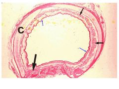
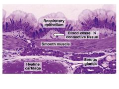
See slide.
Lining respiratory epithelium seen at small blue arrows small black arrows: extensive serious mucus glands in submucosa piece of trachialis muscle which spans ends of hyaline cartilage rings is seen at large black arrow cartilage at C provides flexible mechanical support for trachea |
|
|
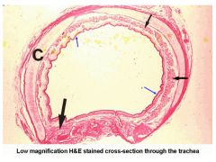
see slide.
Trachea @ higher magnification |
|
|

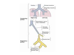
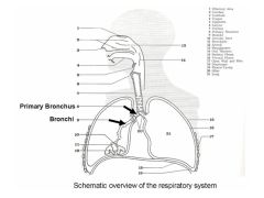
Primary bronchi have same histological characteristics as trachea enter substance of lungs and become intrapulmonary bronchi
within lungs branch/divide in dichotomous fashion (1--> 2--> 4) with total crosssectional area of the lumens of every pair > than mother lumen cartilage rings break up into cartilage plates after entering lung rest is same as trachea |
|
|
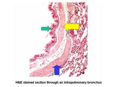
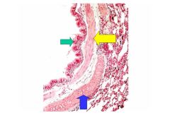
See slide.
GREEN arrow: respiratory epithelium YELLOW: smooth muscle originally derived from trachialis muscle, winds down bronchi in spiral fashion BLUE: piece of hyaline cartilage plate that supports bronchus |
|

|
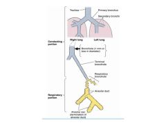
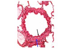
Bronchi branch until they form bronchioles, which have luminal diameter of 1 mm or less, no hyaline cartilage plate support, no glands in underlying lamina propria, and significantly reduced respiratory epithelium with few if any goblet cells.
|
|
|
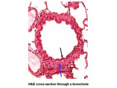
See slide.
-BLACK arrow identifies reduced respiratory epithelium BLUE = smooth muscle **characteristic of bronchioles |
|
|

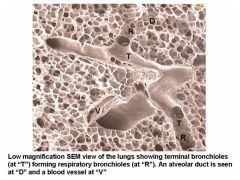
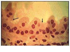
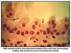
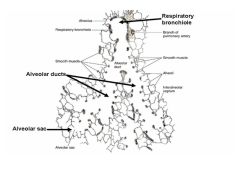
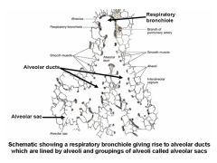
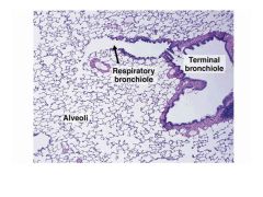
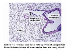
Bronchioles branch until they form smaller bronchioles called terminal bronchioles which are lined by dome shaped secretory cells called clara cells and by shortened ciliated cells. Terminal bronchioles branch to form respiratory bronchioles, the first portion of respiratory tract to have alveoli (exchange of gasses). Respiratory bronchioles are beginning of the respiratory portion of respiratory tract.
Lining epithlium of respiratory bronchioles is simple columnar/cubodial and characterized by short cilia |
|
|
|
|
|
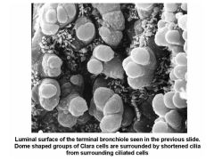
See slide. Note clara cells!!!
|
|
|
|
|
|
|
|
|
|
|
|

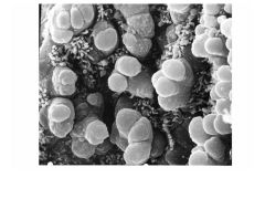
See slide.
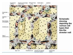
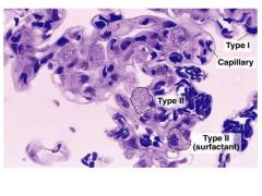
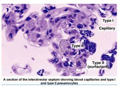
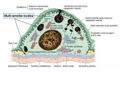
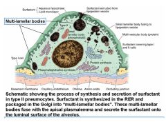
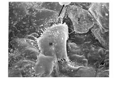
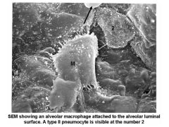
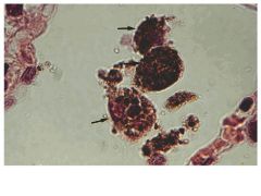
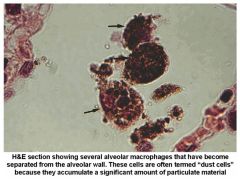
Most of cells are flattened squamous cells termed type I pneumocytes in close proximity to underlying capillaries type II pneumocytes are larger and synthesize and secrete surfactantalso have macrophages lining walls that are derived from monocytes supporting walls are reticular and elastic fibers alveolar pore supports flow between alveoli if airflow has been obstructed |
|
|
|
|
|
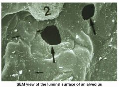
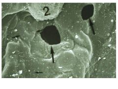
See slide.
Most of cells are thin type I pneumocytes thinness can be appreciated at small black arrows pointing out outline of RBC in underlying capillary 2 indicates a type II pneumocyte two alveolar pores are shown at large black arrows |
|
|
See slide.
Surfactant acts as whetting agent to reduce surface tension and make alveolar walls separate from one another and fill w air |
|
|
|
|
|
|
|

Describe structure of interalveolar septum
|
See slide.
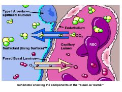
This barrier consists of layer of surfactant, thin type I pneumocyte, alveolar endothelium, and fused basal lamina produced by type I pneumocyte and the alveolar endothelium |
|
|
See slide.
Shows type I pneumocyte (ep) alveolar endothelium (ed) fused basal lamina (BM) – separate blood in alveolar capillaries from air in alveolar lumen |
|
|
See slide.
Both layers are continuous with each other at hilar region of lungs |
|
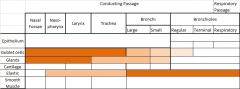
Describe the gradual histological changes in the conducting portion of the respiratory tract
|
|
|
Describe structure of interalveolar septum
|

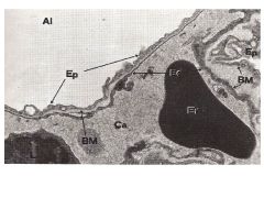
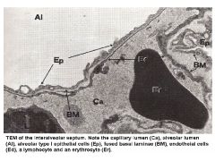
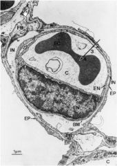
Electron micrograph showing a pulmonary capillary (C) in the alveolar wall. Note the extremely thin blood-gas barrier of about 0.3 µm in some places. The large arrow indicates the diffusion path from alveolar gas to the interior of the erythrocyte (EC) and includes the layer of surfactant (not shown in the preparation), alveolar epithelium (EP), interstitium (IN), capillary endothelium(EN), and plasma. Parts of structural cells called fibroblasts (FB), basement membrane (BM), and a nucleus of an endothelial cell are also seen.
Blood Vessels: whole output of R heart goes to lung diameter of capillaries (7-10 micrometers) thickness of much of blood gas barrier <0.3 micrometers blood spends about .75 seconds in the capillary Blood Gas Interface: Extremely thin (.2-.3 micrometers) 50-100m of surface area large area ~500 million alveoi So thin large increases in capillary pressure can damage the barrier |
|

|
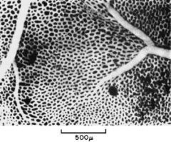
View of an alveolar wall (in the frog) showing the dense network of capillaries. A small artery (left) and vein (right) can also be seen. The individual capillary segments are so short that the blood forms an almost continuous sheet.
|
|

|
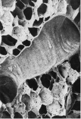
Section of lung showing many alveoli and a small brochiole. The pulmonary capillaries run in the walls of the alveoli. The holes in the alveolar walls are the pores of Kohn
|
|

|
Microscopic section of dog lung shows capillaries in the alveolar walls. The blood gas barrier is so thin that it cannot be identified here. This section was prepared from lung that was rapidly frozen while being perfused.
|