![]()
![]()
![]()
Use LEFT and RIGHT arrow keys to navigate between flashcards;
Use UP and DOWN arrow keys to flip the card;
H to show hint;
A reads text to speech;
35 Cards in this Set
- Front
- Back
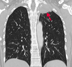
What type of lung cancer occurs in the apex of the lung? What can it do? |

Pancoast tumor: |
|
|
What are the symptoms of Horner syndrome? |
Ipsilateral ptosis, miosis, and anhidrosis |
|
|
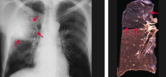
What happens in Superior Vena Cava Syndrome? |
Obstruction of the SVC impairs blood drainage from the head, neck, and upper extremities |
|
|
What are the implications of the blood drainage obstruction of the head, neck, and upper extremities in superior vena cava syndrome? |
- Head → facial plethora |
|
|
What can cause Superior Vena Cava Syndrome? |
Commonly caused by malignancy and thrombosis from indwelling catheters |
|
|
What can Superior Vena Cava Syndrome cause if the obstruction is severe? |
Can raise intracranial pressure → headaches, dizziness, and ↑ risk of aneurysm / rupture of intracranial arteries |
|
|
What are the types of pneumonia? |
- Lobar |
|
|
What are the typical causative organisms responsible for lobar pneumonia? |
- S. pneumoniae most frequently |
|
|
What are the typical causative organisms responsible for bronchopneumonia? |
- S. pneumoniae |
|
|
What are the typical causative organisms responsible for interstitial (atypical) pneumonia? |
- Viruses (influenza, RSV, adenoviruses) |
|
|
What are the characteristics of lobar pneumonia? |
Intra-alveolar exudate → consolidation |
|
|
What are the characteristics of bronchopneumonia? |
- Acute inflammatory infiltrates from bronchioles into adjacent alveoli |
|
|
What is the histologic appearance of bronchopneumonia? |
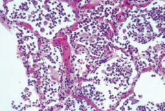
Neutrophils in alveolar space |
|
|
What are the characteristics of interstitial (atypical) pneumonia? |

- Diffuse patchy inflammation localized to interstitial areas at alveolar walls |
|

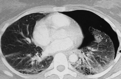
What does this chest x-ray show? |
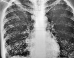
Interstitial Pneumonia: coarse bilateral reticular opacities, worse on the right side |
|
|
What is wrong in a lung abscess? |
Localized collection of pus within the parenchyma |
|
|
What can cause a lung abscess? |
Bronchial obstruction (eg, cancer) or aspiration of oropharyngeal contents (especially in patients predisposed to loss of consciousness [eg, alcoholics or epileptics]) |
|

What does this chest x-ray show? |
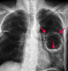
Lung Abscess |
|
|
What are the most common causes of lung abscesses? |
- S. aureus |
|
|
What is wrong in a pleural effusion? |

Excess accumulation of fluid between the two pleural layers → restricts lung expansion during inspiration |
|

What does this chest x-ray show? How would you describe it? |
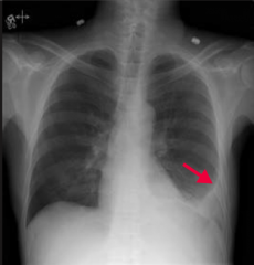
Pleural Effusion |
|
|
What are the types of pleural effusions? |
- Transudate |
|
|
What are the components of transudative pleural effusions? What can cause this? |
- ↓ Protein content |
|
|
What are the components of exudative pleural effusions? What can cause this? |
- ↑ Protein content |
|
|
What are the components of lymphatic pleural effusions / chylthorax? What can cause this? |
- ↑ Triglycerides, milky-appearing fluid |
|
|
What type of pleural effusion needs to be drained? Why? |
Exudative: must be drained due to risk of infection |
|
|
Which type of fluid is found in a pleural effusion caused by CHF, nephrotic syndrome, or hepatic cirrhosis? |
Transudate: ↓ protein content |
|
|
Which type of fluid is found in a pleural effusion caused by malignancy, pneumonia, collagen vascular disease, or trauma? |
Exudate: ↑ protein content |
|
|
Which type of fluid is found in a pleural effusion caused by thoracic duct injury from trauma or malignancy? |
Lymphatic / Chylothorax: ↑ triglycerides (milky-appearing) |
|
|
What is wrong in a pneumothorax? |
Accumulation of air in the pleural space |
|
|
What signs and symptoms occur in patients with pneumothorax? |
All on affected side: |
|
|
What are the types of pneumothorax? |
- Spontaneous pneumothorax |
|
|
What are the characteristics of a spontaneous pneumothorax? Cause? |
- Accumulation of air in the pleural space |
|
|
What are the characteristics of a tension pneumothorax? Cause? |
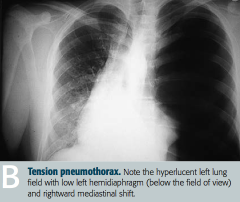
- Air is capable of entering pleural space but not exiting |
|
|
A rupture of an apical bleb in a tall, thin, young male is likely to cause what? |
Spontaneous Pneumothorax: accumulation of air in pleural space |