![]()
![]()
![]()
Use LEFT and RIGHT arrow keys to navigate between flashcards;
Use UP and DOWN arrow keys to flip the card;
H to show hint;
A reads text to speech;
88 Cards in this Set
- Front
- Back
|
Which embryonic structure gives rise to the ascending aorta and pulmonary trunk? |
Truncus arteriosus (TA)
|
|
|
Which embryonic structure gives rise to the smooth parts (outflow tract) of the L & R ventricles?
|
Bulbus cordis
|
|
|
Which embryonic structure gives rise to the trabeculated part of the L & R atria?
|
Primitive atria
|
|
|
Which embryonic structure gives rise to the trabeculated part of the L & R ventricles?
|
Primitive ventricle
|
|
|
Which embryonic structure gives rise to the smooth part of the L atrium?
|
Primitive pulmonary vein
|
|
|
Which embryonic structure gives rise to the coronary sinus?
|
L horn of sinus venosus (SV)
|
|
|
Which embryonic structure gives rise to the smooth part of the R atrium?
|
Right horn of sinus venosus (SV) |
|
|
Which embryonic structure gives rise to the Superior Vena Cava?
|
- Right common cardinal vein
- Right anterior cardinal vein |
|
|
What does the Truncus Arteriosus (TA) give rise to?
|
- Ascending aorta
- Pulmonary trunk |
|
|
What does the Bulbus Cordis give rise to?
|
Smooth parts (outflow tract) of L & R ventricles
|
|
|
What does the Primitive Atria give rise to?
|
Trabeculated part of L & R atria
|
|
|
What does the Primitive Ventricle give rise to?
|
Trabeculated part of L & R ventricles
|
|
|
What does the Primitive Pulmonary Vein give rise to?
|
Smooth part of L Atrium
|
|
|
What does the Sinus Venosus give rise to?
|
- Left horn → Coronary Sinus
- Right horn → Smooth part of R Atrium |
|
|
What do the R Common Cardinal Vein and R Anterior Cardinal Vein give rise to?
|
Superior Vena Cava
|
|
|
What is the first functional organ in vertebrate embryos? When does it start working?
|
Heart - beats spontaneously by week 4 of development
|
|
|
What is the first step in the morphogenesis of the heart?
|
Cardiac Looping
- Primary heart tube loops to establish L-R polarity - Begins in week 4 of gestation |
|
|
What can lead to dextrocardia (apex of heart faces R side of body)?
|
Defect in L-R Dynein (involved in L/R asymmetry)
- Seen in Kartagener Syndrome (primary ciliary dyskinesia) |
|
|
What is the defect in Kartagener Syndrome? What does this lead to?
|
- Defect in L-R Dynein (involved in L/R asymmetry)
- Kartagener Syndrome is a primary ciliary dyskinesia - Causes Dextrocardia |
|
|
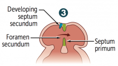
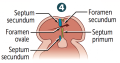
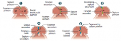
What steps lead to septation of the atria?
|

1. Septum primum grows towards the endocardial cushions, narrowing the foramen primum
2. Foramen secundum forms in septum primum (foramen primum disappears) 3. Septum secundum develops as foramen secundum maintains R→L shunt 4. Septum secundum expands and covers most of the foramen secundum. The residual foramen is the foramen ovale. 5. Remaining portion of septum primum forms valve of foramen ovale. 6. Septum secundum and septum primum fuse to form atrial septum 7. Foramen ovale usually closes soon after birth because of ↑ LA pressure |
|
|
What is the first step in septation of the atria?
|
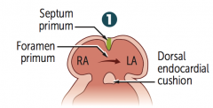
Septum primum grows towards the endocardial cushions, narrowing the foramen primum
|
|

What is the second step in septation of the atria, after the septum primum narrows the foramen primum?
|
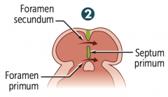
Foramen secundum forms in septum primum (foramen primum disappears)
|
|

What is the third step in septation of the atria, after the foramen secundum forms in the septum primum?
|

Septum secundum develops as foramen secundum maintains R→L shunt
|
|
What is the fourth step in septation of the atria, after the septum secundum develops?
|

- Septum secundum expands and covers most of the foramen secundum.
- The residual foramen is the foramen ovale. |
|
What is the fifth step in septation of the atria, after the septum secundum covers most of the foramen secundum, leaving only the foramen ovale (residual foramen)?
|
Remaining portion of septum primum forms valve of foramen ovale.
|
|

What is the sixth step in septation of the atria, after the remaining portion of the septum primum forms the valve of the foramen ovale?
|
Septum secundum and septum primum fuse to form atrial septum
|
|
|
What is the seventh step in septation of the atria, after the septum secundum and primum fuse to form the atrial septum?
|
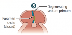
Foramen ovale usually closes soon after birth because of ↑ LA pressure
|
|
|
What forms the foramen ovale?
|

Residual foramen secundum (being closed by septum secundum in step 4)
|
|
|
What forms the valve of the foramen ovale?
|

Remaining portion of septum primum (green in step 4)
|
|
|
What forms the atrial septum?
|
Fusion of septum secundum and septum primum (which previously formed the valve of foramen ovale) |
|
|
What causes closure of the foramen ovale? When?
|
After birth, foramen ovale closes due to increase left atrial pressure
|
|
|
What can cause a patent foramen ovale?
|
Failure of septum primum and septum secundum to fuse after birth
|
|
|
What happens in a patient that has a patent foramen ovale?
|
- Most are left untreated
- Can lead to paradoxical emboli (venous thromboemboli that enter systemic arterial circulation), similar to those resulting from an Atrial Septal Defect |
|
|
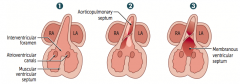
What steps lead to septation of the ventricles?
|

1. Muscular ventricular septum forms; opening is called the interventricular foramen |
|
|
What is the first step in ventricular septation?
|
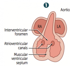
Muscular ventricular septum forms; opening is called the interventricular foramen
|
|

What is the second step in ventricular septation, after formation of the muscular ventricular septum?
|
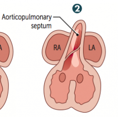
Aorticopulmonary septum rotates and fuses with muscular ventricular septum, closing the interventricular foramen
|
|

What is the third step in ventricular septation, after the aorticopulmonary septum rotates and fuses to the muscular ventricular septum?
|
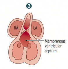
Growth of the endocardial cushions separates atria from ventricles and contributes to both atrial septation and the membranous portion of the interventricular septum
|
|
|
What contributes to both atrial septation and the membranous portion of the interventricular septum?
|
Growth of the endocardial cushions
|
|
|
What are the characteristics of a ventricular septal defect (VSD)?
|
- Most commonly occurs in the membranous septum
- Acyanotic at birth due to L→R shunt |
|
|
How does the outflow tract of the heart form?
|
- Truncus arteriosus rotates |
|
|
What are the possible conotruncal abnormalities?
|
- Transposition of the great vessels
- Tetralogy of Fallot - Persistent truncus arteriosus |
|
|
What do the aortic and pulmonary valves develop from?
|
Derived from endocardial cushions of outflow tract
|
|
|
What do the mitral and tricuspid valves develop from?
|
Derived from fused endocardial cushions of the AV canal
|
|
|
What are the types of valvular anomalies?
|
- Stenosis
- Regurgitation - Atresia (eg, triscuspid atresia) - Displacement (eg, Ebstein anomaly) |
|
|
What are the locations of fetal erythropoiesis? When?
|
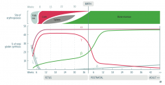
Young Liver Synthesizes Blood:
- Yolk sac (3-8 weeks) - Liver (6 weeks - birth) - Spleen (10-28 weeks) - Bone marrow (18 week s- adult) |
|
|
What are the types of hemoglobin?
|

- Fetal hemoglobin (HbF): α2γ2
- Adult hemoglobin (HbA): α2β2 Always Alpha, Gamma Goes, Becomes Beta |
|
|
What is the difference between HbF and HbA in terms of affinity for O2?
|
- HbF has a higher affinity for O2 d/t less avid binding of 2,3-BPG
- This allows HbF to extract O2 from HbA (maternal Hb) across the placenta |
|
|
Which part of fetal circulation has the highest PO2?
|

Blood in umbilical vein has PO2 of ~30 mmHg and is ~80% saturated w/ O2
(from placenta, going to fetal heart) |
|
|
Which part of fetal circulation has the lowest PO2?
|
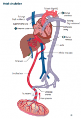
Umbilical arteries (from fetal heart, returns to placenta to mom)
|
|
|
What are the 3 important shunts in fetal circulation?
|

1. Ductus Venosus
2. Foramen Ovale 3. Ductus Arteriosus |
|
|
What is the function of the Ductus Venosus?
|

Blood entering the fetus through the umbilical vein is conducted via the Ductus Venosus (1) into the Inferior Vena Cava to bypass the hepatic circulation
|
|
|
What is the function of the Foramen Ovale?
|

Most highly oxygenated blood reaching the heart via the IVC is diverted through the Foramen Ovale (2) and pumped out the aorta to the head and body
|
|
|
What is the function of the Ductus Arteriosus?
|

- Deoxygenated blood entering the RA rom the SVC goes: RA → RV → main PA → patent Ductus Arteriosus (3) → descending aorta
- Due to high fetal pulmonary artery resistance d/t low O2 tension |
|
|
What happens to the fetal circulation when an infant takes its birth breath after birth?
|
- ↓ Resistance in pulmonary vasculature causes ↑ LA pressure vs RA pressure
- Foramen ovale closes (now called Fossa Ovalis) - ↑ in O2 (from respiration) - ↓ in Prostaglandins (from placental separation) - Closure of Ductus Arteriosus |
|
|
How can you close a Patent Ductus Arteriosus (PDA)?
|
Indomethacin → forms ligamentum arteriosum
|
|
|
How can you maintain a Patent Ductus Arteriosus (PDA)?
|
Prostaglandins E1 and E2 keep PDA open
|
|
|
What is the fetal-postnatal derivative of the umbilical vein?
|
- Ligamentum teres hepatis
- Contained in falciform ligament |
|
|
What is the fetal-postnatal derivative of the umbilical arteries?
|
Medial umbilical ligaments
|
|
|
What is the fetal-postnatal derivative of the ductus arteriosus?
|
Ligamentum arteriosum
|
|
|
What is the fetal-postnatal derivative of the ductus venosus?
|
Ligamentum venosus
|
|
|
What is the fetal-postnatal derivative of the foramen ovale?
|
Fossa ovalis |
|
|
What is the fetal-postnatal derivative of the allantois?
|
Urachus-Median umbilical ligament
- Urachus is the part of the allantoic duct between the bladder and the umbilicus - Urachal cyst or sinus is a remnant |
|
|
What is the fetal-postnatal derivative of the Notochord?
|
Nucleus pulposus of the intervertebral disc
|
|
|
What is the fetal structure that forms the ligamentum teres hepatis? Location?
|
- Derived from umbilical vein
- Contained in falciform ligament |
|
|
What is the fetal structure that forms the medial and median umbilical ligaments?
|
- MediaL: derived from umbiLical arteries
- MediaN: derived from allaNtois |
|
|
What is the fetal structure that forms the ligamentum arteriosum and venosum?
|
- Ligamentum arteriosum: derived from ductus arteriosus
- Ligamentum venosus: derived from the ductus venosus |
|
|
What is the fetal structure that forms the fossa ovalis?
|
Foramen Ovale
|
|
|
What is the fetal structure that forms the nucleus pulposus of the intervertebral disc?
|
Notochord
|
|
|
Which coronary artery supplies the SA and AV nodes?
|

Right Coronary Artery (RCA)
|
|
|
What can an infarct of the Right Coronary Artery cause?
|

Nodal dysfunction (bradycardia or heart block)
|
|
|
How common is right-dominant circulation? Characteristics?
|

85%
- Posterior Descending Artery (PDA) arises from the Right Coronary Artery (RCA) |
|
|
How common is left-dominant circulation? Characteristics?
|

8%
- Posterior Descending Artery (PDA) arises from the Left Circumflex Coronary Artery (LCX) |
|
|
How common is co-dominant circulation? Characteristics?
|

7%
- Posterior Descending Artery (PDA) arises from both Left Circumflex Coronary Artery (LCX) and the Right Coronary Artery (RCA) |
|
|
Where does coronary artery occlusion most commonly occur?
|

Most commonly in the Left Anterior Descending Artery (LAD)
|
|
|
When does coronary artery blood flow peak? Why?
|
Early diastole (the muscles are relaxing so there is more room for blood to flow through arteries)
|
|
|
What is the most posterior part of the heart?
|
Left Atrium
|
|
|
What can enlargement of the heart (specifically most posterior part / LA) cause?
|
- Dysphagia (due to compression of esophagus)
- Hoarseness (due to compression of the L recurrent laryngeal nerve, a branch of vagus) |
|
|
Which artery supplies the lateral and posterior walls of the left ventricle?
|
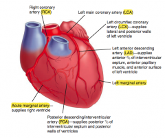
Left Circumflex Coronary Artery (LCX)
|
|
|
Which artery supplies the anterior 2/3 of the interventricular septum, anterior papillary muscles, and the anterior surface of the LV?
|

Left Anterior Descending Artery (LAD)
|
|
|
Which artery supplies the posterior 1/3 of the interventricular septum and the posterior walls of the ventricles? |

Posterior Descending / Interventricular Artery (PDA)
|
|
|
Which arteries supply the interventricular septum?
|

- Anterior 2/3: Left Anterior Descending (LAD)
- Posterior 1/3: Posterior Descending Artery (PDA) |
|
|
what collagen is found in the heart muscle and vessels |
I |
|
|
what part of the heart spans the most anterior part and can be stabbed in the left 4th intercostal leverl |
right ventricle |
|
|
what causes coronory sinus dilatations |
well it drains to right atrium if the right atrium has a lot of pressure due to pulmonary HTN then it will dialte the coronoray artery |
|
|
aortic arch 1:2:3:4:5:6 |
maxilarry aretery stapedial artery common carotid and proximal internal carotid left: airta, R right subclavian 5th degenerates 6th pulmonart artery and ductus arteriosus |
|
|
tetralogy of fallot is due to |
neural crest cells |
|
|
RCA gives rise to |
LAD which can give inferior MI |
|
|
lateral MI |
LCA V5 V6 Vk |