![]()
![]()
![]()
Use LEFT and RIGHT arrow keys to navigate between flashcards;
Use UP and DOWN arrow keys to flip the card;
H to show hint;
A reads text to speech;
20 Cards in this Set
- Front
- Back
|
The head fold
|
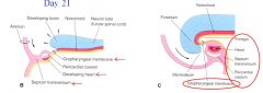
► The oropharyngeal membrane (future month), heart, pericardial cavity and septum transversum move ventrally
|
|
|
The Tail fold |
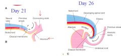
► The allantois - a diverticulum of the yolk sac extending into the connecting
|
|
|
Lateral folding |
► The abdominal walls close ventrally and form the peritoneal cavity
|
|
|
Formation of the amniochorionic sac
|
► Expansion of the amniotic cavity results in
|
|
|
Fate of the pharyngeal arches
|
► The 2nd arch overgrows the 3rd and 4th arches and forms the cervical sinus
|
|
|
What are the muscles derived from the first (mandibular) arch |
Muscles of mastication |
|
|
What are the skeletal structures derived from the first (mandibular) arch? |
Malleus (ear) Incu (ear) Upper Jaw Lower Jaw |
|
|
What are the muscles derived from the second (hyoid) arch? |
Muscles of facial expression |
|
|
What are the skeletal structures derived from the second (hyoid) arch? |
stapes (of ear) |
|
|
Fate of 1st pouch |
pharynngotymapnic (auditory) tube |
|
|
Fate of 2nd pouch |
palatine tonsils (dervived from ectoderm) |
|
|
Fate of 3rd pouch |
Thymus and the inferior parathyroid gland |
|
|
Fate of 4th pouch |
superior parathryoid gland (calcium metabolism) and the ultimobranchial body |
|
|
Fate of the 1st groove |
external acoustic meatus (ear) |
|
|
Fate of the first membrane |
Tympanic membrane |
|
|
DiGeorge Syndrom - what is it and what are the signs? |
Characterized by the absence of the thymus and parathyroid glands as well as
CATCH = Cardiac, Abnormal face, Thymic aplasia, Cleft palate, Hypocalcimia
|
|
|
First Arc Syndrom |
► Caused by insufficient migration of neural crest cells into the first arch
Macrostomia
|
|
|
Development of the 4th week |
► The rostral and caudal neuropores are closing
|
|
|
Development of the 6th and 7t h week |
► 6th week - eyes, eyelids, auricles and upper digital rays are visible
|
|
|
Development of the 8th week |
► The embryo has a distinct human appearance, facial characteristics are clearly visible
|