Reading...
![]()
Play button
![]()
Play button
![]()
Use LEFT and RIGHT arrow keys to navigate between flashcards;
Use UP and DOWN arrow keys to flip the card;
H to show hint;
A reads text to speech;
221 Cards in this Set
- Front
- Back

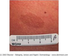
what is this? |
Macula - flat lesion <1 cm
|
|
|
what type of skin lesion is flat lesion <1 cm?
|
macule
|
|
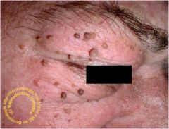
what is this?
|
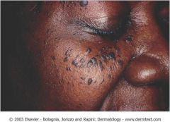
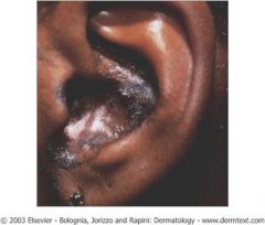
Dermatosis Papulosa Nigra
Sub-type of seborrheic keratosis Numerous brown to black, smooth, dome-shaped papules Most often seen on the head and neck of AAs No treatment is necessary Can be treated with electrodessication Treatment with liquid nitrogen can cause hypopigmentation |
|
|
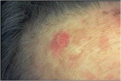
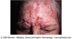
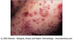
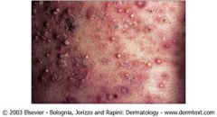
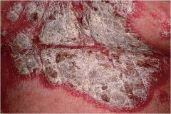
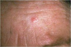
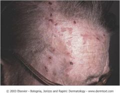
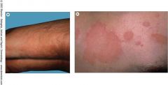
Actinic Keratosis
“Precancerous” growths associated w/ prolonged actinic damage 2 - 3% evolve to SCC Full thickness = Bowen’s disease (SCC in situ) PE = erythematous, scaly, rough papules or plaques +/- adherent yellow/white crust Most often on sun-exposed skin. Often found with palpation >> visualization |
|
how to treat?
|
Treatment of Actinic Keratoses
Liquid N2 Cryotherapy Topical Chemotherapy 5-FU (0.5, 1%, 5%) topical daily - b.i.d. x 3-8 weeks Imiquimod 5% cream 2 - 3X/week x 16 weeks Potentially less irritating. Oral retinoids for immunosuppressed patients Tretinoin cream (0.025 - 0.1%) Doing nothing is NOT an option! |
|
|
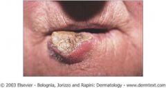
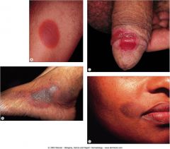
Squamous Cell Carcinoma
2nd most common skin cancer In AAs patients, SCCs are 20% more common than BCCs. (Usually) induced by persistent UVR. Bowen’s disease = SCC in situ Hyperkeratotic, skin-colored to erythematous papule, nodule, or plaque Most common on sun-exposed skin Rate of metastasis = 0.5% - 5.2% More aggressive (i.e. more likely to metastasize) if: Located at site of injury or scar Located on lip, ear, penis, scrotum, anus Immunosuppressed patients |
|
what is the txmt for this?
|
Treatment of SCC
Mohs surgery Recurrent or incompletely excised NMSC Tumors w/ aggressive histologic subtypes (infiltrative, morepheaform/scarring, micronodular BCCs, perivascular or perineural involvement) Tumors w/ poorly defined clinical margins High-risk location (face, eyes, ears, nose) Large tumors (> 1 cm on face; > 2 cm on trunk/extremities) Cosmetically and functionally important areas (groin, hand, foot, nail units) Immunosuppressed patients Tumors w/in previously irradiated skin or scar Genetic conditions w/ increased risk of neoplasms Excision 4mm surgical margins if lesion is <2cm 6mm surgical margins if lesion is >2cm |
|
|
what are the excission guidelines for scc?
|
Excision
4mm surgical margins if lesion is <2cm 6mm surgical margins if lesion is >2cm |
|
|
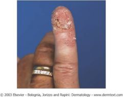
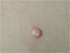
what is Keratoacanthoma?
|
Rapidly (6-8 week) forming SCC
1 - 2.5cm dome-shaped papule or nodule with a central keratin-filled crater Most common on sun-exposed skin May involute on its own leaving a scar Has malignant potential, should be excised |
|
|
Keratoacanthoma
Rapidly (6-8 week) forming SCC 1 - 2.5cm dome-shaped papule or nodule with a central keratin-filled crater Most common on sun-exposed skin May involute on its own leaving a scar Has malignant potential, should be excised |
|
|
what locations does scc more likely to present as more aggressive?
|
More aggressive (i.e. more likely to metastasize) if:
Located at site of injury or scar Located on lip, ear, penis, scrotum, anus Immunosuppressed patients |
|
what is this?
|
Macule = flat lesion <1 cm
|
|
what is this?
|
Patch = flat lesion >1cm
|
|
what is this?
|
Papule = elevated lesion < 1 cm
Papules are circumscribed, solid elevations w/o visible fluid. |
|
what is this?
|
Plaque = elevated lesion >1 cm
|
|
what is this?
|
Nodule = elevated lesion > 1 cm in diameter and depth
Similar to papule (circumscribed, solid); usually centered in the dermis or subcutaneous fat. |
|

what is this?
|
Cyst = lesion filled with fluid or semi-solid material
|
|
what do you see here?
|
Vesicle = fluid-filled blister < 1 cm
Bulla = fluid-filled blister > 1 cm Vesicopustules = seropurulent-filled cavity. |
|
what is this?
|
Pustule = vesicle filled with cloudy or purulent fluid
Papulopustules or vesiculopustules --> pustules. - usu. +erythematous border (as they contain necrotic inflammatory cells). |
|
what is this?
|
Pustular Psoriasis
|
|
what is this?
|
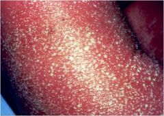
Scale = dry or greasy masses of keratin.
- represents abnormal keratinization with “pathologic exfoliation” of the stratum corneum. |
|
what is this?
|
Crust = dried debris on the skin (serum, pus, blood, epithelial/bacterial remnants)
|
|
what is this?
|
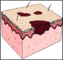
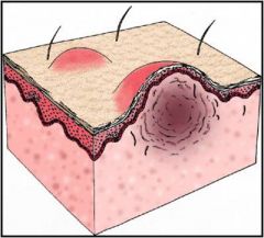
Erosion = loss of the epidermis
Erosion = loss of all or some of the epidermis +/- crust; heals as a scar. |
|
what is this?
|
Fissure = linear cleft through the epidermis
|
|
what is this?
|
Ulcer = loss of skin that extends into the dermis
- Typically heal with scarring. |
|
what is this?
|
Induration = dermal thickening
Ex of clinical significance = PPD test (measured by degree of induration) |
|
what is this?
|
Atrophy = loss of soft tissue
Loss of epidermis leaves skin appearing thin and wrinkled Loss of the dermis/sub-cutaneous tissue leaves a visible depression |
|
what is this?
|
Lichenification = visible and palpable thickening of the epidermis with accentuated skin markings.
|
|
what is this?
|
Wheal = papule or plaque resulting from extravasated fluid (often caused by exposure to allergenic substance)
|
|
what is that?
What is a classic dermatosis this is seen? |
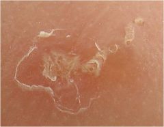
Burrows = linear tunnels or streaks caused by infestation.
Answer = Scabies. |
|
what is this?
|
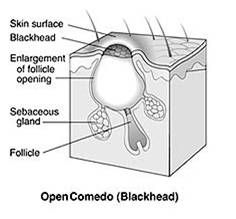
Comedo = non-inflammatory lesion resulting from impaction of keratin material in the outlet of the pilosebaceous canal
Plural = comedones. open comedo = black head Slightly elevated papule w/ dilated ostia filled with oxidized keratin. |
|
what is this?
|
Telangiectasia = dilation of blood capillary or terminal artery
|
|
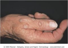
what is this?
|
Verruca Vulgaris
HPV = human papilloma virus Proliferation of virally infected keratinocytes Loss of skin lines/dermatoglyphics Black dots are thrombosed capillaries Distinguishing feature from corns/calluses Transmitted by skin/skin contact, trauma. |
|
|
what is podofilox used to treat and what is the mechanism of action?
|
Podophyllotoxin(a.k.a. podofilox) is a non-alkaloid toxin ligand extracted from the roots and rhizomes of Podophyllum species; arrests cells in metaphase by inhibiting tubulin polymerization.
|
|
|
what are common treatments for verruca vulgaris?
|
Topical salicylic acids
Duofilm Paring Pumice Stone Liquid N2 Cryotherapy Cantharidin Podofilox Excision and cautery CO2 laser Pulse Dye laser |
|
|
what is canthardin?
|
Cantharidin = extract from the “blister beetle”/”spanish fly”.
used to treat verruca vulgaris |
|

what is this?
|

Molluscum contagiosum
Pox virus Transmitted by touch, trauma. Especially contagious if skin is wet 2 - 5mm flesh-colored domed papules with central umbilication Most clear spontaneously within 6-9 months Common in children |
|
|
what disease pathology is described as acanthotic and cup-shaped; w/in cytoplasm of affected epidermal cells containing eosinophilic/basophilic inclusion bodies ?
|
Molluscum Contagiosum
|
|
how is this treated?
|
this is molluscum
txmt: Tretinoin - highest tolerated concentration. Cantharidin (beetle juice) Liquid N2 Cryotherapy Salicylic acid Curettage Imiquimod (Aldara) - limited efficacy. Tape stripping Application of surgical tape daily after bathing x 16 weeks. Do nothing |
|
what is the medical and layman term for this?
|
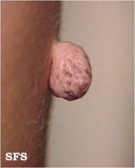
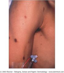
Acrochordon
a.k.a. “skin tag” Flesh-colored to brown colored, polypoid papules attached by a stalk (pedunculated) More common at skin folds/intertriginous areas (i.e. neck, groin, axillae) Predisposing factors = heredity, obesity. Complications rare but can undergo necrosis No treatment necessary. Excision common if symptomatic. |
|
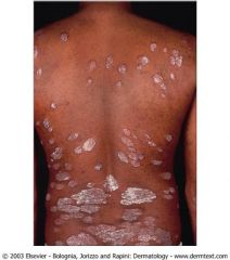
what is this?
|
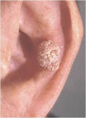
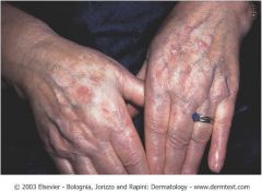
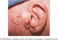
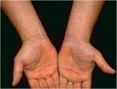
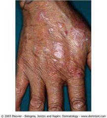
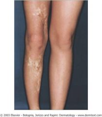
seborrheic keratosis
Benign epidermal “stuck-on”, mamillated/verrucous papules or plaques w/ pseudo-horn cysts (keratin accumulations) Rough, dry, crumbling surface (“barnacles”) No malignant potential BUT can become irritated Sign of Leser-Trelat = sudden appearance or increase in the number and size of SKs due to internal malignancy MC = adenocarcinoma |
|
|
what dermatologic condition is assocated with sign of Leser-Trelat?
|
Seborrheic Keratosis
Sign of Leser-Trelat = sudden appearance or increase in the number and size of SKs due to internal malignancy MC = adenocarcinoma |
|
how is this treated?
|

this is seborrheic keratoses
txmt: Liquid nitrogen ED & C Shave excision Do nothing |
|
what is this?
|
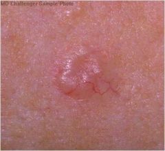
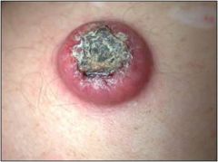
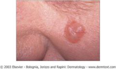
Basal cell carcnoma
Most common form of skin cancer MC site = sun-exposed areas Very rare cases of metastasis PE = (nodular) pearly/translucent papule with telangiectasia and ulceration “Pearly, pink papule” w/ rolled border Most common form PE = (superficial) erythematous macule or patch with fine scale |
|
how is this treated?
|
Excision with 4mm surgical margins
Electrodessication & curretage Superficial Torso Tumors < 6 mm Mohs surgery |
|
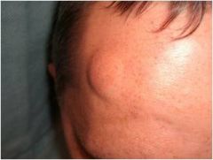
what is this?
|
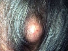
Pilar cyst
Smooth firm nodule found on the scalp No central punctum Contains concentric layers of keratin, no granular layer Firm, thick cyst wall Treat with surgical excision The whole cyst wall must be removed |
|

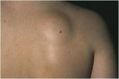
what is this?
|
Mobile nodule with central punctum
Thin, fragile cell wall containing foul-smelling cheesy keratinous material Genital lesions are frequently calcified Treat with surgical excision Whole cyst wall must be removed Gardner syndrome = epidermoid cysts associated with osteomas, fibromas, desmoid tumors, lipomas, fibrosarcomas, leiomyomas and multiple GI polyps AD inheritance APC gene mutation |
|
|
what dermatologic lesion is associated with Gardner synderome?
|
epidermoid cysts associated with osteomas, fibromas, desmoid tumors, lipomas, fibrosarcomas, leiomyomas and multiple GI polyps
AD inheritance APC gene mutatio |
|

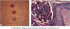
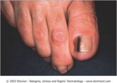
what is this?
|
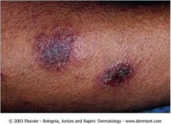
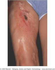
Dermatofibroma
Common, benign lesion Can be pruritic Flesh-colored to hyperpigmented papules “Dimple sign” depresses when squeezed Most common on lower legs, extremities Occurs from fibrous reaction following trauma |
|
how is this treated?
|
this is a dermatofibroma
Treatment: Cryotherapy Excision Do nothing |
|

what is this?
|
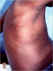
spontaneous keloids
|
|
|
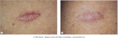
what derm lesion is described as follows?
Extends beyond wound margin Tends to remain the same with time More common in AAs |
keloid
|
|
|
how are keloids treated?
|
Treatments:
IL steroids Topical steroids (Cordran tape) Silicone sheeting (Mederma) Excision Cryotherapy 5-FU CO2 laser excision |
|
|
what derm lesions is decribed as:
Stays w/in wound margins Tends to regress Treatment rarely required |
hypertrophic scars
|
|
|
what lesions is :
Mobile, rubbery, fatty subcutaneous tumor Benign |
lipoma
|
|
|
how are lipomas treated?
|
Excision
Liposuction Do nothing |
|
what is this?
|
hypertrophic scars
|
|
what is this?
|
lipoma
Mobile, rubbery, fatty subcutaneous tumor Benign Treatment: Excision Liposuction Do nothing |
|
what is this?
|
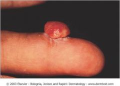
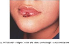
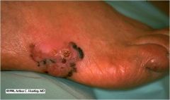
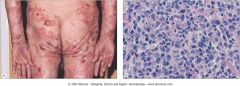
pyrogenic granuloma
Benign, vascular, dome-shaped papule or nodule with moist to scaly surface or collarette Rapidly growing Bleed easily w/ trauma Secondary to trauma of skin or mucous membranes May also be associated with medications: Isotretinoin Capecitabine Indinavir Estrogens (also seen in pregnancy) Treat with saucerization Bx followed by ED&C. Recurrence is common, especially after incomplete removal |
|

how is this treated?
|
pyrogenic granuloma
Treat with saucerization Bx followed by ED&C. Recurrence is common, especially after incomplete removal |
|

what is the ddx for this?
|
pyrogenic granuloma
Differential Diagnosis Amelanotic melanoma Bacillary angiomatosis |
|
how is this treated?
|
pyrogenic granuloma
Treat with saucerization Bx followed by ED&C. Recurrence is common, especially after incomplete removal |
|
what is this?
|
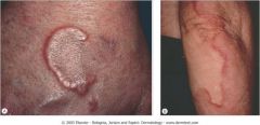
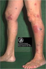
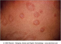
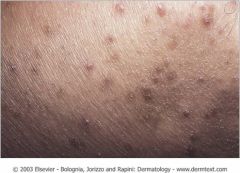
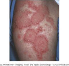
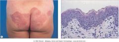
Granuloma Annulare
Slow-growing annular plaque (ring) of small, firm, flesh-colored to red papules Typically begins as a papule that spreads in a centrifugal manner Central involution MC = children and young adult Associated with diabetes Half spontaneously regress Treatment = potent topical, IL steroids, PUVA, tetracyclines. Dapsone, pentoxifylline, SSKI, UVA-1, antimalarials. |
|
|
what derm lesion is:
Slow-growing annular plaque (ring) of small, firm, flesh-colored to red papules Typically begins as a papule that spreads in a centrifugal manner Central involution MC = children and young adult Associated with diabetes |
Granuloma Annulare
|
|
|
how is Granuloma Annulare treated?
|
Treatment = potent topical, IL steroids, PUVA, tetracyclines.
Dapsone, pentoxifylline, SSKI, UVA-1, antimalarials. |
|
|
what derm lesion is described as “ripple in a pond”?
|
Granuloma Annulare
|
|
|
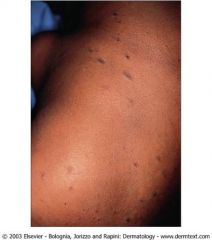
what derm lesion is assocated with young adult women and children after a strep infection?
|
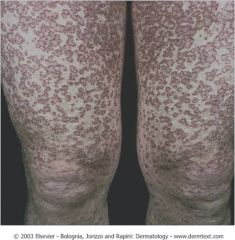
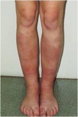
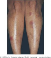
Erythema Nodosum
|
|
|
what derm lesion is assoc with:
Septal panniculitis Erythematous, warm, tender nodules on the extensor extremities Symmetric Most common in young adult women Seen in children following streptococcal infections. |
Erythema Nodosum
|
|
|
what is Lofgren syndrome and what derm lesion is it assocated with?
|
Lofgren syndrome (fever, cough, joint pains, hilar adenopathy w/ sarcoidosis)
Erythema Nodosum |
|
|
what derm lesion is associated with:
Streptococcal infections TB Intestinal infections Sarcoidosis (Lofgren syndome) IBD Hematologic malignancy Behcet syndrome Meds (bromides, iodides, sulfa, OCPs, HRT) |
Erythema Nodosum
|
|
|
what are treatments for Erythema Nodosum?
|
Treatments
Treat inciting factor Bed rest ASA / NSAIDs Colchicine IL corticosteroids + risk of pathergy SSKI + risk of hypothyrodism Antimalarials |
|
|
what form of Karposi's Sarcoma presents on lower legs & feet; elderly Jewish, eastern European, and Mediterranean men; slow progression?
|
classic form of KS
|
|
|
what form of Kaposi's sarcoma is associated with children?
|
Endemic cutaneous or lymphatic; rapidly progressive and fatal; affects young children
|
|
|
what cd4 count is associated with aids related Kaposi's sarcoma?
|
AIDS related rapid progression; may be systemic
CD4 count < 500 |
|
|
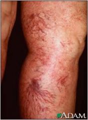
what derm lesion presents as :
Slow to rapidly progressive vascular neoplasm Red to violaceous macules, papules, plaques, & nodules |
Kaposi's Sarcoma
|
|
|
Treatment associated with Kaposi's Sarcoma?
|
HAART
IL vinblastine 0.2 - 0.4 mg/mL Cryotherapy Local irradiation Rx Systemic chemotherapy Symptomatic visceral disease |
|
|
What is the infectious agent associated with KS?
|
Human Herpesviridae - 8.
|
|
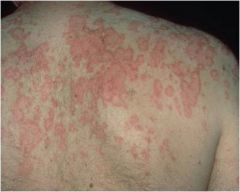
what is this?
|
Disseminated Granuloma Annulare
Slow-growing annular plaque (ring) of small, firm, flesh-colored to red papules Typically begins as a papule that spreads in a centrifugal manner Central involution MC = children and young adult Associated with diabetes Half spontaneously regress Treatment = potent topical, IL steroids, PUVA, tetracyclines. Dapsone, pentoxifylline, SSKI, UVA-1, antimalarials |
|
what is this?
|
Erythema Nodosum
Septal panniculitis Erythematous, warm, tender nodules on the extensor extremities Symmetric Most common in young adult women Seen in children following streptococcal infections. |
|

what is this associated with?
|
this is Erythema Nodosum
Associations Streptococcal infections TB Intestinal infections Sarcoidosis (Lofgren syndome) IBD Hematologic malignancy Behcet syndrome - a form of vasculitis that can lead to ulceration and other lesions. It can be interpreted as a chronic disturbance in the body’s immune system. Specifically attack small bv. Meds (bromides, iodides, sulfa, OCPs, HRT) Spontaneous regression without scars, ulcers, atrophy |
|
how is this treated?
|
Erythema Nodosum
Treatments Treat inciting factor Bed rest ASA / NSAIDs Colchicine IL corticosteroids + risk of pathergy SSKI + risk of hypothyrodism Antimalarials |
|

what is this?
|
Kaposi’s Sarcoma
Slow to rapidly progressive vascular neoplasm Red to violaceous macules, papules, plaques, & nodules Present on skin and mucus membranes Classic form lower legs & feet; elderly Jewish, eastern European, and Mediterranean men; slow progression AIDS related rapid progression; may be systemic CD4 count < 500 Endemic cutaneous or lymphatic; rapidly progressive and fatal; affects young children Iatrogenic due to immune suppression |
|
what treatmet is associated with this?
|
Kaposi’s Sarcoma
HAART IL vinblastine 0.2 - 0.4 mg/mL Cryotherapy Local irradiation Rx Systemic chemotherapy Symptomatic visceral disease |
|
what are these?
|
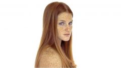
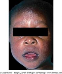
Ephelides
a.k.a. “freckles” Hyperpigmented macule in sun exposed areas Most common in fair-skinned people Fitzpatrick skin types I and II Occur on sun-exposed areas Darken with sun exposure Benign No treatment necessary |
|
What is this?
|
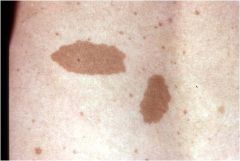
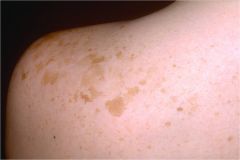
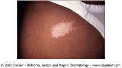
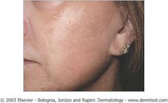
Café au Lait Macules
Uniform, well-demarcated lesions Usually no associated systemic disorder Associated with neurofibromatosis type 1 |
|
|
what are the clinical findings for NF Type I?
|
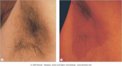
Diagnosis requires 2 or more of the following criteria:
≥ 6 CALMs 5 mm in prepubertal pts; 15 mm in postpubertal pts ≥ 2 neurofibromas Plexiform neurofibroma Axillary (skin fold) freckling Optic glioma Affected 1st degree relative ≥ 2 Lisch nodules Osseous lesion i.e. sphenoid dysplasia, thinning of the long lone cortex |
|
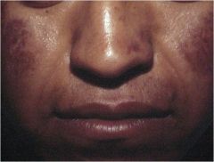
what is this?
|
Melasma
Brown patches on central face, malar area, mandible, nipples, genital area Affects dermis, epidermis, or both More common in Hispanic & AA patients Associated with pregnancy, OCP’s, HRT Treatment Sun avoidance Bleaching agents Tretinoin Glycolic acid peels |
|
what is this?
|
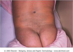
Mongolian Spot
a.k.a. Congenital Dermal Melanocytosis Blue-gray patch Most common in the sacral region More common in Asians, African Americans, Native Americans |
|
|
what is the phenomenon by which light passing through a medium (i.e. the skin) is scattered as it strikes other particles (melanin)
Long wavelength light rays are less scattered so pass down into the skin (visual coloring is absorbed) Shorter wavelength colors (blue, indigo, violet) are scattered to the side and backward, “reflecting” off the skin surface (so discoloration is visually noted) |
Tyndall Effect
|
|
what is this?
|
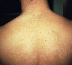
Tinea (Pityriasis) Versicolor
Hyper-to-hypopigmented, tan, or pink oval patches usually on chest (face in infants) More common in young adults Hot weather Sweaty patients Overgrowth of Malassezia furfur KOH shows spores and hyphae (“spaghetti and meatballs”) Treatment: Azoles (i.e.ketoconazole shampoo or cream) Sulfur-based topical agents Ketoconazole 400 mg weekly x 2 weeks; 200 mg qd x 2-5 weeks |
|
what is this?
|
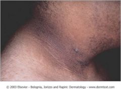
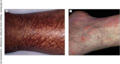
Acanthosis Nigricans
Hyperpigmented, velvety thickening of intertriginous skin Most common on neck and axillae Associated with: Diabetes OCP use Obesity Addison’s disease Cushing’s disease Hypothyroidism Niacin therapy Gastric adenocarcinoma Treatment: Treat underlying condition Ammonium lactate cream |
|
|
type of nevi?
- Macule - Nevus cells at base of epidermis (dermal-epidermal junction) - Normal finding |
Junctional Nevi
|
|
|
what type of nevi?
- Papule - Nevus cells in dermis only - Normal finding |
Dermal Nevi
|
|
|
what type of Nevi?
- Papule or plaque - 1% of newborns |
Congenital nevi
|
|
|
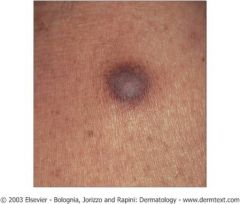
what type of nevi?
- Macular or papular - Hypo- to depigmented border/rim -Inflamed nevus |
Halo Nevi
|
|
|
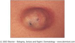
what type of nevi?
- Macular or papular - >5 mm, variegated color, indistinct border - Malignant potential |
Atypical Nevi
|
|
|
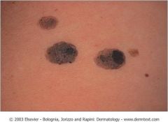
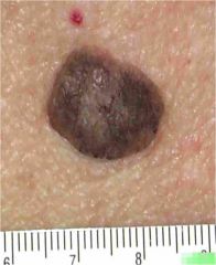
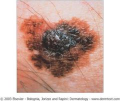
how do melanoma present on exam?
|
PE = Irregular shape, different colors, changing
ANY new, bleeding, itchy, or changing growth MUST be biopsied! ANY ‘ugly duckling’ lesion should be biopsied 10% of melanomas are atypical (amelanotic, etc) and do not abide by the “ABCDEs”… |
|
|
what should the margins be for excisional or punch biopsy for melanoma?
|
All suspected melanomas should have an excisional or punch biopsy with 1-2 mm margins
E.g. 4 mm atypical lesion can be biopsied with a 6 mm punch biopsy tool The aim is to preserve lymphatic flow in case sentinel lymph node biopsy is performed AVOID shave biopsies Staging depends on depth |
|
|
what derm lesion:
Occurs on sun-exposed areas Grows slowly, takes awhile to metastasize Median age: 70 years |
Lentigo Maligna
|
|
|
what is the most common subtype of melanoma?
|
Superficial Spreading Melanoma
Most common subtype of melanoma Median age: 47 years Women = MC site is LEG Men = MC site is BACK Occurs on all surfaces including non-sun-exposed skin Pre-metastatic for 1-7 years |
|

what type of melanoma is this image?
|
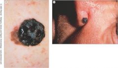
Nodular Melanoma
Occurs on all surfaces Including non-sun-exposed Median age : 50 years Pre-metastatic for months to 2 years |
|
what type of melanoma is this image?
|
Nodular Melanoma
Occurs on all surfaces Including non-sun-exposed Median age : 50 years Pre-metastatic for months to 2 years |
|
what type of melanoma is associated with:
Occurs on palms, soles, nail beds Most common in Asians and Blacks Median age: 61 years Pre-metastatic for months to 8 years |
Acral Lentiginous Melanoma
|
|
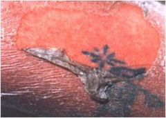
what is the palthognomic clinical sign seen here?
|
Hutchinson’s Sign
Melanonychia with pigmentation involving the proximal nailfold. |
|
|
what derm lesion is assoc with:
Hutchinson’s Sign Melanonychia with pigmentation involving the proximal nailfold. |
Asteatotic Dermatitis
|
|
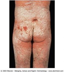
what is chronic, pruritic, erythematous patches, papules and thin plaques
+/- lichenification (thickening of skin with increased skin markings) Papular variant often seen in AAs |
atopic dermatitis
|
|
|
what is the atopic triad?
|
atopic dermatitis, asthma, allergic rhinitis
|
|
|
how is atopic dermatitis treated?
|
dry skin care
topical steroids Class IV – VII (mid/low potency) steroids for face & intertriginous areas Class I – III (high/super high potency) steroids for body or thick areas Anti-histamines Phototherapy “Bleach baths” |
|
|
What dermatits is widespread herpes simplex infection involving eczematous skin?
|
eczema herpeticum
|
|
|
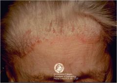
what type of dermatitis is chronic, superficial inflammation of hairy areas
Most common on scalp, eyebrows, glabella, nasolabial creasesface Yellow-to-erythematous patches with greasy or powdery fine scale Hair loss is uncommon Waxing and waning course? |
Seborrheic dermatitis
|
|
|
what is the etiology of seborrheic dermatitis?
|
Likely etiology is yeast Pityrosporum ovale
Common, affects 3-5% of the population “Cradle cap” = seborrheic dermatitis in infants |
|
|
what is the txmt for seborrheic dermatitis?
|
Treatment:
zinc pyrithione, selenium sulfide, ketoconazole, topical steroids, sulfur based products |
|
|
what dermatitis is due to increased hydrostatic pressure and capillary damage -> extravasated fluid and RBCs
Inflammation and deposition of heme -> eczematous lesions |
Stasis dermatitis
|
|
|
what dermitis presents as:
hyperpigmented, lichenified usually circumferential induration of the legs. May be “weepy”, warm or red at onset or with flares MC site = caudal to the medial malleolus |
Stasis dermatitis
|
|
|
how is stasis derm txmt?
|
Treatment:
Compression Leg elevation Weight reduction Topical steroids Treatment of infections |
|
|
what dermatitis is described as:
Round to oval erythematous thin plaques or patches Often “weepy” Intensely pruritic MC location = trunk and extremities Idiopathic How is it txed? |
Nummular Dermatitis
Treatment: Topical steroids |
|
|
what lesions is assoc with:
Chronic lesions due to scratching Always occurs within reach of hands or scratching implement PE = hyperpigmented, lichenified papules and plaques Occasionally, surrounding erythema. |
Lichen Simplex Chronicus
|
|
what is this?
|
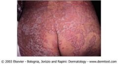
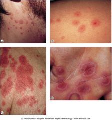
pityriasis rosea
Idiopathic self-limiting inflammatory eruption May be associated with viral infection. Most common in young adults Typically in cooler months PE= oval, minimally elevated, scaling patches, papules, and plaques + Herald patch = initial, large, single lesion followed by large outbreak “Christmas tree” pattern (along skin lines) on trunk +/- pruritus Symptomatic treatment for pruritus |
|
what type of dermatitis is assoc with:
Inflammatory dermatosis with increased epidermal proliferation PE = Sharply-demarcated erythematous plaques with silver scale Most common on scalp, extensor surfaces Mild to moderate pruritus Nail involvement in 50% of patients Inverse variant typically without scale since areas are moist |
Psoriasis
|
|
what is the txmt?
|
Treatment:
Topical steroids Tar Anthralin Retinoids Vitamin D derivatives Phototherapy Methotrexate Cyclosporine Biologics |
|
|
what is Auspitz sign?
|
pinpoint bleeding upon removal of scale
|
|
|
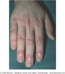
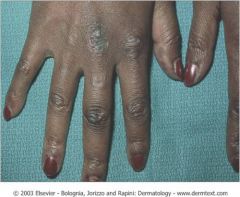
what derm lesions presents as:
“purple polygonal papules”, flat papules covered with subtle fine white scale. Always check the mouth: +/- fine, reticulated, white patches on the buccal mucosa. Arranged in linear groups due to trauma (koebnerization) |
Lichen Planus
-Idiopathic inflammatory disorder |
|
What other common inflammatory dermatoses exhibit this phenomenon, Arranged in linear groups due to trauma (koebnerization)
? |
picture shows lichen planus
PE= purple polygonal papules, flat papules coverd wth subtle fine white cale also seen in psoriasis, PRP, lichen nitidus, vitiligo |
|

how is this treated?
|
Topical steroids, antihistamines, topical retinoids
|
|
|
what is koebnerization?
|
Arranged in linear groups due to trauma
-assoc with Lichen Planus |
|
|
What is the name of the white streaks found overlying lichen planus?`
|
Wickham’s striae
|
|
|
what is the most common organism assoc with tinea capitis?
|
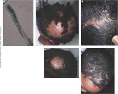
T. tonsurans
|
|
|
what is Kerion?
|
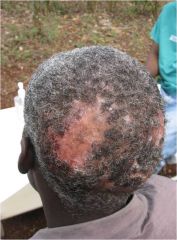
severe inflammatory form of tinea capitis
|
|
|
what derm lesion presents as patchy alopecia, broken hair shafts, scale?
|

Tinea capitis
|
|

how is this dx and txed?
|
tinea capitis
Diagnose with KOH scraping Must be treated with oral meds - topical medications cannot reach deep within follicle. Griseofulvin is 1st line treatment Terbinafine (Lamisil), itraconazole (Sporanox), fluconazole (Diflucan) |
|
|
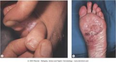
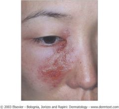
what is this?
Superficial fungal infection (dermatophytosis) of body, groin, and feet PE = annular thin plaques with central clearing and erythematous border + flaking scale + maceration between toes |
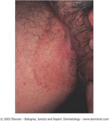
Tinea Corporis, Cruris, Pedis
|
|
how is this dx and tx?
|
Tinea Corporis, Cruris, Pedis
Diagnose with KOH scraping Treatment: Limited areas can be treated with topical anti-fungals. Hair & nail involvement require systemic meds Do NOT treat with corticosteroids! Tinea incognito = atypical presentation of tinea following treatment with topical steroids. |
|
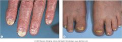
what are the 2 most common causes of Onychomycosis?
|

T. rubrum and T. mentagrophytes
|
|
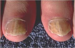
how is this dx and tx?
|
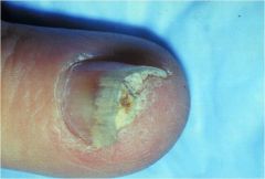
Onychomycosis
Diagnose with KOH scraping, fungal culture, PAS (H&E screening) Treatment = Oral antifungals |
|
|
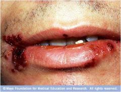
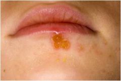
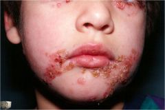
what organism is assoc with Intact or broken bullae (very fragile) with honey-colored crust?
|
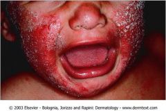
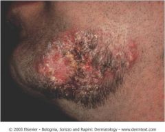
Bullous Impetigo
Caused by infection with S. aureus Most common around nose and mouth |
|
|
how is Bullous Impetigo treated?
|
Treat with topical mupirocin 2% ointment applied 3x per day
cephalexin (Keflex) erythromycin (Iloson) docloacillin (Dynapen) |
|
what is this?
|
Bullous Impetigo
|
|
what is this?
|
Milaria Rubra
Commonly called “heat rash”, “prickly heat” Very pruritic red papules Most common in areas where sweat ducts are prone to occlusion Patient may have fever Self-limited eruption Symptomatic treatment for pruritus |
|
|
what characteristics are assoc with herpes simplex?
|
Grouped vesicles on an erythematous base, becomes crusted
-erosion w/ “scalloped” border |
|
|
what tests are used to dx herpes simplex?
|
Tzank smear
DFA viral culture DFA = direct fluorescent antibody |
|

What is this and how is it treated?
|
Herpes simplex
Tx: acyclovir, valacyclovir, famciclovir |
|
|
what derm lesions is assoc with ‘Dew drop on a rose petal’
PE = small vesicles on erythematous base + crusting? |
varicella
aka chicken pox |
|
what is this and how is it treated?
|

varicella
Treat with antivirals (e.g. acyclovir, valacyclovir), symptomatic treatment for fever and pruritus |
|
|
What is Hutchinson’s sign ?
|
Herpes zoster ophthalmicus
|
|
|
what syndrome is assoc with
Tinnitus, facial palsy, deafness, vertigo? |
Ramsay Hunt syndrome
- involvement of CN 7 and 8 |
|
what is this and how is it treated?
what other conditions is associated with it? |

Zoster
Treat with valacylovir, acyclovir Post-herpetic neuralgia difficult to treat gabapentin, “Pain Team” consult |
|
|
what is the Nikolsky sign and what is it assoc with?
|
superficial trauma to unaffected skin will cause blisters
assoc with pemphigus vulgaris |
|
|
what is the Asboe-Hansen sign and what is it assoc with?
|
pressure on intact bulla forces fluid to spread under skin
assoc with pemphigus vulgaris |
|
|
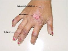
what derm lesions is an autoimmune disease with antibodies against intercellular adhesion molecules and flaccid blisters on skin, shallow erosions in mouth?
|
pemphigus vulgaris
|
|

what is this and how is it treated?
|
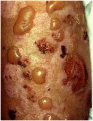
Pemphigus vulgaris
Autoimmune disease with antibodies against intercellular adhesion molecules Flaccid blisters on skin, shallow erosions in mouth Nikolsky sign superficial trauma to unaffected skin will cause blisters Asboe-Hansen sign pressure on intact bulla forces fluid to spread under skin Onset in middle age, can be FATAL! Treat with systemic steroids, immunosuppressants, and antibiotics for secondary infections |
|
|
what Autoimmune disease with antibodies against hemidesmosome?
|
bullous pemphigoid
|
|
|
what skin lesion is assoc with:
Tense blisters on skin Oral involvement rare Negative Nikolsky sign |
bullous pemphigoid
|
|
|
what treatment does bullous pemphigoid respond better to?
|
steroids
|
|
what is this?
|
bullous pemphigoid
|
|
|
what diease is assoc with Defect in heme biosynthesis pathway
Uroporphyrinogen decarboxylase enzyme deficiency? |
Porphyria cutanea tarda
|
|
|
what disease has:
Bulla in areas of trauma Scars and milia over old blisters Associated with liver disease, alcoholism, hepatitis C, AIDS, estrogen supplementation |
Porphyria cutanea tarda
|
|
|
What is the treatement of Porphyria cutanea tarda?
|
Treat with phlebotomy, antimalarials
|
|
what is this?
|
Porphyria Cutanea Tarda
|
|
|
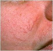
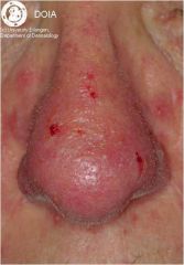
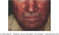
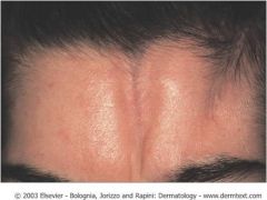
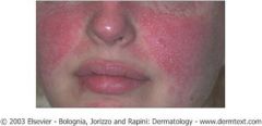
what condition is an Inflammatory acneiform eruption, present with flushing and papules, & is most common in middle aged Caucasian people?
|
Rosacea
|
|
|
what derm condition is assoc with rhinophyma?
|
Rosacea
|
|

what is this and how is it treated?
|

rosacea
Treat with metronidazole, sulfur, tetracycline |
|
|
what is this and what is it assoc with?
|
rhinophyma and rosacea
|
|
what is this and how is it treated?
|
rosacea
Treat with metronidazole, sulfur, tetracycline |
|
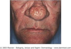
what is this?
|
rhinophyma seen in rosacea pt
|
|
what is this?
|
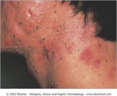
folliculitus
Papules & pustules involving hair follicles Gram (+) >> Gram (–) bacteria Can also be fungal or viral Eosinophilic |
|
|
what is assoc with Papules & pustules involving hair follicles ? and how is it treated?
|
folliculitis
Gram (+) >> Gram (–) bacteria Can also be fungal or viral Eosinophilic Treat with topical or systemic antibiotics Avoid re-infecting with combs, razors |
|
|
what is assoc with Beefy red erythematous patches with satellite pustular and papular lesions
Very pruritic Whitish plaques on mucosal surfaces, can be scraped off |
candidiasis
|
|
what is this?
|
candidiasis
Beefy red erythematous patches with satellite pustular and papular lesions Very pruritic Whitish plaques on mucosal surfaces, can be scraped off Treat with antifungals |
|
|
what derm condition is assoc with Hyperpigmented to erythematous patch, most common in axilla, groin, toe webs?
|
Erythrasma
|
|
|
what bacteria is assoc with erythrasma?
|
Etiology is Corynebacterium minutissimum
|
|
|
how is erythrasma dx and how is it tx?
|
dx: Fluoresces coral red on Wood’s lamp exam
Treat with erythromycin, clindamycin, miconazole |
|
what is this?
|
Erythrasma
Not a blister or papule! Hyperpigmented to erythematous patch Most common in axilla, groin, toe webs Etiology is Corynebacterium minutissimum Fluoresces coral red on Wood’s lamp exam Treat with erythromycin, clindamycin, miconazole |
|
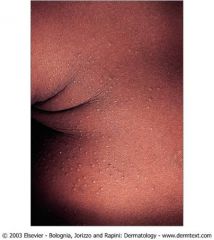
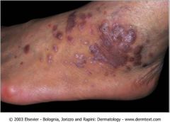
what is this?
|
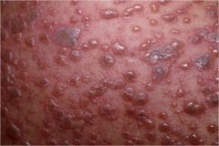
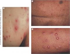
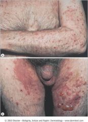
scabies
Species specific (can’t blame the dog!) Female mites burrow under skin, lay eggs and deposit feces Burrows & scaly papules on physical exam Most common on hands (web spaces), umbilical area, genitals, perianal Diagnose by scraping with mineral oil look for feces, eggs, mites Treat patient and contacts |
|
|
what are the most common locations to find scabies?
|
Most common on hands (web spaces), umbilical area, genitals, perianal
Female mites burrow under skin, lay eggs and deposit feces Burrows & scaly papules on physical exam |
|
what is this?
|
Flea bites
Common after contact with animals, exposure in wooded areas Most common on lower extremities Can be vesicular or bullous Treat symptomatically |
|
|
where are pediculosis capitis and pubis most often found:
|
Occurs on scalp, pubic hair, axillary hair, eyelashes, eyebrows
Nits attach to hair shaft |
|
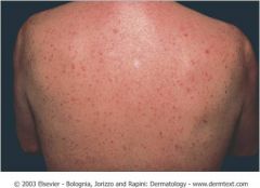
|
what drugs are most assoc with Exanthematous Drug Eruptions?
|
Most common causes include sulfa, PCN & derivatives, anticonvulsants, allopurinol
|
|
what is this?
|
Exanthematous Drug Eruptions
Most common manifestation of drug reactions Most common causes include sulfa, PCN & derivatives, anticonvulsants, allopurinol Skin may peel as rash is resolving Other forms of drug eruptions: Fixed drug eruption, urticaria, lupus-like eruption, photosensitivity, erythema multiforme (EM), Stevens-Johnson syndrome, toxic epidermal necrolysis (TEN) |
|
|
what derm lesions...
Occurs in same place each time a patient is challenged with a particular drug -Round or oval lesion, gray to slate blue center -May be bullous |
Fixed Drug Eruption
|
|
|
what drugs are assoc with Fixed Drug Eruption?
|
Common causes include laxatives, NSAIDs, sulfa drugs, tetracyclines (doxycycline, lymecycline and minocycline)
|
|
what is this?
|
Fixed Drug Eruption
Occurs in same place each time a patient is challenged with a particular drug Round or oval lesion, gray to slate blue center May be bullous Common causes include laxatives, NSAIDs, sulfa drugs, tetracyclines |
|
|
what derm lesion is most assoc with herpes simplex infection and discribed as Erythematous targetoid lesions?
|
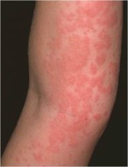
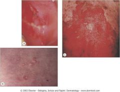
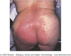
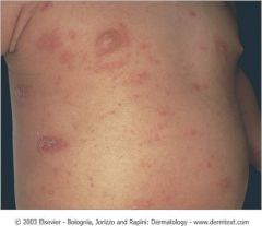
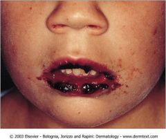
Erythema Multiforme (EM)
Many etiologies, most commonly due to herpes simplex infection May be due to drug ingestion Erythematous targetoid lesions Self-limited, often recurrent Major variant (Stevens-Johnson syndrome) requires hospitalization and immediate treatment |
|
what is this?
|
Erythema Multiforme (EM)
Many etiologies, most commonly due to herpes simplex infection May be due to drug ingestion Erythematous targetoid lesions Self-limited, often recurrent Major variant (Stevens-Johnson syndrome) requires hospitalization and immediate treatment |
|
|
what derm disease is described as a more serious eruption, usually due to drug ingestion
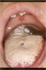
-Target-like skin lesions -At least 2 mucosal sites involved with necrosis -<10% body surface area involvement of skin sloughing (although erythema may be more widespread) -Low mortality |
Stevens-Johnson Syndrome
|
|
what is this?
|
Stevens-Johnson Syndrome
More serious eruption, usually due to drug ingestion Target-like skin lesions At least 2 mucosal sites involved with necrosis <10% body surface area involvement of skin sloughing (although erythema may be more widespread) Low mortality |
|
|
what derm disease is commonly caused by NSAIDs, antibiotics, anticonvulsants?
|
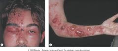
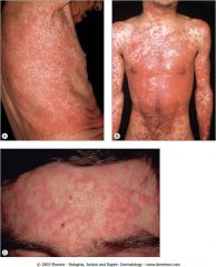
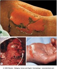
Toxic Epidermal Necrosis
Common causes include NSAIDs, antibiotics, anticonvulsants More severe than EM & SJS Rapid course, large sheets of necrotic skin, can involve mucosa Minimal inflammation of biopsy Can be fatal! Treat patient in burn unit, administer IV Ig, plasmapheresis, use of corticosteroids is very controversial |
|
|
how is Toxic Epidermal Necrosis treated?
|
Treat patient in burn unit, administer IV Ig, plasmapheresis, use of corticosteroids is very controversial
|
|
what is this?
|
Toxic Epidermal Necrolysis
|
|
|
what are some causes of Urticaria?
|
Many causes include: drugs, food, pressure, exercise, temperature changes
|
|
|
what derm disease/lesion is assoc with angioedema?
|
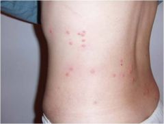
Urticaria
Many causes include: drugs, food, pressure, exercise, temperature changes Transient lesions (<24 hours, usually only a few hours) Treat by removing offending agent, administer anti-histamines Angioedema = severe form of urticaria with subcutaneous swelling Can affect airway Most commonly due to C1 complement deficiency |
|
|
what is the term for severe form of urticaria with subcutaneous swelling?
|
Angioedema = severe form of urticaria with subcutaneous swelling
Can affect airway Most commonly due to C1 complement deficiency |
|
what is this?
|
Urticaria
Many causes include: drugs, food, pressure, exercise, temperature changes Transient lesions (<24 hours, usually only a few hours) Treat by removing offending agent, administer anti-histamines Angioedema = severe form of urticaria with subcutaneous swelling Can affect airway Most commonly due to C1 complement deficiency |
|
|
what is violaceous edematous eruption around eyes?
|
Heliotrope rash assoc with Dermatomyositis
-Combination of proximal muscle pain & weakness, fever, and skin manifestations Difficulty lifting arms about head or getting up from chair |
|
|
what is violaceous poikiloderma on upper trunk?
|
Shawl sign assoc with Dermatomyositis
-Combination of proximal muscle pain & weakness, fever, and skin manifestations Difficulty lifting arms about head or getting up from chair |
|
what is this? what is it assoc with?
|
Heliotrope rash
-assoc with Dermatomyositis |
|
|
what derm lesion is described as lichenoid papules over the knuckles and what disease is it assoc with?
|
Gottron’s papules = lichenoid papules over the knuckles
Dermatomyositis |
|
|
what derm lesion is decribed as violaceous discoloration of knuckles, elbows, knees? and what disease is it assoc with?
|
Gottron’s sign = violaceous discoloration of knuckles, elbows, knees
Dermatomyositis |
|
what is this?
|
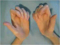
Gottron’s Sign & Papules assoc with Dermatomyositis
Gottron’s papules = lichenoid papules over the knuckles Gottron’s sign = violaceous discoloration of knuckles, elbows, knees Proximal nail fold atrophy & telangiectasia (“ragged cuticles”) |
|
|
what derm disease is assoc with inflammatory scar-like sclerosis, Local or widespread skin involvement, With or without systemic manifestation
-Can be severe and rapidly fatal Renal disease and pulmonary sclerosis CREST syndrome |
Scleroderma
|
|
|
what are the key words for CREST syndrome?
|
C = calcinosis = Ca deposits in skin
R = Raynaud's Phenomenon -spasm of blood vessels in response to cold or stress E = Esophageal dysfunction -acid reflux and dec in motility of esophagus S = Sclerodactyly -thickening and tightening of skin on the finger and hands T = Telangiectasias -dilation of capillaries causing red markes on surface of skin |
|
what is this?
|
Scleroderma
Inflammatory scar-like sclerosis Local or widespread skin involvement With or without systemic manifestation Can be severe and rapidly fatal Renal disease and pulmonary sclerosis CREST syndrome Up to 50% of body can be involved and still have negative ANA titers |
|
|
what is localized scleroderma?
|
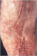
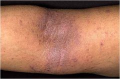
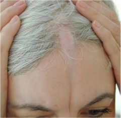
Morphea
Localized scleroderma Can occur anywhere on body May have joint contracture, limited mobility En coup de sabre = linear morphea of head |
|
|
what is the term for linear morphea of head?
|
En coup de sabre
|
|
what is this?
|
Linear Morphea
Localized scleroderma Can occur anywhere on body May have joint contracture, limited mobility |
|

what is this?
|
En Coup De Sabre Morphea
-Localized scleroderma of head |
|
what is this?
|

En Coup De Sabre Morphea
-Localized scleroderma of head |
|
|
what is the acute form of ?Cutaneous Lupus Erythematosus
|
typical photosensitive malar “butterfly” rash
|
|
|
what is the subacute form of Cutaneous Lupus Erythematosus?
|
psoriaform or papulosquamous eruption, photosensitive (sun-exposed areas)
|
|
|
What is the criteria for SLE?
|
hematologic changes, renal problems, ANA titers, anti-DNA or anti-Sm, neurologic pathology, etc
many of the Cutaneous Lupus Erythematosus patients meet the criteria for sle |
|
|
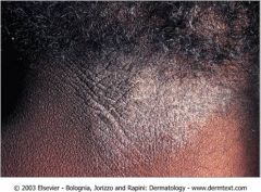
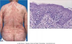
what derm disease is described as:
-Scarring and chronic form -Scarred pink to whitish plaques with scale -Active lesions have violaceous periphery -Can lead to scarring alopecia |
Discoid Lupus Erythematous
|
|
|
what form of Lupus Erythematous has patients with many of the sle criteria?
|
Cutaneous Lupus Erythematosus
SLE criteria include: hematologic changes renal problems ANA titer anti-DNA anti-Sm |
|
|
what form of Lupus Erythematous has patients with few of the sle criteria?
|
Discoid Lupus Erythematous
|
|
|
what is the txmt for Discoid Lupus Erythematous?
|
Treat with intralesional or topical steroids, sun avoidance, antimalarials if severe or large areas involved
|
|
what is this?
|

Acute Lupus Erythematosus
Acute = typical photosensitive malar “butterfly” rash Many of these patients will meet criteria for SLE (hematologic changes, renal problems, ANA titers, anti-DNA or anti-Sm, neurologic pathology, etc) |
|
what is this?
|
Subacute Lupus Erythematosus
-Subacute psoriaform or papulosquamous eruption, photosensitive (sun-exposed areas) Many of these patients will meet criteria for SLE (hematologic changes, renal problems, ANA titers, anti-DNA or anti-Sm, neurologic pathology, etc) |
|

what is this?
|
Discoid Lupus Erythematosus
Scarring and chronic form of cutaneous lupus Scarred pink to whitish plaques with scale Active lesions have violaceous periphery Can lead to scarring alopecia Few patients meet criteria for SLE Treat with intralesional or topical steroids, sun avoidance, antimalarials if severe or large areas involved |
|
|
what is the criteria for neurofibromatosis?
|
Criteria (need at least 2):
2 or more neurofibromas 6 or more café au lait macules Axillary or inguinal freckles (Crowe’s sign) 2 or more Lisch nodules in eyes Optic gliomas Bony abnormalities 1st degree relative |
|
|
what is Mycosis FungoidesS?
|
Most common cutaneous T-cell lymphoma
Middle-aged men Patches, plaques, tumors, erythroderma Often in sun-protected sites Eczematous, wrinkling atrophy (“cigarette paper”), poikiloderma Treat with steroids, UVB, nitrogen mustard, PUVA, methotrexate, retinoids, interferons |
|
|
how is Mycosis fungoides treated?
|
Treat with steroids, UVB, nitrogen mustard, PUVA, methotrexate, retinoids, interferons
|
|
what is this?
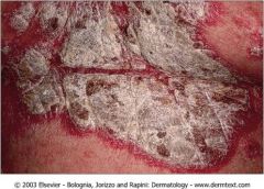
|
MF: Patch Stage
MF = Mycosis Fungoides -most common cutaneious T-cell lymphoma |
|
what is this?
|

MF: Tumor Stage
|