Reading...
![]()
Play button
![]()
Play button
![]()
Use LEFT and RIGHT arrow keys to navigate between flashcards;
Use UP and DOWN arrow keys to flip the card;
H to show hint;
A reads text to speech;
40 Cards in this Set
- Front
- Back
|
What does the thorax include?
a. location relatively b. contents (4 main ones) c. primary divisions |
a. area between neck and abdomen (not chest)
b. thoracic cavity and thoracic wall surroundings Include: primary organs of respiratory and circulatory system (heart, great vessels, lungs, trachea) c. i. Two pulmonary cavities (contain lungs and pleurae) ii. Mediastinum, located centrally and contains (Heart, thoracic great vesses, trachea, thoracic esophagus, and thymus) |
|
|
Describe the following by what it includes, and functions to do...
thoracic wall a. What it includes b. functions (3 main) |
a. Includes: Thoracic cage, myofascial structures, neurovasculature structures
b. 1. protects thoracic and abdominal viscera 2. resists pressure of elastic recoil from lungs and inspiratory movements 3. muscular attachment for upperlimbs, abdomen, neck, and back |
|
|
Describe the following by what it includes, and functions to do...
thoracic cage boney components a. general shape and function b. includes 2 parts c. supported by (two things)? d. made up of (4 components) |
a. shaped like a domed bird cage that provides rigidity absorbs external blows and compression without fracture and changes shape for breathing
b. includes superior and inferior thoracic aperture c. Sternum and 12 thoracic vertebrae d. Made of: i. 12 ribs, thin and flexible ii. costal cartilages iii. joints iv. intercostal spaces |
|
|
Describe what the musculature of the thoracic wall includes...
a. Inspiration (principal muscles) |
Two divisions for Breathing (Expiration and Inpiration)
a. Inspiration 1. Principal i. External intercostal muscles- are the principal muscles in inspiration...point anterio-inferiorly elevate ribs, increase width of thoracic ii. interchondral part of internal intercostal muscles- same as above iii.diaphragm- domes descend and increase longitudinal dimension of thoracic cavity also helps elevate lower ribs |
|
|
What does the musculature of the thoracic wall include... (2 general categories)
|
Out into two groups (inspiration muscles and expiration muscles)
1. Inspiration a. Accessory muscles b. Principal muscles 2. Expiration a. quiet breathing (no specific muscles just passive recoiling of lungs) b. active breathing muscles |
|
|
Describe what the musculature of the thoracic wall includes...
a. Inspiration (accessory muscles) |
a. stenorcleidomastoid- elevates sternum
b. (Middle, anterior, posterior scalene)- elevate and fix upper ribs |
|
|
Describe what the musculature of the thoracic wall includes...
a. expiration (active breathing muscles) |
a. internal inercostals (except interchondral part)- lower ribs and decrease width of thoracic cavity
b. (Abdominals) -rectus abdominus, external oblique, internal oblique, transvers abdominis - depress ribs and compress abdominal contents and push up diaphragm |
|
|
Describe Dyspnea and the use of additional musculature to assist with respiration
What is a position someone would be in that is showing signs of dyspnea and why? |
- difficult breathing- causes use of accessory respiratory muscles (Pectoralis major/minor, serratus anterior, scalene
muscles) to help expand thoracic cavity - leaning foward hands on knees to fix pectoral girdle so muscles can expand thoracic wall - |
|
|
Describe how surgeons get to extra pleura intrathoracic structures in the thorax?
|
Endothoracic fascia is opened to gain access to extrapleural intrathoracic structures- such as arteries and satellite veins
|
|
|
Differentiate intercostal membranes from endothoracic fascia
|
intercostal- completes thoracic wall in areas where intercostal muscles are absent and fascia has continued
Endo- thin fibroareolar layer between internal aspect of thoracic cage and lining of pulmonary cavities (used for surgical access) |
|
|
What do the nerves of the thoracic wall come from?
What are the groups of Anterior Rami? |
- from spinal nerves
- 12 pairs- both sensory and motor - Anterior rami- a. T1-11 "intercostal" nerves in intercostal spaces b. T12- "subcostal nerve" |
|
|
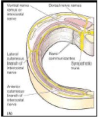
What are the branches of intercostal nerves (5)
|
LAM RC
1. Lateral cutaneous 2. Anterior cutaneous 3. Muscular branches- intercostal, subcostal, transverse thoracic, levatores constarum, serratus posterior muscles 4. Rami communicantes- (communicating branches) connect to ipsilateral sympathetic trunk 5. Collateral branches- arise around rib angle, course along superior aspect of rib below |
|
|
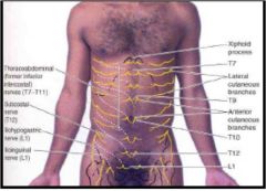
Which branches of the posterior and anterior ramus does the cutaneous branches make the dermatomes?
What are reference points? |
anterior and lateral branches supply skin
T4- around nipples T7- Xiphoid T10- umbilicus T12- Superior inguinal ligament |
|
|
What does herpes zoster have to do with dermatome?
How is it activated? |
Herpes is spinal ganglia so vesicular rash and sharp pain is in dermatome distribution
- reactivation of chickenpox (varicella zoster virus) |
|
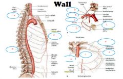
Describe the course of the intercostal nerves posteriorly to laterally (typical intercostal nerves 1-6) and laterally to anteriorly
|
Posteriorly-
Start--> a. middle intercostal space within endothoracic fascia, b. move between internal intercostal innermost intercostal muscles (near rib angle) c. Moves within the costal grooves on inferior surface of rib Laterally- a. Becomes lateral cutaneous branches at midaxillary line b. Divides to anterior/ posterior branches Anteriorly starts - On the internal surface of the internal intercostal muscles a. Near sternum passes between the costal cartilages anteriorly becomes anterior cutaneous branches finally Divides into medial/ lateral branches |
|
|
What is course of atypical intercostal nerves?
|
give off lateral cutaneous branch, cross the costal margin and continue on the abdomen to become thoracoabdominal nerves
- Supply abdominal skin and muscles via muscular and anterior cutaneous branches |
|
What is course of atypical intercostal nerves?
|
give off lateral cutaneous branch, cross the costal margin and continue on the abdomen to become thoracoabdominal nerves
- Supply abdominal skin and muscles via muscular and anterior cutaneous branches |
|
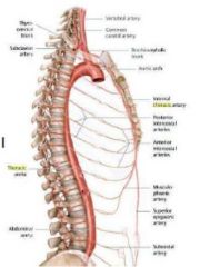
Label A-L
|
a. Subclavian artery
b. Thoracic aorta c. Posterior intercostal arteries d. Superinter-costal artery e. Costocervical trunk f. Subclavian artery g. Second posterior inter-costal artery h. Posterior inter-costal artery i. Thoracic aorta j. Collateral branch k. Posterior intercostal arteries l. First posterior intercostal artery |
|
What are the three primary arteries of the thoracic wall and what do they include?
What does the vasculature of the Thoracic wall supply? |
Supply the intercostal m.,
pectoral m., breasts, skin 1. Thoracic aorta- includes Posterior intercostal and subcostal a. 2. Subclavian a.- includes Internal thoracic and supreme intercostal a. 3. Axillary a. Superior and lateral thoracic a. |
|
|
In the posterior intercostal arteries where does the following vasculature arise from and how?
a. 1st and 2nd ribs b. 3-11 and subcostal |
a. Arise from the supreme (superior) intercostal a.
--> Subclavian a. costocervical trunk supreme intercostal a. b. Arise from the thoracic aorta -->Right intercostal a. cross over the vertebrae |
|
|
What 4 things do all the posterior intercostal arteries have in common?
|
1. Accompany the intercostal n. along its path in the costal groove
2. Give off posterior branches that accompanies the posterior ramus 3. Give off collateral branches that courses on superior surface of rib 4. Anastamose with anterior intercostal a. |
|
|
Where are internal thoracic arteries located and where do they orginate from? What are they used for?
|
located lateral to sternum on internal surface of sternum
- from subclavian a. - used to supply breasts - used for Coronary Artery Bypass Graft (CABG) surgery (most common) |
|
|
Which artery supplies blood to pericardium?
|
pericardiophrenic artery
|
|
|
Where do the anterior intercostal arteries anastomose with the posterior intercostal arteries?
|
around anterior axillary line
|
|
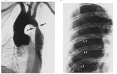
Explain pathology in slide
|
Rib Notching
Caused by coarctation of the aorta causes an increase in blood flow through the internalthoracic arteries and anterior intercostal a. along with a decrease in blood flow to the posterior intercostal a. This leads to dilation of the anterior intercostal arteries causing indentations in the inferior margin of the anterior part of the ribs. |
|
|
What does coarctation of the aorta cause?
|
"rib notching"
causes an increase in blood flow through the internal thoracic arteries and anterior intercostal a. along with a decrease in blood flow to the posterior intercostal a. This leads to dilation of the anterior intercostal arteries causing indentations in the inferior margin of the anterior part of the ribs. |
|
|
Describe the course of the phrenic nerve and possible pathalogies associated with phrenic nerve problems...
|
starts from in front of anterior scalene muscle and It courses in front of the root of the lung.
C3,4,5 keeps us alive Problems: One half diaphragm can become paralyzed from injury to one phrenic n. Causes: Malignancy of the lung compressing the nerve (Most common) – Trauma/brachial plexus injury – Cervical spondylosis – Herpes zoster – idiopathic |
|
|
If the diaphragm does not form correctly during embryogenesis what could likely happen? Which pleural cavity would we see this?
What does this lead to? |
Pleural cavities will not become separated from the peritoneal cavities resulting in congenital diaphragmatic hernia
– Usually the left pleural cavity – Abdominal viscera herniates up into the thorax – Leads to pulmonary hypoplasia (no utero lung development) |
|
|
If someone comes in with chest pain described as "sharp, stabbing, especially with increased exertion and depth of breathing" and you ausculatate and hear something like a clump of hair being rubbed together... Diagnosis?
What may this problem lead to? |
pleuritis- inflammation of the lung
May cause pleural adhesions (Parietal and visceral layers adhere) |
|
|
Patient comes in with a Penetrating wound of parietal pleura or another patient comes in with a Rib fracture that tears visceral pleura and lung... What is differential diagnosis
|
pneumothorax
The surface tension between the visceral and parietal pleura is broken and causes the lung to collapse The surface tension between the visceral and parietal pleura is broken and causes the lung to collapse – Tension pneumothorax can cause displacement of mediastinum (mediastinal shift) |
|
|
What is hydro, hemo, and hemopneumo thorax and causes...
|
hydrothorax- accumulation of fluid in pleural cavity
hemo- accumulation of blood (stab) hemopneumo- accumulation of blood and air |
|
|
If someone has a pneumothorax or hemothorax
|
Thoracostomy
small incision in 5th or 6th intercostal space (make that it does not connect to diaphram), on superior margin of rib, inferiorly so to drain fluid and superiorly to drain air, |
|
|
Someone is stabbed in the neck just above the clavicle and cannot breathe in a kid or infant
|
I would be worried about a pneumothorax
• The cervical pleura reaches a relatively higher level in infants and children because their necks are short, so they are especially vulnerable to injury during the first few years after birth |
|
|
This is the trachea's last ditch effort to prevent aspiration..
|
carina located between the orifices of the main bronchi
|
|
|
Why might you be more likely to get a foreign body stuck on one side that the other bronchi?
|
Right side- shorter, wider, and runs more vertically
|
|
|
Patient respiratory distress, pregnant, and other hypercoaguable factors shown in history, acute onset also has thigh pain
|
pulmonary embolism
Forms when a blood clot, fat globule, or air bubble travels from a leg vein to the lungs (after it passes through the right side of the heart) • If large causes respiratory distress • If deprives blood to visceral pleura it gets inflamed and irritates parietal pleura pleuritic pain • If deprives blood to visceral pleura it gets inflamed and irritates parietal pleura pleuritic pain |
|
|
Where do the bronchial arteries arise from?
Anastomose with? What about bronchial veins |
R. bronchial from posterior intercostal a.
2 Left bronchial artery from thoracic aorta anastamose distally with pulmonary branches Veins anastomose with anzygous (right side), and accessory hemizygous v (left side) |
|
|
When you have pain coming from the parietal pleura what might we see?
Why might someone have neck pain when they have injury to diaphragm part of parietal pleura? |
pain could be local or distribution to different dermotome, pain could be felt in other areas phrenic (which is c3,c4,c5) innervates such as the neck
|
|
|
What contents does the superior mediastinum include?
|
- Thymus- on top of trachea (fatty pad)
– Trachea- anterior to esophagus – Esophagus – Great vessels of heart – Vagus n., recurrent laryngeal n., phrenic n., cardiac plexus – Thoracic duct and lymphatic trunks |
|
|
Person comes in without ventilation and you need to put a tube down to open airway... what is it called and how do you place it?
Where does pressure need to be placed to intubate? |
Endotracheal tube
into airway at level of above of carina, - amateurs put it in the stomach on accident - apply pressure on cricoid cartilage to compress esophagus between trachea and vertebral column ensuring correct placement and no gastric regurgitation |