Reading...
![]()
Play button
![]()
Play button
![]()
Use LEFT and RIGHT arrow keys to navigate between flashcards;
Use UP and DOWN arrow keys to flip the card;
H to show hint;
A reads text to speech;
101 Cards in this Set
- Front
- Back
|
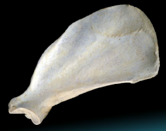
Scapula (Fig 2.3 -2.4)
|
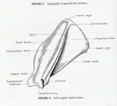
flat roughly triangular bone possessing two surfaces (lateral and ), three borders (Dorsal, Causal, Cranial), and three angles (Cranial, Caudal, Ventral).
|
|
|
Acromion
|
truncated process at the distal end of the scapula spine
where part of the deltoideus muscle arises |
|
|
Supraspinous fossa
|
the entire lateral surface cranial to the spine of the scapula
the supraspinatus arises from all but the distal part of this fossa |
|
|
Intraspinous fossa
|
caudal to the spine of the scapula, is a triangular, with the apex at the neck.
the infraspinatus arises from |
|
|
Serrated face
|
small proximal, cranial rectangular area of the medial/costal surface. serves as insertion for the serratus ventralis muscle.
|
|
|
Subscapular fossa
|

large remaining part of the medial/costal surface of the scapula. Nearly flat and usually presents three straight muscular lines that converge distally.
the subscapularis arises from the whole surface |
|
|
cranial border
|
thin, near the ventral angle the border is concave as it enters into the formation of the neck.
thickens at the dorsal end until the cranial angle (then continues as the dorsal border) |
|
|
Scapular notch
|
notch formed when the cranial border concaves at the neck of the scapula
|
|
|
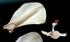
Glenoid Cavity
|
at the ventral angle/ the distal or articular end of the scapula
shallow, articulates with the head of the humerus |
|
|
Intrafraglenoid Tubercle
|
on the caudal border
where the teres minor and long head of the tricepts arises |
|
|
Supraglenoid Tuercle
|

eminence at the cranial part of the glenoid cavity. Shows a slight medial inclination on which a small tebercle, the coracoid process, can be distinguished.
where the bicept brannchii arises from |
|
|
Coracod Process
|

small tebercle lateral to the supraglenoid tubracle.
where the coracobrachialis arises from |
|
|
Humerus (Fig 2.5)
|
located in the arm/brachium. Enters into the formation of the shoulder (articulation of scapula and humerus) and elbow joint (articulation of the ulna and radius and humerus).
proximal extremity includes the head, neck and greater and lesser tubercles. distal extremity (condyle) includes the trochlea, capitulum, and the radial and olecranon fossae which communicate proximal to the trochlea through the supratrochlear foramen. Medial and lateral epicondyles on the sides of the condyle. the body lies between the promimal extremity and condyle |
|
|
Intertubercular groove (humerus)
|
begins at the cranial end of the articular area. Lodges the tendon of origin of the biceps branchii and is defeflected toward the medial plane by the greater tubercle
|
|
|
thoracic griddle
|
consists of paired scapulae and clavicles
scapulae is large while the clavicles are reduced in canine |
|
|
Clavicle (Fig 2.10)
|
small oval plate located cranial to the shoulder within the clavicular tendon in the brachiocephalicus muscle (Fig 2-12)
First bone to show a center of ossification in the fetal dog, but in the adult is partly or completely cartilaginous (frequently visable in Dorsoventral radiographs of the trunk medial to the shoulder joint) |
|
|
Scapula Neck
|
constricted part that units with the expanded blade of the scapula
|
|
|
Scapula Spine
|
shelf of bone that divides the scapula into nearly equal fossae.
most prominant feature of bone. begins at dorsal border as a thick, low ridge and then becomes thinner and wider toward the neck. In all breeds the free border is slightly thickened, and in some breeds it is everted caudally |
|
|
Fibrous Joint (Synarthroses)
|
Suture
bones are held together by a thin layer of dense fibrous connective tissue a slight movement or immovable joint ie skull |
|
|
Syndesmosis
|
amphiathrosis joint/cartilaginous joint - allow some movement
occurs when two bones join by a section of cartilage no joint cavity ex. between the tibia and fibula |
|
|
synchondrosis
|
amphiarthrosis/ primary joint
union b/w two bones formed by HYALINE cartilage ex. sternocostal joint (where the ribs meet the sternum) |
|
|
Symphysis
|
amphiarthrosis joint/ secondary
fibrocartilage sandwiched b/w hyaline cartilage each vertabrae is divided by a cartilaginous joint, which provides protectin and cushioning b/w vertebrae ex. b/w the pubic symphyss & vertebrae |
|
|
Synovial joints
|
Ball and Socket
Encapsulated, fluid, cartilage on contact surface. Will allow rotational movement and can move in all planes three types: Hinge- freely oving joint that can only bend in one direction (knee, elbow joint) Plane- opposing surfaces are nearly plane and there is only a slight, gliding motion (carpal joint-wrist) Pivot : allow limited rotating movements (head-neck) |
|
|
Subcutaneous tissue
|
soft tissue immediately underlying the skin or epidermis
ex. areolar tissue, supraficial and deep fascia, adipose tissue, dense connective tissue |
|
|
Areolar Tissue
|
loose connective tissue that containspockets for fat and fluid
superficial to fascia, less fibrous compared to fascia subcutaneous |
|
|
Superficial fascia
|
deep to areolar tissue
conncetive tissue enveloping, seperating or binding together muscle,organs, and other soft structures of the body occupies the subcutaneous space between deep fascia and the skin |
|
|
Deep fascia
|
firmly attached to the muscle that it encloses
found among the muscles and bones of the trunk and limbs |
|
|
Adipose Tissue
|
used for cushioning, thermal insulation, energy storage (fat)
|
|
|
Dense Connective tissue
|
forms strong, rope-like structures such as tendons and ligaments.
Tendons attach skeletal muscle to bones; ligaments connect bones to bones at joint |
|
|
Origin (muscle attachment)
|
proximal attachment
the part that moves the least direct attachment of muscle cells to bone |
|
|
Insertion (muscle attachment)
|
distal attachment,
the part that moves the MOST often is by tendon or aponeurosis extending from the muscle cells to the bone |
|
|
Cutaneous trunci
|
thin sheet of muscle covering most of the thorax and abdomen.
twitches the skin innervation: lat thoracic n. males; muscle fibers form preputial m. |
|
|
Extrinsic Muscles of the thoracic limb
|
Extrinsic muscles have one attachment on the limb and one attachment on the trunk or neck/head.
Their actions depend on weight distribution. They move the limb relative to the body, but if the limb is anchored by body weight, the body is moved relative to the limb. (In contrast, intrinsic muscles are attached totally to limb bones and thus they move only joints of the limb.) 8 extrinsic muscles of the thoracic limb: Superficial pectoral – Deep pectoral – Brachiocephalicus – Sternocephalicus – Omotransversarius – Trapezius – Rhomboideus – Latissimus dorsi – Serratus ventralis |
|
|
Platysma
|
cutaneous muscle of neck and head
|
|
|
Dorsal border (scapula)
|
extends from the cranial to the caudal angles of the scapula
in life it is capped by a narrow band of cartilage the rhomboideus attaches to this border |
|
|
Caudal border (scapula)
|
extends from the caudal to ventral angle.
Thick. bears the infraglenoid tubercle from which arises the teres minor and the long head of the tricept subscapularis and long head of the tricept arise from the broad and smooth middle third portion of the caudal border |
|
|
Ventral angle (scapula)
|
forms the expanded distal end of the scapula
clinically the most important part of the scapula because it enters the formation of the shoulder joint |
|
|
greater tubercle (humerus)
|
a
|
|
|
lesser tebercle (humerus)
|
a
|
|
|
neck (humerus)
|
a
|
|
|
cranial surface (humerus)
|
a
|
|
|
crest of the greater tubercle (humerus)
|
a
|
|
|
lateral surface (humerus)
|
a
|
|
|
deltoid tuberosity (humerus)
|
a
|
|
|
tricipital line (humerus)
|
a
|
|
|
tuberosity of the teres minor (humerus)
|
a
|
|
|
brachialis groove (humerus)
|
a
|
|
|
lateral supracondylar crest (humerus)
|
a
|
|
|
teres major tuberosity (humerus)
|
a
|
|
|
humeral condyle
|
a
|
|
|
trochlea (humerus)
|
a
|
|
|
capitulum (humerus)
|
articulates only with the head of the radius
|
|
|
lateral epicodyle (humerus)
|
a
|
|
|
medial epicodyle (humerus)
|
a
|
|
|
olecranon fossa (humerus)
|
-deep excavation of the caudal part of the humerus condyle.
-It receives the anconeal process of the ulna during extension of the elbow |
|
|
radial fossa (humerus)
|
on the cranial surface of the humeral condyle, which communicated with the olecranon fossa by the supratrochlear foramen.
|
|
|
supratrochlear foramen (humerus)
|
opening on the humeral condyle
no soft structures pass through |
|
|
antebrachium
|
forearm. bones consist of the radius and ulna
|
|
|
Ulna
|
caudal part of the antebrachium (forearm). Proximal end is medial to the radius (articulate at the ulna trochlear notch and the articular circumfrance of the radius)and the dsital end is lateral to the radius.
tapers proximal to distal end |
|
|
Radius
|
bone of the antebrachium (forearm). Shorter than the ulna. Articulates proximally with the humerus and caudal surface of the ulna. Articulates distally with the carpus, and lateral border of the ulna.
|
|
|
Head (radius)
|
widest medial to lateral. forms an oval, depressed srticular surface called the FOVEA CAPTIS which articulates with the capitulum of the humerus.
|
|
|
Articular circumference (radius)
|
smooth caudal border of the radius head.
for articulation with the radial notch of the ulna |
|
|
radial tuberosity (radius)
|
lies distal to the neck on the medial border of the radius.
the biceps brachii and brachialis insert in part on the tubercle |
|
|
Body (radius)
|
compressed so that it possesses cranial and caudal surfaces and lateral and medial borders. Slightly convex cranially. Caudal surface= concave and roughened.
Has a ligamentous attachment to the ulna. |
|
|
Trochlea (radius)
|
distal extremity of the radius.
the carpel articular surface is concave. |
|
|
ulnar notch
|
on the lateral surface of the distal extremity (trochlea). Has facet for articulation with the ulna
|
|
|
styloid process (radius)
|
roughened projection on the medial surface of the distal extremity (trochlea) of the radius
the medial collateral ligament of the carpus attaches proximal to the process |
|
|
cranial surface (radius distal extremity)
|
a
|
|
|
Olecranon (ulna)
|
proximal extremity of the ulna. Includes the olecranon tuber and anconeal process.
Serves as a level arm for the extensor muscles of the elbow |
|
|
Olecranon tuber (ulna)
|
proximal end of the olecranon. Grooved cranially and enlarged and rounded caudally
the triceps branchii, anconeus, and tensor fasciae antebrachii attach to the caudal part |
|
|
Trochlear notch (ulna)
|
smooth, vertical, half-moon shaped cncavity facing cranially
it articulates with the trochlea of the humerus |
|
|
anconeal process (ulna)
|
sharp-edged, slighly hooked at the proximal end.
Fits into the alecrannon fossa of the humerus when the elbow joint is extended |
|
|
Medial and lateral coronoid process (ulna)
|
articulate with the humerus and radius
|
|
|
Deltoideus (Fib 2-13, 2-15 *, 2-17)
|
composed of two parts (scapular and acromial) that are fused and act in common across the shoulder
scapular part - arises as a wide aponeurosis the length of the scapular spine, covers the infraspinatus acromial part- arises from the acromion and has a fusiform shape Both portions fuse before they insert on the deltoid tubrosity of the humerus Action = to flex the shoulder joint |
|
|
Infraspinatus (Fig 2-13, 2-16*, 2-17)
|
fusiform, lies principally in the infraspinous fossa
sebtendinous synovial burse - found between the tendon of interstion and the greater tubercle of the humerus Orgin: infraspinous fossa Insertion: small, circumscribed area on the lateral side of the greater tubercle of the humerus |
|
|
sebtendinous synovial bursa-
|
found between the tendon of interstion of the infrasoinatus and the greater tubercle of the humerus
bursa = closed connective tissue sac containing synovial fluid --> which reduces friction |
|
|
Teres Minor (fig 2-13, 2-16)
|
small wedge shaped muscle caudal to the shoulder joint , covered by the deltoideus
O: infraglenoid tubercle and distal third of the caudal border of the scapula I: teres minor tubrosity of the humerus A: flex the shoulder joint, rotate shoulder laterally, and prevent medial rotation when bearing weight |
|
|
Supraspinatus (Fig 2-12, -13, -14, -16, -17, - 19, -27)
|
wider and larger than the infraspinatus, deep to the cervical part of the trapezius and osmotransversarius
O: supraspinous fossa (extends of the cranial border of the scapula- part of the muscle is closley united with the subscapularis medially) I: Greater tubercle of the humerus by a thick tendon A: to extend and laterally stabilize the shoulder joint |
|
|
Subscapularis (Fig 2-12, -13, - 14, -17, -19)
|
o: occupies the entire subscapular fossa
closley associated with the supraspinatus cranally and teres major caudally I: the lesser tubercle of the humerus A: adduct, extend, and medially stabalize the shoulder joint.... (p 26) |
|
|
Teres Major (Fig 2-13, -14, -16, -17, -19, -27)
|
caudal to the subscapularis. Not round, has three surfaces
O: caudal angle and adjacent caudal border of the Iscapula; the caudal surface of the subscapularis I: teres major tubrosity of the humerus (medial surface of the upper half of the body of the humerus) A: flex the shoulder, rotate the shoulder medially, and prevent lateral rotation when weight bearing |
|
|
Coracobrachialis (Fig 2-14, 2-19)
|
O: coracoid process of the scapula (by a relatively long tendon)
I: crest of the lesser tubercle of the of the humerus proximal to the teres major tubrosity (The conjoined tendon of the teres major and the latissimus dorsi cross the insertion) A: to adduct, extend, and stabalize the shoulder joint Crosses the medial surface of the shoulder obliquely, small spindle shaped muscle. Courses craniallyto the center of the shoulder. Courses caudodistally across the lesser tubercle (provided with a synovial sheath), crosses the tendon of insertion of the subscapularis. The muscle belly is distal to the lesser tubercle and medial to the medial head of the tricept. |
|
|
Tensor fasciae antebrachii (Fig 2-14, 2-18, 2-19)
|
thin strap the extends from the latissimus dorsi to the medial fascia of the forarme and the olecranon. It lies on the long head of the tricepts.
O: fascia covering the lateral side of the latissimus dorsi I: olecranon A: to extend the elbow |
|
|
Triceps brachii
|
consists of four heads (instead of the usual three) with a common tendon to the olecranon tuber (ulna).
Only the long head arises from the scapula, the other three arise from the proximal head of the humerus. (other flashcards for each) |
|
|
Long head Triceps brachii (Fig 2-13 through 2-19, 2-27)
|
O: caudal border of the scapula
I: Olecranon tuber (ulna) A: extend the elbow and flex the shoulder completely bridges the humerus, has two bellies |
|
|
Medial head Triceps brachii (Fig 2-14, -18, -19, -23, -27)
|
O: crest of the lesser tubercle near the teres major tubrosity
I: olecranon A: extend the elbow lies caudally on the humerus, lateral and caudal to the biceps branchii |
|
|
Lateral head triceps brachii (Fig 2-12, -13, -15, -18, 20 -27)
|
O: tricipital line of the humerus
I: olecranon tuber A: extend the elbow lies distal to the long head, caudal to the acromial part of the deltoideus, and lateral to the accessory head (which it covers) |
|
|
accessory head of the triceps brachii
|
lies between the lateral and medial heads
O: neck of the humerus I: olecranon bone A: extend the elbow |
|
|
biceps brachii (p 30)
|
O: Supraglenoid tubercle
I: Ulnar and radial tuberosities A: to flex the elbow and extend the shoulder long, fusiform muscle that lies on the medial and cranial surface of the humerus. Covered by the pectoral muscles |
|
|
brachialis (p 30)
|
O: the proximal third of the lateral surface of the humerus
I: ulnar and radial tubrosities A: to flex the elbow long thin muscle that lies in the brachialis groove of the humerus. From the proximal third of this groove, the brachialis curves laterally and cranially as it courses distally, crosses the elbow, and inserts by a terminal tendon on the medial side of the proximal end of the ulna. |
|
|
Foot pads
|
carpal pad- small pad that protrudes palmar to the carpus
metacarpal pad- largest, triangular, on the palmar side of the metacarpophalangeal joint digital pads- ovoid and flattened , located palmar to the distal interphalangeal joint. |
|
|
Anconeus
|
O: lateral supracondylar crest and the lateral and medial epicodyles of the humerus
I: lateral surface of the proximal end of the ulna (olecrannon) A: to extend the elbow |
|
|
Extensor carpi radialis
|
O: Lateral supracondylar crest
I: The small tuberosities on the dorsal surface of the base of metacarpals I and II Longest of the craniolateral antebrachial muscles , lies on the cranial lateral surface of the radius. 2 branched Tendons held in by the extensor retinaculum |
|
|
Common digital extensor
|
O: Lateral epicondyle of the humerus
I: Extensor processes of the distal phalanges of digits II, III, IV and V Caudal to the extensor carpi radialis. 4 tendons leave the muscle – held into the lateral groove of the radius by the extensor retinaculum |
|
|
Lateral digital extensor
|
O: Lateral epiconfyle of the humerus
I: Proximal end of the phalanges if digits III, IV, V and mainly the extensor processes of the distal phalanges of the digits Half the size of the common digital extensor, medial to the ulnaris lateralis. Begins in the middle of the forearm, passes under the extensor retinaculum in a groove b/w the radius and ulna and spits into three branches. |
|
|
Ulnaris Lateralis
|
O: Lateral epicondyl of the humerus
I: Lateral aspect of the proximal end of metacarpal V and the accessory carpal bone. Larger than the lateral digital extensor. Bounded deeply by the ulna and the large flexor group of muscles which lie caudal and medial to it. Only flexor to arise on the lateral epicondyl. |
|
|
Supinator
|
O: Lateral epicondyl of the humerus
I: Cranial surface of the proximal fourth of the radius Short, broad and flat. Obliquely placed across the lateral side of the flexor surface of the elbow joint. Covered by the extensor carpi radialis and the common digital extensor. Lies on the proximal 4thof the radius. |
|
|
Abductor digiti I longus
|
O: Lateral border and cranial surface of the ulna; interosseous membrane
I: Proximal end of metacarpal I Triangular, lies primarily in the groove b/w the ulna and radius. |
|
|
Pronator teres
|
O: Medial epicondyl of the humerus
I: Medial border of the radius b/w the proximal and middle thirds Extends obliquely across the medial surface of the elbow. Round (transverse) at origin to flat at insertion. Lies b/w the extensor carpi radialis and the flexor carpi radialis. |
|
|
Deep digital flexor - humeral head
|
O: Medial epicondyl of the humerus.
I: Flexor tubercle on the palmar surface of the distal phalanx of each digit Larger than other heads and has several bellies |
|
|
Deep digital flexor - ulnar head
|
O: Proximal ¾ of the caudal border of the ulna
I: Flexor tubercle on the palmar surface of the distal phalanx of each digit Small |
|
|
Deep digital flexor - radial head
|
O: Middle 1/3 of the of the medial border of the radius I: Flexor tubercle on the palmar surface of the distal phalanx of each digit
Smallest, forms single tendon with other two heads. Tendon held in place in the carpal canal by the flexor retinaculu |