![]()
![]()
![]()
Use LEFT and RIGHT arrow keys to navigate between flashcards;
Use UP and DOWN arrow keys to flip the card;
H to show hint;
A reads text to speech;
25 Cards in this Set
- Front
- Back
|
Blood Circulation |
To deliver nutrients, salt, and hormones to tissue and to take away waste products. Diffusion only works over short distances, so our body has developed a vast pumping network. |
|
|
Basic Plumbing |
Pump is the heart, pumps blood to the arterial system, which branches into the capillaries where transfer of materials occurs. Capillaries fuse back into venous system which brings blood back to the heart. |
|
|
Capillaries |
Contain walls that are very thin and fluid is forced out. Fenestrations in the wall allow for small cells like macrophages to crawl out. This fluid is the lymph and collected via the lymphatic system. |
|
|
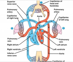
Heart |
2 separate pumps, on the left and on the right. Each of these controls a separate circulation. Right pump circulates the blood for oxygenation, the left sends blood through the rest of the body. Blood leaves the heart through the aorta. Blood is sent out and divides into arterioles turn into capillaries, joins together into venules and then finally to the large vena cava that delivers blood back to the heart. |
|
|
Mechanism Pump |
Heart is a mechanism pump that moves liquid much like a well. Contains muscular chambers. The atrium has thin walls and is a low pressure pump, while the ventricle has thick walls and is a high pressure pump. Atrium pumps blood into the ventricle, and ventricle pumps to the rest of the body. |
|
|
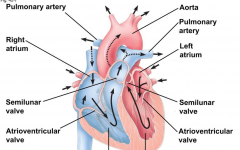
Valves |
2 valves in each chamber, 4 in each heart. The valves flop open or closed depending on which has higher hydrostatic pressure. If it's higher on one side they are open and blood flows through. AV is between the atrium and ventricle. |
|
|
Heart Pressures |
If pressure in atrium is less than the pressure in the ventricle, the valve is closed. when the ventricle contracts and AV valve is closed, the pulmonary valve open because it's pressure is greater than aorta. |
|
|
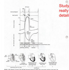
Ventricle Filling |
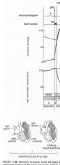
The AV valve is open and blood is flowing from the atrium into the ventricle. Low pressure because AV valve is open. AV valve closes and Atrium contracts |
|
|
Isovolumetric Contraction |
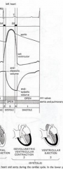
The pressure in atrium increases with contraction and the pressure shoots up until the aeortic or pulmonary valve opens. When these open the blood flows into the aeorta or pulmonary artery. |
|
|
Isovolumetric Relaxation |
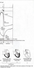
Pressure drops as the atrium relaxes and blood flows out. Atrium contracts a bit to top off the ventrical and the cycle repeats. |
|
|
Right AV Valve |
3 cusps (flaps) or RAT (Right AV-Tricusp) |
|
|
Left AV Valve |
2 cusps or LAMB (Left AV-mitral, Bicuspid) |
|
|
Aortic and Pulmonary Valve |
Outlet valves, or semilunor valves. |
|
|
Systole |
Contraction |
|
|
Diostole |
Relaxation |
|
|
Heart Contraction |
Electric fibers propogate down cells and are in the shape of fibers, they make the muscles contract. |
|
|
Heart Cells |
Joined side by side by gap junctions, these allow for action potentials to spread from cell to cell. Heart cells can generate their own action potentials simultaneously. |
|
|
Nodes |
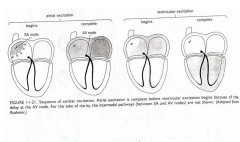
Two nodes, the sinoatrial node (SA node) and atriventricular node (AV node) SA is at the top of the atrium and AV is at the base of the right atrium. Emerging from the AV node is a thick bundle of fibers that split into R and L branches as they head for the bottom of each ventricle. These fibers are called Purkinje fibers which are specialized with big, fat gap junctions. |
|
|
Electronic Signalling |
The SA node starts cycle by sending a wave of excitement to the AV node. The action potential arises in the AV node and spreads down the Purkinje fibers and excite the rest of the mucle. The delay in a heart beat is produced by the conduction pathway. |
|
|
Heart Beating |

Controlled by the nervous system that can change the heart rate. Also controlled by a blood hormone called adrenaline. The parasympathetic nervous system releases acetylcholine to slow the heart beat down. |
|
|
Blood Pressure |
The blood pressure is determined by cardiac output. Capillaries are surrounded smooth muscle that contracts or relaxes under the control of the autonomic nervous system. |
|
|
Control System |
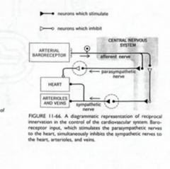
Set of detectors, a control system, and effectors. The detectors are called baroreceptors (pressure receptors) They are found right outside the aorta and two in the carotid arteries supplying the L and R side of the brain. The pressure changes during every heartbeat and receptors signal this by changing their rate of firing action potentials. (If pressure is low theres less action potentials fired, if pressure is higher then more action potentials are released.) Info is sent to the central nervous system where if pressure is high, parasympathetic nerves fire faster (which releases acetylcholine, making them fire faster. and slowing heart right.) |
|
|
High Pressure |
When pressure is high parasympathetic nerves fire faster, and the nerves release acetychololine. This slows the heartbeat, and dilates the blood vessels, which in turn reduces the blood pressure. |
|
|
Low Pressure |
The parasympethic system is not turned on and sympathetic system is not turned off. So the heart rate goes up and the blood vessels constrict. |
|
|
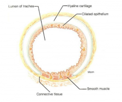
Capillary walls |
Simple squamous epithelium with a basal lamina. (Specialized to let different blood components pass.) Some protein leaks, and this fluid leakage due to the pressure inside is collected via lymphatic system and returned to the blood near the heart. |