![]()
![]()
![]()
Use LEFT and RIGHT arrow keys to navigate between flashcards;
Use UP and DOWN arrow keys to flip the card;
H to show hint;
A reads text to speech;
82 Cards in this Set
- Front
- Back
|
Central Nervous System |
comprised of the brain and spinal cord |
|
|
Peripheral Nervous System |
any part of the nervous system outside the central nervous system (includes nerves) |
|
|
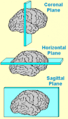
Transverse Plane |
frontal cross section coronal section |
|
|
Horizontal Plane |

|
|
|
Sagittal Plane |
|
|
|
Dorsal |
toward the top of the head |
|
|
Ventral |
surface facing the ground |
|
|
Lateral |
away from the middle |
|
|
Medial |
toward the middle |
|
|
Anterior |
rostral-- the front end, toward the head |
|
|
Posterior |
caudal- toward the tail end |
|
|
Neuroaxis |
imaginary line that runs along the length of the CNS |
|
|
The Meninges (3) |
tough connective tissues surrounding the brain Consists of: Dura matter Arachnoid membrane Pia Matter |
|
|
Dura Matter |
outer layer, thick, tough and unstretchable |
|
|
Arachnoid Membrane |
middle, soft spongy, web-like |
|
|
Pia Matter |
layer closest to the brain, contains blood vessels |
|
|
Subarachnoid space |
filled with cerebral-spinal fluid similar to salt-water blood plasma, lymph |
|
|
Blood-Brain Barrier |
semipermeable barrier between the blood and brain produced by the cells in walls of brain's capillaries capillaries in all of the body, except the brain, contain gaps that permit free passage of substances |
|
|
Area Postremia |
region of the medulla where blood-brain barrier is weak poisons can be sensed here and initiate vomiting |
|
|
Ventricular System |
interconnected hollow spaces filled with CSF |
|
|
Lateral Ventricles |
1st and 2nd ventricles, largest of the four |
|
|
Third Ventricle |
located between the two thalamic nuclei |
|
|
Cerebral Aquedcut |
connects the third and fourth ventricle |
|
|
Fourth Ventricle |
sits between the pons and cerebellum (base of the brain) |
|
|
Cerebral-Spinal Fluid |
produced continuously by the choroid plexus, protrudes into ventricles Half life of three hours Leaves 4th ventricle and flows in subarachnoid space |
|
|
Forebrain |
Ventricle: 1 and 2 (lateral), third Subdivision: Telencephalon and Diencephalon Principal Structures: Telencephalon: Cerebral cortex, Limbic System, Basal ganglia Diencephalon: thalamus and hypothalamus |
|
|
Midbrain |
Ventricle: cerebral aqueduct Subdivision: Mesencephalon Principal Structures: Tectum and Tegmentum |
|
|
Hindbrain |
Ventricle: fourth Subdivision: Metencephalon and Myelencephalon Principal Structures: cerebellum, pons, medulla |
|
|
Thalamus |
sensory info goes to thalamus first and is then relayed to the appropriate primary sensory cortex area in the cerebral cortex |
|
|
Cerebral Cortex Development |
inside-out, last cells must go through the previously developed layer of cells. Glial cells help guide migration of newly formed cells |
|
|
Cerebral Cortex |
Part of the telecaphalon subdivision surrounds hemisphere like tree-bark sulci and fissures |
|
|
Sulci |
small grooves in the cerebral cortex |
|
|
Fissures |
large grooves (convolutions) in the cerebral cortex |
|
|
Gyri |
ridges between the sulci and fissures |
|
|
Grey Matter |
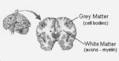
outermost portion. High concentration of cell bodies |
|
|
White Matter |

beneath grey matter: consists of high concentration of myelenated axons |
|
|
Longitudinal Fissure |
separates the two hemispheres |
|
|
Lateral Fissure |
separates the frontal from the temporal brain Insular (gustatory) info |
|
|
Central Sulcus |
separates the anterior (rostral) from the posterior (caudal) hemispheres |
|
|
Primary Motor Cortex |
Located in the frontal lobe. Info sent down spinal cords, to the muscles |
|
|
Somatosensory Cortex |
Parietal lobe receive touch info. Different regions receive different info from specific areas (somatosensory homunculus) |
|
|
Primary Visual Cortex |
Occipital Lobe |
|
|
Primary Auditory Cortex |
temporal lobe |
|
|
Insular Cortex |
lateral fissure, gustatory information |
|
|
Sensory Association Cortex |
located adjacent to the relevant primary sensory cortex. Receives info from only one primary cortex |
|
|
Limbic System |
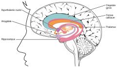
Limbic Cortex: located at medial edges of hemispheres Part of the telencephalon subdivision Consists of amygdala, hippocampus, cingulate gyrus Emotion formation, information processing, learning |
|
|
Frontal Lobe |
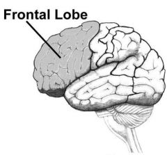
primary motor cortex |
|
|
Parietal Lobe |
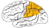
somatosensory cortex |
|
|
Temporal Lobe |
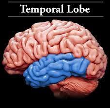
auditory info amygdala |
|
|
Occipital Lobe |
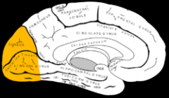
visual info |
|
|
Neocortex |
most recently evolved potion of the cortex: primary sensory areas, association areas, primary motor cortex |
|
|
Amygdala |
emotional processing ventral to the hippocampus temporal lobe |
|
|
Hippocampus |
memory temporal lobe |
|
|
Cingulate Gyrus |

encircles the corpus callosum |
|
|
Septum |

passes info from the hippocamus to the amygdala |
|
|
Fornix |
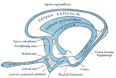
passes info between the septum and hippocampus |
|
|
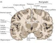
Basal Ganglia |
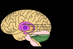
Telencephalon subdivision lays beneath anterior portion of lateral ventricles control of voluntary movements, procedural learning, emotion caudate nucleus |
|
|
Caudate Nucleus |
portion of the Basal Ganglia: striatum-- associated with Parkinson's Disease |
|
|
Putamen |
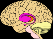
Portion of the basal ganglia: striatum |
|
|
Telencephalon Subdivision |
Cerebral Cortex Basal Ganglia Limbic System |
|
|
Diencephalon |
3rd ventricle Hypothalamus Thalamus |
|
|
Hypothalamus |
closely related to the endocrine system/gland- stimulate pituitary gland to release hormones two-lobed, several nuclei organize behaviours related to survival-- reproductive and physiological behaviour |
|
|
Hormones |
non-synaptic long distance chemical communication. released by endocrine gland and has effects in target organs |
|
|
Thalamus |
two-lobed and divided into several nuclei sensory relay widespread cortical projections |
|
|
Midbrain |
Cerebral Aqueduct Mesencephalon Tectum & Tegmentum |
|
|
Tectum |
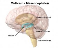
"roof" two bumps on dorsal surface of brain stem superior and inferior colliculi |
|
|
Superior and Inferior Colliculi |
part of the tectum, dorsal to the brain stem superior = visual info inferior= auditory info |
|
|
Tegmentum |
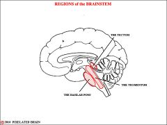
"covering" involved in sleep, arousal, attention, movement, reflexes |
|
|
Hindbrain |
fourth ventricle Metencephalon & Myelencephalon cerebellum, pons, medulla |
|
|
Cerebellum |
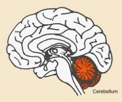
coordinate and regulate muscular activity integration of sensory perception modifies coordinating and smoothing effects on movements Damage: impairs standing, walking, coordinated movements |
|
|
Pons |
large bulge in brain stem relay messages between cerebral cortex and the cerebellum sleep, arousal |
|
|
Medulla (oblongata) |
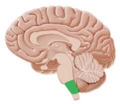
most caudal part of the brain stem vital to respiration and cardiovascular system |
|
|
Afferent |
towards the CNS |
|
|
Efferent |
away from the CNS motor signals from the CNS to the muscles |
|
|
Somatic System |
part of peripheral nervous system interacts with external environment-- afferent-- carries signals from eyes, ears etc. TO the CNS efferent carries motor signals from CNS to skeletal muscles |
|
|
Autonomic system |
- regulates the body's internal environment - afferent carries info from the organs to the CNS - sympathetic and parasympathetic |
|
|
Sympathetic |
system supports activities associated with increased energy expenditure (ex. heart rate) |
|
|
Parasympathetic |
system supports body in relaxed state, involved in increasing body's energy stores ex. digestion, increased blood flow to gastrointenstinal system |
|
|
Spinal Cord |
distributes motor fibres to the effector organs of the body (those that interact with the environment) collects somatosensory info to be passed onto the brain (CNS) |
|
|
Sensory Neuron |
detects changes in the external or internal environment and sends info to the CNS |
|
|
Motor Neuron |
located within the CNS, controls contraction of a muscle or secretion of a glad |
|
|
Interneuron |
located entirely within the CNS axons sent very locally within a brain area |