Reading...
![]()
Play button
![]()
Play button
![]()
Use LEFT and RIGHT arrow keys to navigate between flashcards;
Use UP and DOWN arrow keys to flip the card;
H to show hint;
A reads text to speech;
51 Cards in this Set
- Front
- Back
|
neural circuits
|
- organization of neurons
- receive sensory information - integrate information - control effector cells |
|
|
simple sensory motor circuit
|
- sensory neuron synapsing directly on motor cell
- interneuron added = sensory interneuron motor - can by polyneuronal |
|
|
nervous system of animals
|
- networks of sensory, inerneuron, and motor neurons
|
|
|
CNS
|
- brain controls motor behavior
- the cerebellum and basal nuclei are the ultimate planners and coordinators of complex motor activities sending input to the primary motor cortex - cerebral cortex is the highest level of conscious motor pathways - thalamus is sensory relay center and is important in motor control - motor control exerted by lover levels in involuntary and is mediated by reflex arcs and fixed action patterns or stereotypical, sequential motor actions triggered by an appropriate stimulus |
|
|
3 levels of cerebellar and basal nuclei motor control
|
- segmental level
- projection level - pre-command level - develop a motor program for the specific voluntary task - pull the appropriate pattern in the primary motor cortex to bring about the sequence of necessary contractions |
|
|
segmental level
|
- located in cerebellum and basal nuclei
- spinal cord circuits that activate a network of anterior/ventral horn neurons to a single cord segments causing them to stimulate a specific group of muscle fibers |
|
|
organization of vertebrate CNS
|
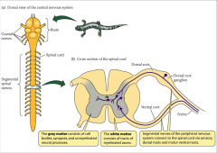
- gray matter = consists of cell bodies, synapses, and unmyelinated neural processes
- white matter = consists of tracts of myelinated axons - segmental nerves of PNS connect to the spinal cord via sensory dorsal roots and motor ventral roots |
|
|
spinal cord tracts
|
- white column = bundles of myelinated axons that carry signals up and down to and from brainstem
- 3 pairs of columns: dorsal, lateral, and anterior columns - each column is filled with named tracts or fasciculi |
|
|
fasciculi
|
- fibers with a similar origin, destination, and function
|
|
|
ascending and descending tracts
|
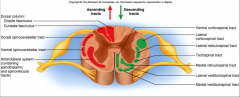
- ascending and descending tract head up or down while decussation means that the fibers cross sides
- contralateral means origin and destination are on opposite sides while ipsilateral means on same side |
|
|
central pattern generators (CPG)
|
- those circuits that control locomotion and other specific, often repeated motor activities
- neural circuit in which CNS can endogenously generate a sequential patterned activation of motor neurons to antagonistic muscles that underlies a behavior pattern - a group of neurons organized in CNS that produces a complex series of neural activity patterns responsible for motor actions - sensory timing information can reset the CPG |
|
|
control of flight in locust
|
- depressor and levator muscles show alternating bursts of muscle potentials, resulting in rhythmic wing movements
- wing-hinge stretch receptor is activated when the wing has been elevated by contraction of the levator muscle - when the wing has be lowered by contraction of the depressor muscle, depression proprioceotors are activated |
|
|
two hypotheses for motor pattern of wing muscles
|
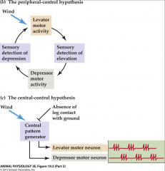
- sensory feedback resulting from a movement triggers the next movement
- CPG produces the motor pattern without requiring moment-to-moment sensory input |
|
|
projection level
|
- located in cerebellum and basal nuclei
- spinal cord is under the direct control - consists of upper motor neurons of the motor cortex - synapse on anterior/ventral motor horn cells that regulate fast and fine motor movements - or brain stem nuclei that then send synapse with neurons in anterior horn = regulate muslce tone, mediate head movements used in vision, and control CPG during locomotion - convey information to lower motor neurons and send a copy to higher command levels |
|
|
pre-command level
|
- precisely start or stop movements
- control the outputs of the cortex and brain stem - cerebellum is ultimate target of ascending motor tracts but doesn't act directly on spinal cord - acts on brain stem and motor cortex through hypothalamus |
|
|
basal nuclei
|
- receive inputs from all coritcal areas and send output to premotor and prefrontal motor cortical areas
- appear to be involved in most complex aspects of motor control |
|
|
basal ganglia/nuclei
|
- inhibition of muscle tone
- coordination of slow sustained movements - suppression of useless patterns of movement - parkinson's disease affects the basal ganglia associated with a dopamine deficiency |
|
|
basal ganglion
|
- receive output from a number of cortical areas
- these structures provide positive feedback to SMA and PMA - may help in initiation of a behavior |
|
|
cerebellum
|
- receives cortical input and provides primary motor cortex with information on the status of a behavior as well as information on its feasibility
|
|
|
preparing for movement
|
- conscious decision to move info is relayed from frontal lobes to supplemental motor association areas
- supplemental motor association areas then relay info to the cerebellum and basal nuclei - at same time, cerebellum receives info from proprioceptors and visual and equilibrium pathways - cerebellar cortex calculates best way to coordinate force, direction, extent of muscle contraction - cerebellum send to the cerebral motor cortex its blueprint of coordination - cerebellum send to the brain stem information that influence motor neurons of the spinal cord |
|
|
interaction of brain areas in the planning, execution, and control of voluntary movement
|
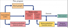
- two loops from the association cortex are involved in pre-programming a movement
- basal ganglia involved in selection and initiation - cerebrocerebellum involved in initial programming - information passes to the primary motor cortex for executing the movement - sensory information from the spinal cord and the cerebellum gives integrated feedback information to the primary motor cortex |
|
|
performing a movement
|
- as the movement begins the supplemental motor association areas send instructions to the primary motor cortex
- feedback from the cerebellum and basal nuclei modify those commands - outputs directs involuntary adjustments in position and muscle tone |
|
|
spinal cord
|
- relays information to and from the brain
- central pattern generators - ascending and descending paths in the spinal cord - reflexes - not merely a pathway for sensory and motor connections between the brain and peripheral nervous system - responsible for sensory-motor coordination particularly reflex arcs |
|
|
spinal reflex/reflex arc
|
- simple automatic response to a sensory stimulus
|
|
|
spinal reflexes
|
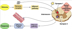
- receptors in skin, muscles, tendons, and joint synapse with dorsal root ganglia fibers/afferent fibers through the dorsal root ganglia
- action potential travels afferent fiber to the dorsal root of the spinal cord - afferent fiber synapses with interneurons - interneurons synapse with motor neurons in the ventral horn and neurons in higher centers of the CNS - motor fibers/efferent fibers leave the CNS through the ventral horn - motor fibers synapse with effector |
|
|
stretch reflex, myotactic reflex
|
- essential for posture and coordination of movements
- involves muscle spindles - absent in people with chronic diabetes, neurosyphilis, and injury to the 2,3,4 lumbar segments |
|
|
characteristics of knee jerk reflex
|
- ipsilateral = response on the same side of the body
- intrasegmental = only one segment of the spinal cord is involved - monosynaptic = one synapse within the spinal cord |
|
|
muscle spindle
|
- an internal mechanoreceptor associated with the musculoskeletal system
- monitors length of skeletal muscles - in parallel to muscle fibers - embedded along with the muscle fibers - associated with the 1 afferent sensory fiber-1a which is largest and fastest conducting sensory fiber in the body - 1a axon enters spinal cord and makes direct excitatory synaptic contact with motor neurons to the same muscle |
|
|
vertebrate muscle spindle stretch receptors
|
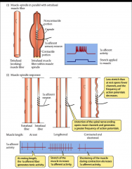
- at resting length, 1a afferent fiber generates tonic activity
- stretch of muscle increases 1a afferent activity - shortening of muscle during contraction decreases 1a afferent activity |
|
|
stretch reflex mediates load compensation in voluntary movement
|
- simplest manifestation of stretch reflex involves only a 1a fiber and a motor neuron
- stretch muscle spindle - action potential along 1a fiber - synapse with alpha motor neuron in the ventral horn that controls same muscle fiber as muscle spindle - motor neuron output EPSPs to extrafusal muscle fiber - contraction |
|
|
reciprocity
|
- neural circuit of stretch reflex
- muscles tend to be arranged in antagonist pairs that oppose each other - extensor = muscle that increases the angle between two articulating bones - flexor = muscle that decreases the angle between two articulating bones |
|
|
principle of reciprocity
|
- any signal that activates movement is sent to a pair of muscle-agonist and the antagonist
- causing the agonist to contract and the antagonist to relax - reciprocal inhibition prevents muscles from working against each other |
|
|
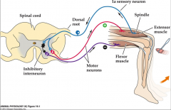
stretch reflex
|
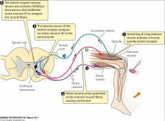
- stretching of a leg extensor muscle activates a muscle spindle stretch receptor
- sensory neuron of the stretch receptor synapses on motor neurons (E) to the same muscle - stretch receptor sensory neuron also activates inhibitory interneurons that inhibit the motor neurons (F) to antagonistic muscle fibers |
|
|
reflex pathway
|
|
|
|
renshaw cell
|
- interneuron that inhibits or prevents prolonged motor neuron activity in an inhibitory feedback loop
|
|
|
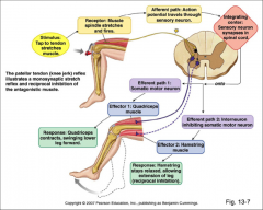
patellar tendon reflex arc
|
- extensor muscle stretched
- muscle spindle stimulated - primary afferent neuron excited - primary afferent neuron stimulates alpha motor neuron to extensor muscle - alpha motor neuron stimulates extensor muscle to contract - primary afferent neuron stimulates inhibitory interneuron - interneuron inhibits alpha motor neuron to flexor muscle - flexor muscle (antagonist) reflexes |
|
|
gamma motor neurons
|
- small motor neurons that innervate intrafusal muscle fibers
- activation of the gamma motor neurons excites 1a sensory neuron by contracting the contractile end of the intrafusal fiber - 2 ways to increase muscle spindle receptor activity: passive stretch of the muscle and gamma motor neuron activity |
|
|
stretch reflex mediates load compensation in a voluntary movement
|
- CNS estimates the force necessary to pick up a load
- descending command co-activates the alpha and gamma motor neurons |
|
|
no load present
|
- extrafusal fibers shorten to flex the arm
- intrafusal shorten due to gamma signal - intrafusal fiber shortening decreases its tension and lessens the activation of the stretch receptor |
|
|
load present
|
- prevents muscle from shortening
- coactivation of alpha and gamma fiber - extrafusal fibers don't shorten because the load is to great - intrafusal fiber shortens due to gamma activation - creating an error signal that activates the alpha motor neuron = train of action potentials - adds to proportional excitation and tension in the muscle to help overcome the load - creates a load-compensating servo loop (failure to shorten-more force) |
|
|
circuit diagram of ventral horn of mammalian spinal cord
|
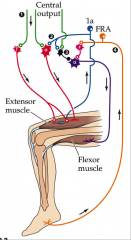
- motor neurons are activated primarily by CNS input rather that spinal reflexes
- motor output neurons of spinal cord are alpha motor neurons to flexor and extensor muscles and gamma motor neurons to the intrafusal fibers of muscle spindle - input pathways to motor neurons and interneurons from the ventral horn include - descending pathways from the brain = green -1a afferent excitatory neuron = blue - 1a inhibitory inter neuron/inhibitory pathways from the muscle spindle = black |
|
|
additional proprioceptors of vertebrates
|
- golgi tendon organs
- joint receptors |
|
|
golgi tendon organ
|
- collects information about differences in tension among tendons
- relay data to the CNS where they are processed - help to coordinate fine muscular contractions - in series with working muscle fibers |
|
|
golgi tendon reflex
|
- 1mm long
- encapsulated nerve bundle - excessive tension on tendon inhibits motor neuron = muscle contraction decreased - functions when muscle contracts unevenly |
|
|
joint receptors
|
- embedded in connective tissue at the joint
- respond over certain range of joint positions and movments |
|
|
flexor reflex
|
- reflex afferents have endings in skin muscles and joints
- make excitatory synapses with interneurons in the CNS that excite motor neurons in flexor muscles = not direct connections as seen in stretch reflex - occur along with cross extensor reflex - polysynaptic reflex = 3 or more neurons are involved - ipsilateral = response on same side of body - intersegmental = involves more than one segment |
|
|
flexor withdrawal reflexes
|
- occurs during withdrawal of foot from pain
- polysnaptic reflex arc - neural circuitry in spinal cord controls sequence and duration of muscle contractons |
|
|
cross extension reflex
|
- contralateral reflex
- uses commissional, interneurons in spinal cord to relay information to opposite side - painful stimulus to left foot can elicit withdrawal of the left and extension of the right to support body |
|
|
neural circuit of flexion reflex and crossed extension reflex
|
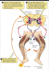
- noxious stimulation of skin excites flexion reflex afferent neurons
- afferent neurons excite interneurons to synaptically excite flexor motor neurons and inhibit extensor motor neurons on the stimulated side - flexion-reflex afferents also synapse onto interneurons that cross the midline of the spinal cord and indirectly stimulate extension of the opposite leg |
|
|
crossed extensor reflexes
|
- maintains balance by extending other leg
-intersegmental reflex extends up and down spinal cord - contralateral reflex arcs explained by pain at one foot causes muscle contraction in other leg |
|
|
flexor reflex and crossed extension reflex
|
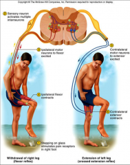
- stepping on glass stimulates pain receptors in foot
- sensory neuron activates multiple interneurons - ipsilateral motor neurons to flexor excited - ipsilateral flexor contracts - contralateral motor neurons to extensor excited - contralateral extensor contracts |