![]()
![]()
![]()
Use LEFT and RIGHT arrow keys to navigate between flashcards;
Use UP and DOWN arrow keys to flip the card;
H to show hint;
A reads text to speech;
272 Cards in this Set
- Front
- Back

Front (Term) |
Know |
|
|
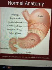
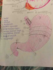
Normal anatomy of esophagus |
Expands through thoracic cavity and diaphragm to the stomach |
|
|
Stomach is a |
Large smooth muscular organ |
|
|
Three parts of stomach |
Fundus body pilorus |
|
|
The small intestines is _____ _____ |
Long coiled |
|
|
Three parts of small intestines |
Duodenum jejunum ileum |
|
|
What is the measurement of the small intestine and large intestine |
5 m long 4 cm in diameter |
|
|
The head of the pancreas is in the |
Second portion of the duodenum |
|
|
Inferior thyroid branch of subclavian supplies the |
Upper esophagus |
|
|
Descending thoracic aorta supples the |
Midesophagus |
|
|
Harris branch of celiac axis and the inferior phrenic artery of the abdominal aorta supply the |
Lower end of the esophagus |
|
|
Varices may be seen to rise from the |
Gastroesophageal arteries |
|
|
Stomach: vascular supply if provided by |
Right gastric arterial branch Pyloric and right gastroepiploic branches of hepatic artery Left gastroepiploic __________ |
|
|
The small intestines: What lines the small intestine and contains the SMV nerves lymphatic and fat |
Mesenteric |
|
|
Supplies the duodenum through the right gastric Gastroduodenak abd superior pancraticoduodenal branches |
Celiac axis |
|
|
The large intestine: What supply both the small and large intestines |
Celiac SMA IMA |
|
|
The _____ supplies the intestine from the left border of the trv colon |
SMA |
|
|
The ______ tract is the largest endocrine ..... |
Gastrointestinal tract |
|
|
Food is swallowed and goes to |
Mouth, chewed, swallowed Nutrients pass through wall of intestine into blood or lymph |
|
|
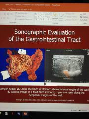
What is rugae |
Grooves that stretch out |
|
|
Contractions of stomach help |
Mix food up |
|
|
Three layers of the smooth muscle in the stomach wall enable |
To mash and churn food and move it along the peristalsis |
|
|
Food is converted into |
Chyme |
|
|
What is chyme |
What is broken down. Liquid and food (soupy mixture) |
|
|
The villi within the small intestine |
Increase its surface area for digestion and absorption of nutrients |
|
|
The _________ are found btw the villi and secret large amount of fluid that serve as a medium for digestion and absorption of nutrients |
Intestinal glands |
|
|
The hormone, ______, which if released by the stomach mucosa stimulates the gastric glands to secrete |
Gastrin |
|
|
Most of the digestion occurs with the |
Duodenum |
|
|
____ and _____ from the liver and pancreas are secreted into the duodenum to act on chyme and break own food particles |
Bile and enzymes |
|
|
______ is released by presences of fat in the intestine and regulates gallbladdde contraction and gastric emptying |
Cholecystokinin |
|
|
The most common laboratory data is |
Blood in stool |
|
|
_____ may present as a result of chronic loss |
Anemia |
|
|
Infection would show an elevation of the |
White blood count |
|
|
Increase in the _________ is found in patients with inflammatory bowel disease |
Carcinoembryonic antigen |
|
|
Clinical signs and symptoms of gastrointestinal problems |
Nausea Vomiting Diarrhea |
|
|
Colitis bowel abscess acute diverticulitis and appendicitis may be associated with |
Gastrointestinal problems |
|
|
Bowel layers : Mucosa |
Directly contacts intraluminal contents |
|
|
Bowel layers Submucosa |
Contains blood vessels and lymph channels |
|
|
Rim of lucency is |
Represents the wall and its periserosal fat produces the outer echogenic order of the tract wall |
|
|
The _______ represents the wall and its periserosal fat produces the outer echogenic border of the tract wall |
Rim of lucency |
|
|
The bowel wall have how many layers |
Five |
|
|
The odd number of layers of bowel wall are |
Echogenic |
|
|
The even numbered layers of bowel layers are |
Hypoechoic and measure 3-5 mm |
|
|
The ____ is seen on sag scan to left of midline as a bulls eye or target shaped structure anterior to aorta, posterior to the left lobe of liver and inferior to the hemidiaphragm |
Gastroesophagal junction |
|
|
The ____ is seen on sag scan to left of midline as a bulls eye or target shaped structure anterior to aorta, posterior to the left lobe of liver and inferior to the hemidiaphragm |
Gastroesophagal junction |
|
|
The gastro Antrum |
..... |
|
|
The ____ is seen on sag scan to left of midline as a bulls eye or target shaped structure anterior to aorta, posterior to the left lobe of liver and inferior to the hemidiaphragm |
Gastroesophagal junction |
|
|
The gastro Antrum |
..... |
|
|
If the patient has a cystic mass in the LUQ you should |
Determine whether the mass is the fluid filled stomach |
|
|
To determine if a cystic mass in LUQ is the stomach you can |
Give them a soft drink (bubbles) Watch for change in shape or size Alter patients positions by scanning in an upright or left or right lateral decubitus |
|
|
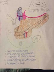
The duodenum four parts |
Superior portion Descending portion Transverse Ascending |
|
|
The ____ is seen on sag scan to left of midline as a bulls eye or target shaped structure anterior to aorta, posterior to the left lobe of liver and inferior to the hemidiaphragm |
Gastroesophagal junction |
|
|
The gastro Antrum |
..... |
|
|
If the patient has a cystic mass in the LUQ you should |
Determine whether the mass is the fluid filled stomach |
|
|
To determine if a cystic mass in LUQ is the stomach you can |
Give them a soft drink (bubbles) Watch for change in shape or size Alter patients positions by scanning in an upright or left or right lateral decubitus |
|
|
The duodenum four parts |
Superior portion Descending portion Transverse Ascending |
|
|
The duodenum serves as an excellent land mark for |
Head of pancreas |
|
|
Small bowel cannot usually be seen with |
Us but can be seen with Ct |
|
|
The _____ may be seen as a linear echo densities spaces 3-5 mm apart |
Valvulae conniventes |
|
|
The vulvulae is called the |
Keyboard sign |
|
|
You will see loops of bowel in |
Pelvic area |
|
|
The ____ is seen on sag scan to left of midline as a bulls eye or target shaped structure anterior to aorta, posterior to the left lobe of liver and inferior to the hemidiaphragm |
Gastroesophagal junction |
|
|
Vermiform appendix is |
Long tubular structure that extends from the cecum |
|
|
Located in the abdominal wall under McBurney's point |
Vermiform appendix |
|
|
Scan around umbilicus to scan the |
Appendix |
|
|
If the appendix is ruptured you will see |
Fluid around it |
|
|
The appendix measures |
1-9 inches in length Average 3 inches |
|
|
The cellular layers of the appendix |
Serosa Adventitia ..... |
|
|
The colon is a prominent |
Fluid filled |
|
|
______ should be used to help determine whether the mass is within the colon seperate form the colon of colon itself |
Water enema technique |
|
|
You can see a ________ or ______ sign also viewing the stomach |
Colon |
|
|
The gastro Antrum |
..... |
|
|
If the patient has a cystic mass in the LUQ you should |
Determine whether the mass is the fluid filled stomach |
|
|
To determine if a cystic mass in LUQ is the stomach you can |
Give them a soft drink (bubbles) Watch for change in shape or size Alter patients positions by scanning in an upright or left or right lateral decubitus |
|
|
The duodenum four parts |
Superior portion Descending portion Transverse Ascending |
|
|
The duodenum serves as an excellent land mark for |
Head of pancreas |
|
|
Small bowel cannot usually be seen with |
Us but can be seen with Ct |
|
|
The _____ may be seen as a linear echo densities spaces 3-5 mm apart |
Valvulae conniventes |
|
|
The vulvulae is called the |
Keyboard sign |
|
|
You will see loops of bowel in |
Pelvic area |
|
|
The ____ is seen on sag scan to left of midline as a bulls eye or target shaped structure anterior to aorta, posterior to the left lobe of liver and inferior to the hemidiaphragm |
Gastroesophagal junction |
|
|
Vermiform appendix is |
Long tubular structure that extends from the cecum |
|
|
Located in the abdominal wall under McBurney's point |
Vermiform appendix |
|
|
Scan around umbilicus to scan the |
Appendix |
|
|
If the appendix is ruptured you will see |
Fluid around it |
|
|
The appendix measures |
1-9 inches in length Average 3 inches |
|
|
The cellular layers of the appendix |
Serosa Adventitia ..... |
|
|
The colon is a prominent |
Fluid filled |
|
|
______ should be used to help determine whether the mass is within the colon seperate form the colon of colon itself |
Water enema technique |
|
|
You can see a ________ or ______ sign also viewing the colon |
Whirlpool or bulls eye |
|
|
The gastro Antrum |
..... |
|
|
If the patient has a cystic mass in the LUQ you should |
Determine whether the mass is the fluid filled stomach |
|
|
To determine if a cystic mass in LUQ is the stomach you can |
Give them a soft drink (bubbles) Watch for change in shape or size Alter patients positions by scanning in an upright or left or right lateral decubitus |
|
|
The duodenum four parts |
Superior portion Descending portion Transverse Ascending |
|
|
The duodenum serves as an excellent land mark for |
Head of pancreas |
|
|
Small bowel cannot usually be seen with |
Us but can be seen with Ct |
|
|
The _____ may be seen as a linear echo densities spaces 3-5 mm apart |
Valvulae conniventes |
|
|
The vulvulae is called the |
Keyboard sign |
|
|
You will see loops of bowel in |
Pelvic area |
|
|
A tube, 8 m long, extending from the mouth to the anus |
Digestive (alimentary tract) |
|
|
The part of the digestive system that is below the diaphragm |
Gastrointestinal |
|
|
Accessory of digestive glands |
Salvitary glands Liver Pancreas |
|
|
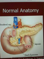
Label |
1) superior duodenum 2) descending duodenum 3) transverse duodenum 4) ascending duodenum |
|
Label |
1) superior duodenum 2) descending duodenum 3) transverse duodenum 4) ascending duodenum |
|
|
Front (Term) |
Know |
|
|
Front (Term) |
Know |
|
Front (Term) |
Know |
|
|
The SMA supplies the gut from halfway down the second part of the duodenum to the distal third of the |
Transverse colon |
|
|
The branches of the IMA |
Left colic artery Sigmoid artery Superior rectalbartery |
|
|
_____ is released form the small bowel to stimulate the secretion of bicarbonate to decrease the axis content of the intestine |
Secretin |
|
|
Is a thin loose layer of connective tissue |
Serosa |
|
Front (Term) |
Know |
|
Front (Term) |
Know |
|
|
Know |
|
Front (Term) |
Know |
|
|
Alimentary tract is a table that is ___ long traveling from the mouth to ____ |
8 m long Mouth to anus |
|
|
Alimentary tract is a table that is ___ long traveling from the mouth to ____ |
8 m long Mouth to anus |
|
|
Is part of the digestive system that is below the diaphragm |
Gastrointestinal tract |
|
|
What are the three types of accessory digestive glands |
Salivary Liver Pancreas |