![]()
![]()
![]()
Use LEFT and RIGHT arrow keys to navigate between flashcards;
Use UP and DOWN arrow keys to flip the card;
H to show hint;
A reads text to speech;
13 Cards in this Set
- Front
- Back
|
Identify the parts of a microscope and use it properly. List the parts and function |
Eyepiece: The lens the viewer looks through to see the specimen. The eyepiece usually contains a 10X or 15X power lens. Diopter Adjustment: Useful as a means to change focus on one eyepiece so as to correct for any difference in vision between your two eyes. Body tube (Head): The body tube connects the eyepiece to the objective lenses. Arm: The arm connects the body tube to the base of the microscope. Coarse adjustment: Brings the specimen into general focus. Fine adjustment: Fine tunes the focus and increases the detail of the specimen. Nosepiece: A rotating turret that houses the objective lenses. The viewer spins the nosepiece to select different objective lenses. Objective lenses: One of the most important parts of a compound microscope, as they are the lenses closest to the specimen. slide: The specimen is the object being examined. Most specimens are mounted on slides, flat rectangles of thin glass. Stage: The flat platform where the slide is placed. Stage clips: Metal clips that hold the slide in place. Stage height adjustment (Stage Control): These knobs move the stage left and right or up and down. Aperture: The hole in the middle of the stage that allows light from the illuminator to reach the specimen. On/off switch: This switch on the base of the microscope turns the illuminator off and on. Illumination: The light source for a microscope. Older microscopes used mirrors to reflect light from an external source up through the bottom of the stage; however, most microscopes now use a low-voltage bulb. Iris diaphragm: Adjusts the amount of light that reaches the specimen. Condenser: Gathers and focuses light from the illuminator onto the specimen being viewed. Base: The base supports the microscope and it’s where illuminator is located.
|
|
|
Draw detailed images of objects that you'd see under the microscope and draw to scale. |
|
|
|
Describe how to measure objects under the microscope in mm and convert to um. Also describe how to calculate total magnification |
to measure the length of an object note the number of ocular divisions spanned by the object. Then multiply by the conversion factor for the magnification used. The conversion factor is different at each magnification. |
|
|
List the 3 parts of the cell theory |
1. cell is the structural unit of life. |
|
|
Describe the roles that van Leewenhoek, Hooke, Schleiden, Schwann, and Virchow had with regards to the microscope and the cell |
Leeuwenhoek improved magnification of microscopes by polishing lenses
Hooke discovered the cellular composition of cork and introduced the word cell to science
Schleiden discovered the plants were made up of cells
Schwann discovered that animals were made up of cells
Virchowstated that all living things come from other living things Add a new card (or press TAB) |
|
|
Distinguish between the 2 types of electron microscope- SEM & TEM. What are the advantages and disadvantages |
SEM = three-dimensional images
TEM = two-dimensional
|
|
|
Distinguish between prokaryotes and eukaryotes. |
Eukaryotic cells contain membrane-bound organelles, such as the nucleus, while prokaryotic cells do not. |
|
|
Describe the structure and function of the cell parts |
Chromatin composed of continuous DNA molecules wrapped around clusters of proteins called histones.
cell memebrane membrane mainly composed of protein and lipid molecules. function: maintains integrity of the cell. controls passage of material into and out of the cells.
ribosomes particles composed of protein and RNA molecules. functions: synthesizes proteins.
endoplasmic reticulum complex of connected, membrane-bound sacs, canals, and vesicles. function: transports material in the cell, provides attachment for ribosomes, and synthesizes lipids.
vesicles membranous sacs. function: contain substances that recently entered the cell. store and transport newly synthesized molecules.
golgi apparatus group of flattened, membranous sacs. function: release energy from food molecules, and concert the energy into usable form.
mitochondria membranous sacs with inner partitions. function: release energy from food molecules and convert the energy into usable form.
lysosomes membranous sacs. function: contains enzymes capable of digesting worn cellular parts of substances that enter cells.
nucleolus dense, nonmebranous body composed of protein and RNA molecules. (function): site of ribosome formation.
|
|
|
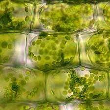
Compare and contrast plant and animal cells. Lost several differences |
animal cells do not have a cell wall or chloroplasts but plant cells do. Animal cells are round and irregular in shape while plant cells have fixed, rectangular shapes. |
|
|
Explain the structures found in a prokaryotic cell |
generally have a single chromosome: a piece of circular, double-stranded DNA located in an area of the cell called the nucleoid. |
|
|
Water accounts for 70% of the weight cell |
None |
|
|
Identify the components to the phosphilipid bilayer. Draw a cell membrane and label its parts |
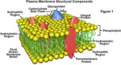
Phospholipids and proteins make up the composition of the bag. plasma membrane is phospholipids. Proteins surround the holes and help by moving molecules in and out. The organization is called the fluid mosaic model. |
|
|
Explain cholesterols role in the plasma membrane |
Cholesterol is required to build and maintain cell membranes; it regulates membrane permeability and fluidity over a wide range of temperatures. |