![]()
![]()
![]()
Use LEFT and RIGHT arrow keys to navigate between flashcards;
Use UP and DOWN arrow keys to flip the card;
H to show hint;
A reads text to speech;
58 Cards in this Set
- Front
- Back
- 3rd side (hint)
|
Enlargements of spinal cord are caused by |
Amount of gray matter in segment |
|
|
|
Cervical enlargement |
Nerves of shoulders and upper limbs |
|
|
|
Lumbar enlargement |
Nerves of pelvis and lower limbs |
|
|
|
What does a dorsal root carry? |
Incoming sensory info (Sensory-Afferent-Dorsal) SAD |
|
|
|
What does a ventral root carry? |
Outgoing motor commands (Motor-Efferent-Ventral) MEV |
|
|
|
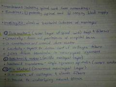
Dura mater |
Outer layer of spinal cord |
|
|
|
Arachnoid mater |
Middle layer |
|
|
|
Pia mater |
Innermost layer |
|
|
|
Epidural space |
Between spinal dura mater and walls of vertebral canal; anesthetic injection site |
|
|
|
Subarachnoid space |
Between arachnoid mater and pia mater; filled with cerebrospinal fluid |
|
|
|
Location of anesthetic injection site |
Epidural space |
|
|
|
What method withdraws CSF? |
Spinal tap |
|
|
|
Spinal tap |
Method to withdraw cerebrospinal fluid |
|
|
|
Denticulate ligaments |
Extend from pia mater to due mater; stabilize side to side movement |
|
|
|
What prevents horizontal movement of the spine? |
Denticulate ligaments |
|
|
|
White matter |
(Superficial) contains myelin at ed and un myelin a ted axons |
|
|
|
Gray matter |
Around central canal of spinal cord; has unmyelinated axons |
|
|
|
Posterior gray horns |
Contain somatic and visceral sensory nuclei (SAD) |
|
|
|
Anterior gray horns |
Contain somatic motor nuclei (VEM) |
|
|
|
Gray commissures |
Next to central canal; axons that cross from one side of cord to the other before reaching gray matter |
|
|
|
Sensory nuclei |
Dorsal, connect to peripheral receptors |
|
|
|
Motor nuclei |
Ventral, connect to peripheral effectors |
|
|
|
Ascending tracts |
Carry sensory info upward |
|
|
|
Descending tracts |
Carry motor info commands to spinal cord downward |
|
|
|
3 connective tissue layers of peripheral nerve |
Epineurium, perineurium, endoneurium |
|
|
|
Epineurium |
Outer layer of nerve |
|
|
|
Perineurium |
Middle layer, divides nerve into fascicles (axon bundles) |
|
|
|
Endoneurium |
Inner layer, surrounds individual axons |
|
|
|
Nerve plexus |
Complex interwoven networks of nerve fibers |
|
|
|
What is formed from blended fibers of ventral rami of adjacent spinal nerves? |
Nerve plexus |
|
|
|
Cervical plexus |
Includes ventral rami of spinal nerves c1-c5; innervates neck, thoracic cavity, diaphragmatic muscles (phrenic nerve) |
|
|
|
Phrenic nerve |
Controls diaphragm |
|
|
|
Brachial plexus |
Includes c5-t1; innervates pectoral girdle and upper limbs (axillary, musculotaneous, median, radial, ulnar) |
|
|
|
Axillary nerve |
Innervates Deltoid and teres major |
|
|
|
Musculotaneous nerve |
Innervates anterior arm muscles |
|
|
|
Median nerve |
Innervates anterior forearm muscles |
|
|
|
Radial nerve |
Innervates Posterior arm and forearm muscles |
|
|
|
Ulnar nerve |
Innervates hand muscles |
|
|
|
Lumbar plexus |
T12-L4; innervates anterior thigh (femoral, obturator) |
|
|
|
Femoral nerve |
Innervates anterior thigh muscles |
|
|
|
Obturator nerve |
Innervates adductor muscles |
|
|
|
Sacral plexus |
L4-S4; innervates Posterior thigh, leg and foot (pudental, sciatic, fibular, tibial) |
|
|
|
Pudental nerve |
Innervates pelvic floor |
|
|
|
Sciatic nerve |
Innervates thigh, leg and foot; branches into fibular and tibial nerves |
|
|
|
Fibular nerve |
Innervates anterolateral leg and foot |
|
|
|
Tibial nerve |
Innervates Posterior thigh, posterior leg, foot |
|
|
|
Neuronal pools |
Functional groups of interconnected (inter)neurons; may stimulate or depress parts of the brain or spinal cord, each w/ limited input sources and output destinations |
|
|
|
Neural reflex |
Rapid, automatic responses to specific stimuli |
|
|
|
Reflex arc |
Wiring of a reflex; begins at receptor, ends at peripheral effector |
|
|
|
5 steps in a neural reflex |
1. Arrival of stimulus, activation of receptor (physcial or chemical changes) 2. Activation of sensory neuron (graded depolarization) 3. Info processing at postsynaptic cell (triggered by neurotransmitters) 4. Activation of motor neuron (action potential) 5. Response of peripheral effector (triggered by neurotransmitters) |
|
|
|
Structures in neural reflex |
Receptor, sensory neuron, postsynaptic cell, motor neuron, peripheral effector |
|
|
|
Innate reflex |
(Formed before birth) basic neural reflexes |
|
|
|
Acquired reflex |
(Learned motor patterns) rapid automatic |
|
|
|
Monosynaptic reflex |
Sensory neuron synapses directly onto motor neuron |
|
|
|
Postsynaptic reflex l |
At least one neuron between sensory neuron and motor neuron |
|
|
|
Patellar reflex |
Monosynaptic reflex, stretch reflex, have least delay between sensory input and motor output |
|
|
|
Reciprocal inhibition |
The stretch reflex of antagonistic (extensor) muscle must be inhibited by interneurons in spinal cord |
|
|
|
What outcome of the Babinski reflex indicted CNS injury? |
Plantar reflex => curling of toes = seen as healthy; absence of descending inhibition => lack of plantar reflex = injury in CNS |
|