![]()
![]()
![]()
Use LEFT and RIGHT arrow keys to navigate between flashcards;
Use UP and DOWN arrow keys to flip the card;
H to show hint;
A reads text to speech;
28 Cards in this Set
- Front
- Back
|
What are the four main functions of the cerebellum? |
- Balance and posture - Coordination of voluntary movement - Motor learning (procedural) - Cognitive functions (fluidity) |
|
|
What are the four anatomical divisions of the cerebellar cortex? |
- Anterior lobe - Posterior lobe - Vermis - Flocculonodular lobe |
|
|
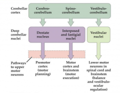
What are the four cerebellar nuclei, from the medial to the the lateral? |
- Fastigial - Interposed (globose and emboliform) - Dentate - Vestibular (external) |
|
|
What is the output of the cerebellum? With what exception? |
Deep Cerebellar Nuclei (DCN) are the sole output of the cerebellum, except for the flocculonodular cortex, that synapses directly to the vestibular nuclei |
|
|
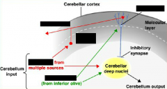
What are the three pathways that input the cerebellar nuclei (with associated neurotrasmitters) ? |
- Purkinje from cerebellar cortex (GABA) - Climbing Fibers from Inferior Olive (Glut) - Mossy Fibers from anywhere (Glut) |
|
Input of the cerebellum. Complete the following elements (4 elements). |
Cerebellar cortex and deep nuclei both receive input from the mossy fibers (from different areas) and the climbing fibers (inferior olive) |
|
Output of the cerebellum. Complete the following elements (4 elements). |
Vestibular nuclei are the only nuclei that receive input directly from the CC via ICP.
|
|
Functional output of the cerebellum Complete the following elements (6 elements). |
|
|
|
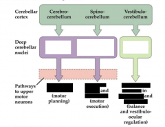
What does the vestibulocerebellar division of the cerebellum comprise? What is its function? |
Anatomy: Flocculonodular lobe Projections: To the lateral vestibular nuclei. Function: It is involved in vestibular reflexes (such as the vestibulo-ocular reflex, VOR) and in postural maintenance. |
|
|
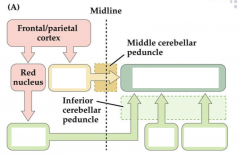
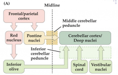
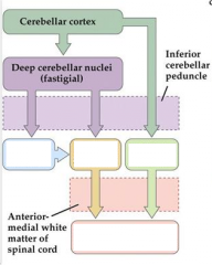
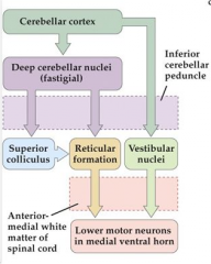
What does the spinocerebellar division of the cerebellum comprise? What are its projections? What is its function? |
Anatomy: Vermis and the intermediate zones of the cerebellar cortex. Fastigial and interposed nuclei Projections: It receives major inputs from the spinocerebellar tract. Its output projects to rubrospinal, vestibulospinal, and reticulospinal tracts. |
|
|
What does the cerebebrocerebellar division of the cerebellum comprise? What are its projections? What is its function? |
Anatomy: It is the largest functional subdivision of the human cerebellum, comprising the lateral hemispheres and the dentate nuclei. Projections: Connected to the cerebral cortex via the pontine nuclei and MCP (afferents) and the VL thalamus via SCP (efferents). Function: It is involved in the planning and timing of movements and higher-order cognitive functions. |
|
|
What is the Crossed Cerebellar Diaschisis (CCD)? |
Crossed cerebellar diaschisis (CCD) refers to a depression of blood flow (hypofusion) and metabolism affecting the cerebellar hemisphere occurring as a result of a contralateral focal, supratentorial (cerebral) lesion. |
|
|
What is the Charcot's triad? What is diagnostic of? hint: N.I.D |
- Nystagmus - Intention tremor (NOT resting tremor) - Dysarthria Diagnostic of cerebellar multiple sclerosis. |
|
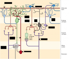
General connectivity of the cerebellum. |
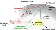
Except for the vestibular nuclei, that receive input directly from the cerebellar cortex, the DCN are the sole cerebellar output and Purkinje cells are the sole cortical output (GABAergic , inhibitory) |
|
Complete: (11 elements) |
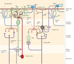
Review the connections with the respective neurotransmitters Granule: Glut Golgi: GABA Basket Cells: GABA |
|
|
Describe the following effect of cerebellar damage and the clinical test to elicit them - Decomposition of movement - Intention tremor |
Decomposition of movement: Some cerebellar patients may not be able to coordinate smooth complex movement and break them down into subcomponents. Test: finger to nose a break-down of movement can be noticed is not the same as dysmetria) and the "British Constitution" speech test. - Intention tremor: Subtype of kinetic tremor amplified as the target is reached. Test should distinguish between the various action tremors (postural, kinetic and intentional) and resting |
|
|
Describe the following effect of cerebellar damage and the clinical test to elicit them - Dysdiadochokinesia - Rebound |
Dysdiadochokinesia: Patients have difficulty performing rapidly alternating movements, such as hitting a surface rapidly and repeatedly with the palm and back of the hand. Test is alternate palm up/down Rebound: Increased range of movement with a lack of normal recoil to original position is seen in cerebellar disease. Test: Protect the patient chest and face with an arm; pull their arm toward you and release it abruptly. |
|
|
Describe the following effect of cerebellar damage and the clinical test to elicit them: - Dysmetria - Cerebellar Ataxia |
Dysmetria: Undershoot/Overshoot intended movement. Heel to shin and finger to nose. (also with eyes open an closed, see below) Cerebellar ataxia: Ataxia is a term for a group of disorders that affect co-ordination, balance and speech. Romberg test distinguishes cerebellar from sensory ataxia. - Ask the patient to stand up, with feet together. and be ready to catch them. Ask them if they feel unsteady. If so, it may not be suitable to perform the test. - If steady, ask the patient to close their eyes, and hold their arms out in front of themselves. - Watch and see if the patient is able to stay steady, or if they begin to wobble. In cerebellar ataxia – the patient should be no more unsteady than with their eyes open. If they wobbles more when they close their eyes is sensory ataxia (positive Romberg). |
|
|
Describe the effect of cerebellar damage in terms of deficit in motor learning (example with the VOR, vestibulo-ocular reflex) |
When a human or animal subject rotates the head, eyes rotate in an equal and opposite direction in order to keep the image stable on the retina. The vestibular system provides the input regarding the head movement, and the motor system has to learn the precise output commands in order to keep the image stable. When magnifying glasses are placed on an animal, the eyes do not move fast enough to compensate for the increased speed of movement of the magnified image, and thus the image moves along the retina (termed “retinal slip”) in the direction opposite to the movement of the head. Over time, however, the motor system learns to move the eyes faster (e.g., the gain of the eye movement command is increased), and the image becomes stable again. When the goggles are removed, the eyes now move too quickly, causing retinal slip in the same direction as head movement. With time, the system will learn to calibrate the VOR again. Patients and experimental animals with damage to the vestibulocerebellum are not able to adapt their VOR to the addition and removal of the goggles, demonstrating the role of the cerebellum in this form of motor learning. |
|
|
Describe the projections to/from the Superior Cerebellar Peduncle (SCP) |
The SCP is almost exclusively an efferent pathway. dentate and interposed nuclei --(through SCP)--> mediodorsal thalamus (MD) --> premotor/motor cortex fastigial nuclei --(through SCP)--> vestibular nuclei |
|
|
Describe the projections to/from the Middle Cerebellar Peduncle (MCP) |
The SCP is an afferent pathway. cerebral cortex --(through corticopontine fibers)--> contralateral pontine nuclei --(through MCP)--> deep cerebellar nuclei/cerebellar cortex |
|
|
Describe the projections to/from the Inferior Cerebellar Peduncle (ICP) |
The ICP is an afferent/efferent pathway. climbing fibers from inferior olive -(through ICP)--> deep cerebellar nuclei/cerebellar cortex deep cerebellar nuclei -(through ICP)--> vestibular nuclei and reticular formation |
|
|
Describe the main features of the Cerebellar Cognitive Affective Syndrome |
A. It can be seen with a variety of etiology (tumour, stroke, cerebellitis, neurodegenerative, etc.) 1. Executive dysfunctions (planning, abstract thinking, working memory, set-shifting, etc.) 2. Language (agrammatism, dysprosody) 3. Affect (blunting or inappropriateness) |
|
|
What is the role that computational models ascribe to Purkinje cells, climbing fibers and parallel fibers (Albus and Marr) ?
|
The high convergence of parallel fibers on Purkinje cell suggests that some kind of pattern recognition may be take place at this synapse (PF/PC). This synapse is known to undergo both long term depression (LTD), in the presence of climbing fiber (CF) signals and long term potentiation (LTP), in its absence. Thus, the CF from the olivary nuclei may control the direction of plasticity (Albus, 1971; Marr, 1969). |
|
|
What is the role that computational models ascribe to the granule cells (generating parallel fibers and receiving input from mossy fibers) in terms of patterns. |
The immense number of granule cells and the few connections of each granule cell often led to the suggestion that the granular layer plays the role of pattern separation, which has been buttressed by recent studies on the coding capacities of the granule layer. (Billings, Piasini, Lorincz, Nusser, & Silver, 2014). |
|
|
Golgi cells inhibits granule cells. What is their putative oscillatory role? |
Some models suggest that the negative feedback to Golgi cells promote oscillatory gating of mossy fiber activity. In vitro studies have verified that resonant frequencies tend to exist, particularly in the beta band (~13-30 Hz) (Solinas et al., 2010; Vervaeke et al., 2012). |
|
|
What is the morphological peculiarity of Parallel fibers? What kind of functional role could this imply? (Eccles & Braitenberg) |
Parallel fibers are unusually long, thin, unmyelianated axons, with low conduction speeds (0.1- 0.3m/s). Eccles & Braitenberg to suggest that PFs could act as delay lines and PNs, would respond as integrators of coincidence, leading to Braitenberg's 'tidal wave hypothesis': "Mossy fibers activated in a linear sequence along the parallel fiber axis contribute to a wavefront that would either accumulate to activate Purkinje cells and inhibitory interneurons" |
|
|
In terms of information processing, what does cerebellum compute? Explain the putative processes behind eyelid conditioning. |
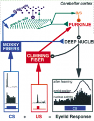
Purkinje cells of the cerebellar cortex project to the deep nuclei (output). Input reaches the cerebellum viatwo excitatory pathways: mossy fibers and climbing fibers. Mossy fibersoriginate from various brain stem nuclei that receive information from cerebral cortex and spinal cord. They also influence cerebellar output through direct excitatory connectionsonto the deep nuclei cells (mf→nuc synapses) and through a more complex pathway (GC, PF, PC) inthe cerebellar cortex that culminates in Purkinje cell inhibition of deep nuclei neurons.This pathway begins with mossy fiber excitation of both granule and Golgi cells, includesthree types of inhibitory interneurons (Golgi, basket and stellate cells), and involves theabundant excitatory synapses that granule cells make onto Purkinje cells (gr→Pkj synapses). There are about 200,000 for each Purkinje cell, and given that Purkinje cells convergeonto deep nucleus cells, ∼100 million gr→Pkj synapses contribute to the output of a singlenucleus cell). The other input pathway, the climbing fibers, is strikingly different. Second, the US is conveyed by activating the climbing fiber input. Third, increases in the activityof cerebellar output cells in the anterior interpositus nucleus drive the expression of the learned eyelidresponse.Learning this conditioned response involves two sites of plasticity: gr→Pkj synapses in the cerebellarcortex, and in the cerebellar nucleus, possibly at mf→nuc synapses. (Medina & Mauk, 2000) |