![]()
![]()
![]()
Use LEFT and RIGHT arrow keys to navigate between flashcards;
Use UP and DOWN arrow keys to flip the card;
H to show hint;
A reads text to speech;
23 Cards in this Set
- Front
- Back
|
The nervous system |
A communication network that enables an individual to respond or adjust to changes in internal and/or external environment |
|
|
Components of Nervous System |
Sensory- detect changes
|
|
|
Neuron Cell |
A Cell Body: Containing a nucleus, ER, mitochondria and other typical organelles |
|
|
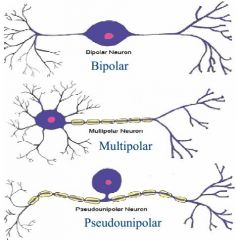
Types of Neurons |
|
|
|
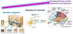
Somatic sensations |
Sensation from the skin, muscles, bones, tendons and joints
|
|
|
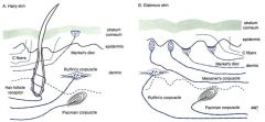
Somatic sensations Diagram |
|
|
|
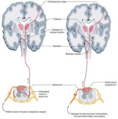
Somatic receptors tracts |
The Anterolateral Tract
|
|
|
Somatic receptors tracts Diagram |
|
|
|
Somatic receptors destiation |
Somatic receptors on left side of body go to the right cerebral hemisphere and vice-versa, both for anterolateral and dorsal tracts. |
|
|
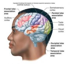
Somatosensory cortex |
|
|
|
Receptor types |
|
|
|
Pacinian corpuscles |
- Found deep in the dermal layers of both hairy and glabrous skin - Have relatively large endings that are widely spaced. |
|
|
Ruffini’s endings |
Found deep in the dermal layers of both hairy and glabrous skin. |
|
|
Meissner’s corpuscles |
approx. 1/10 the size of Pacinian corpuscles.
|
|
|
Merkel’s disk |
located in the epidermis. Consist of a nerve terminal and a flattened non-neural epithelial cell. Small receptive field. |
|
|
Hair follicle receptors |
hairy skin only. Can be either rapidly |
|
|
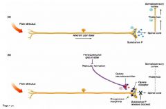
Pain sensation |
Receptors, “nociceptors” |
|
|
|
Mechanoreceptors, thermal receptors, chemoreceptors, and polymodal receptors (these respond to all 3 stimuli).
|
|
|
|
|
|
|
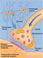
Neuron Communication |
An electrical impulse travels down an axon and to the synaptic terminal Synapse |
|
|
Neuron Communication Diagram |
|
|
|
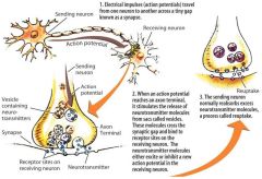
Events in Neuromuscular Transmission |
1. Depolarization of motor neuron and generation of action potential |
|
|
Neuromuscular Transmission Diagram |
|