![]()
![]()
![]()
Use LEFT and RIGHT arrow keys to navigate between flashcards;
Use UP and DOWN arrow keys to flip the card;
H to show hint;
A reads text to speech;
8 Cards in this Set
- Front
- Back
|
Staining |

|
|
|
Organelles |

|
|
|
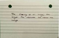
Resolution |

|
|
|
Magnification |
A number of times larger and image appears compared to the actual size of the object |
|
|
Electron micrograph |
A photograph of an image seen under an electron microscope |
|
|
Magnification formula |
Magnification = image size/object size |
|
|
Laser scanning microscopes |
– Use laser light– Scan object point by point– Construct an image of it on the computer using pixel information– Images = high-res, high contrast– Depth selectivity– Use for whole living specimens and cells |
|
|
Scanning electron |
Electrons do not pass through the specimen– Electron been because secondary electrons to bounce off specimen surface– Focused on to a onto the screen– 3D image– Magnification times 15×200,000 resolution 2.05– Greyscale colour and in computers and false colour imaging– Specimen coated in fine film metal and put into vacuum |

