![]()
![]()
![]()
Use LEFT and RIGHT arrow keys to navigate between flashcards;
Use UP and DOWN arrow keys to flip the card;
H to show hint;
A reads text to speech;
33 Cards in this Set
- Front
- Back
- 3rd side (hint)
|
Magnification |
This tells you how many times bigger the image produced by the microscope is than the real-life object you are viewing |
|
|
|
Resolution |
The ability to distinguish between objects that are close together. |
|
|
|
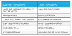
Optical (Light) Microscopes |
Uses light to form an image, this limits the resolution to around 0.2 micrometers due to the wavelength of light. They can be used to observe eukaryotic cells, their nuclei and potentially mitochondria and chloroplasts. The maximum magnification is about x1500. |
|
|
|
Electron Microscopes |
They use electrons to form an image, this greatly increases the resolution of electron microscopes giving a more detailed image. They have a maximum resolution of 0.0002 micrometers and maximum magnification of about x1,500,000. There are two types : Transmission Electron Microscopes (TEMs) and Scanning Electron Microscopes (SEMs) |
|
|
|
Transmission Electron Microscopes (TEMs) |
They use electromagnets to transmit a beam of electrons through the specimen. Denser parts of the specimen absorb more electrons , making them appear darker. |
|
|
|
Advantages of TEMs |
High resolution images, allows the internal structures within cells/organelles to be seen. |
|
|
|
Disadvantages of TEMs |
They can only be used on thin specimens, they cannot be used on live specimens due to vaccum, they do not produce a colour image. |
|
|
|
Scanning Electron Microscopes (SEMs) |
They scan a beam of electrons across the specimen and as the electrons bounce off they are detected to form an image. |
|
|
|
Advantages of SEMs |
They can be used on thick or 3D specimens, they allow the external 3D structure to be observed |
|
|
|
Disadvantages of SEMs |
They give a lower resolution than TEMs, they cannot observe live specimes due to vaccum, they do not produce a colour image. |
|
|
|
Laser Scanning Confocal Microscopes |
The cells being viewd are stained with fluorescent dyes, the tissue is then scanned with a laser beam and the beam is reflected by the dye producing an image. |
|
|
|
Advantages of Laser Scanning Confocal Microscopes |
They can be used on thick or 3D specimens, they allow the external structure to be observed, they produce a high resolution. |
|
|
|
Disadvantages of Laser Scanning Confocal Microscopes |
It is a slow process, the laser has potential to cause photodamage to the cells. |
|
|
|
Magnification Formula |
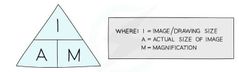
|
"I AM" good at Biology |
|
|
Light vs Electron Microscope Table |
|
|
|
|
Cell Surface Membrane (Eukaryotic Cell Structure) |
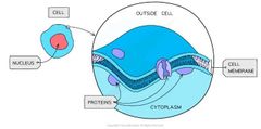
All cells are surrounded by a cell surface membrane which controls the exchange of materials between the internal cell environment and external environment. It is formed from a phospholipid bilayer and is described as being 'partially permeable'. |
|
|
|
Cell Wall (Eukaryotic Cell Structure) |
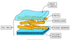
Found in plant cells but not in animal cells. They are formed outside the cell membrane and offer structural support provided by the celluolse (plants) or peptidoglycam (bacteria). Narrow threads of cytoplasm called plasmodesmata connect the cytoplasm of neighbouring plant cells. |
|
|
|
Nucleus (Eukaryotic Cell Structure) |
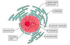
Present in most eukaryotic cells, the nucleus contains chromatin which forms chromosomes. Chromosomes are made of sections of linear DNA tightly wound around histones. The Nucleus is relatively large and is seperated from the cytoplasm by the nuclear envelope which contains pore to allow mRNA and ribosomes to travel out. |
|
|
|
Mitochondria (Eukaryotic Cell Structure) |
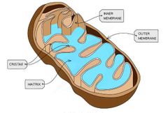
The site of aerobic respiration within all eukaryotic cells. It is surrounded by double-membrane with the inner membrane folded to form cristae. The matrix formed by the cristae contains the enzymes needed for aerobic respiration to produce ATP. |
|
|
|
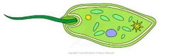
Chloroplasts (Eukaryotic Cell Structure) |
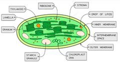
Found in plant cells, they contain a double membraine. They are the site of photosynthesis. Membrane bound compartments called thylakoids containing chloropyll stack to form grana which are joined by lamellae. |
|
|
|
Ribosomes |
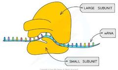
Found in all cells either freely in the cytoplasm or a part of the rough endoplasmic reticulum. They are the site of translation (protein synthesis). They are a complex of rRNA and protines. 80s ribosomes are found in eukaryotic cells, 70s ribosomes in prokaryotes. |
|
|
|
Rough Endoplasmic Reticulum (RER) |

Found in plant and animal cells, their surface is covered in ribosomes. They process the protiens made by the ribosomes. |
|
|
|
Smooth Endoplasmic Reticulum (SER) |
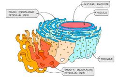
Found in plant and animal cells, does not have ribosomes on the surface unlike the RER. Involved in the production, processing and storage of lipids, carbohydrates and steroids. |
|
|
|
Golgi Apparatus |
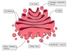
Found in plant and animal cells, flattened sacs of membrane similar to the smooth endoplasmic reticulum. Modifies proteins and lipids before packaging them into Golgi Vesicles. The vesicles then transport the proteins and lipids to their required destination. |
e.g Lysosomes production |
|
|
Large Permanent Vacuoles |
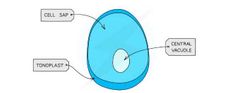
A sac in plant cells surrounded by tonoplast, selectively permeable membrane. Vacuoles in animal cells are not permanent and small. |
|
|
|
Microvili |
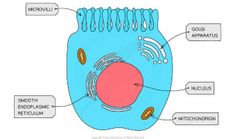
Found in specialised animal cells, they are cell membrane projections used to increase surface area in order to increases the rate of exchange of substances |
|
|
|
Flagella |
Found in specialised cells they are made of microtubules. Contract to provide cell movement e.g Sperm Cells. |
|
|
|
Cytoskeleton |
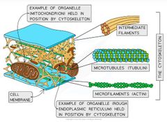
An extensive network of protein fibres within the cytoplasm. It is made of two main types of protein fibre: microfilaments and microtubules. |
|
|
|
Microfilaments |
Solid strands mostly made of the protein actin. These fibres can cause cell movement and the movement of organelles by moving against each other. |
|
|
|
Microtubules |
Hollow strands that are mostly made of tubulin. Organelles and other cell contents are moved along these fibres using ATP> |
|
|
|
The importance of the cytoskeleton |
Strength and support, intracellular movement, cellular movement. |
|
|
|
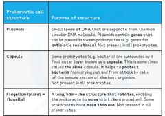
Structures unique to Prokaryotic Cells |
|
|
|
|
Prokaryotic & Eukaryotic Cells Comparison |
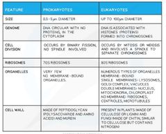
|
Size, Cell Division, Ribosomes etc |