Reading...
![]()
Play button
![]()
Play button
![]()
Use LEFT and RIGHT arrow keys to navigate between flashcards;
Use UP and DOWN arrow keys to flip the card;
H to show hint;
A reads text to speech;
207 Cards in this Set
- Front
- Back
|
True or False. The etiology of cardiomyopathy is unknown.
|
True.
|
|
|
What other names is cardiomyopathy referred to?
|
It is referred to as idiopathic cardiomyopathy or primary cardiomyopathy.
|
|
|
There are three different types of cardiomyopathy. What are they? Which is the most common?
|
Dilated, hypertrophic and restrictive.
Dilated cardiomyopathy is the most common. |
|
|
What % of cardiomyopathies are dilated?
|
90% of cases
NOTE: 80% was mentioned in class lecture. |
|
|
True or False. Regarding dilated cardiomyopathy, there is two-chamber hypertrophy and dilation of the heart in a football shape
|
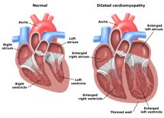
False. Regarding dilated cardiomyopathy, there is FOUR-chamber hypertrophy and dilation of the heart in a GLOBOID shape.
|
|
|
True or False. Regarding dilated cardiomyopathy, it becomes floppy, thin-walled and unable to contract properly.
|
True.
|
|
|
What percentage of dilated cardiomyopathy is genetic?
|
20%
|
|
|
Regarding dilated cardiomyopathy, the cause is...
|
idiopathic
|
|
|
What are four risk factors/associations of dilated cardiomyopathy?
|
Metabolic - weak thyroid, which weakens myocardium
Toxins - alcohol and poison Viral childhood infections Previous myocarditis |
|
|
What are some symptoms of dilated cardiomyopathy?
|
Dyspnea
Edema Arrhythmia Fatigue Pulmonary circuit problems Bed ridden Left heart problems DEAF PBL Deaf pebble |
|
|
What percentage of dilated cardiomyopathy patients die within 2 years from heart failure or arrhythmia?
|
50%
|
|
|
What is the only treatment for dilated cardiomyopathy?
|
Heart transplant.
|
|
|
Which cardiomyopathy is like the opposite of dilated?
|
Hypertrophic cardiomyopathy is like the opposite of dilated.
|
|
|
True or False. With hypertrophic cardiomyopathy, the heart is heavy and hypercontracts and has diastolic dysfunction.
|
True.
|
|
|
What is the cause of hypertrophic cardiomyopathy?
|
Idiopathic
|
|
|
What are the symptoms of hypertrophic cardiomyopathy? What is the most common?
|
Sudden Death
Septum thicker than free wall in athletes Arrhythmia (most common) Young Males Narrow obstructed outflow SSAYN Sayin' |
|
|
What percentage of hypertrophic cardiomyopathy is genetic?
|
50%
|
|
|
Regarding hypertrophic cardiomyopathy cardiac failure is due to...(list 5)
|
Decreased diastolic filling
Reduced chamber size Reduced stroke volume Abnormally thick walls Poor compliance of the muscle DRRAP |
|
|
True or False. A characteristic finding of hypertrophic cardiomyopathy is assymetric hypertrophy of the inferior portion of the interventricular septum.
|
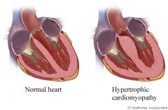
False. A characteristic finding of hypertrophic cardiomyopathy is assymetric hypertrophy of the SUPERIOR portion of the interventricular septum.
|
|
|
What outcome is common for hypertrophic cardiomyopathy patients?
|
Sudden death
|
|
|
What is the classic example of sudden death for hypertrophic cardiomyopathy patients?
|
The classic example is a young athlete collapsing during competition.
|
|
|
What type of breakfast food does they heart resemble with hypertrophic cardiomyopathy?
|
A nice warm bagel with a hole in the center, maybe with lox?
|
|
|
Two terms that describe an off-center bagel hole, as with hypertrophic cardiomyopathy are...
|
eccentric and symmetric
The opposite: concentric and asymmetric |
|
|
What would an ECG look like in an a hypertrophic cardiomyopathy patient?
|
A very high QRS spike.
|
|
|
True or False. Restrictive cardiomyopathy prevents contraction of the heart for diastolic filling.
|
False. Restrictive cardiomyopathy prevents RELAXATION of the heart for diastolic filling.
|
|
|
How does the heart appear in restrictive cardiomyopathy?
|
Normal chambers
Normal thickness No dilation Abnormal decomposition of myocardium (histologically) Small ECG pump Stiff myocardium NNNASS |
|
|
Would you say there is intramuscular fibrosis restrictive cardiomyopathy?
|
Yes, I would say usually there is intramuscular fibrosis.
|
|
|
Several disease processes can give rise to fibrosis in the heart, name two.
|
Amyloidosis and Endomyocardial Fibrosis.
|
|
|
What is the least common type of cardiomyopathy?
|
Restrictive cardiomyopathy. It is extremely rare.
|
|
|
When is restrictive cardiomyopathy diagnosed?
|
Post mortem
|
|
|
What does restrictive cardiomyopathy progresses to?
|
Congestive heart failure
|
|
|
What is the cause of restrictive cardiomyopathy?
|
Idiopathic
|
|
|
What are the most common complications of all three cardiomyopathies?
|
Pump failure
Arrhythmia Both lethal |
|
|
What is the treatment for all three cardiomyopathies?
|

Heart transplant.
|
|
|
People with hemochromatosis may have which type of of cardiomyopathy?
|
Dilated - the iron is considered a toxin
|
|
|
People with amyloidosis have hearts with a consistency of glue. Their hearts bounce like a basketball they are so rubbery. These patients may have which type of of cardiomyopathy?
|
Restrictive
|
|
|
The two categories of trauma are...
Which is most common? |
Nonpenetrating (blunt) - MOST COMMON
Penetrating. Both can damage the heart and its internal structure, pericardium, or great vessels. |
|
|
A blunt nonpenetrating injury is the result of either an external or internal force. Which is it?
|
External force.
|
|
|
What are two types of blunt force?
|
1) Sudden deceleration or acceleration
2) Forceful blow to the sternum |
|
|
Why is sudden deceleration or acceleration bad?
|
It ruptures great vessels
These forces causes shear force and torsion on the great vessels, rupturing them. |
|
|
Where is a common site for great vessel rupture caused by sudden deceleration/acceleration blunt force?
|
The aorta
|
|
|
What is the most severe case for great vessel rupture?
|
Complete transection
|
|
|
What results from blunt force to the sternum?
|
Heart crushed against the sternum or vertebrae.
|
|
|
At the least, nonpenetrating trauma results in...
|
myocardial contusion (a bruised heart).
|
|
|
At the worst, nonpenetrating trauma results in...
|
rupture of the myocardium or great vessels.
|
|
|
What is a penetrating injury?
|
A sharp force, such as a knife or bullet, that penetrates into the heart or great vessels.
|
|
|
Which part of the heart do most penetrating injuries occur?
|
Right ventricle, the most anterior chamber
|
|
|
What are COMMON complications of penetrating injuries?
|
Hemothorax (blood in the chest cavity)
IVC Rupture Rupture of Rt. Atria Luxation (rotation great vessels) HIRL |
|
|
True or False. There is extremely high mortality due to the massive hemothorax that results.
|
True.
|
|
|
If the object leaves a closed wound in the pericardial sac, bleeding from the heart fills the sac and creates...
|
a cardiac tamponade
|
|
|
In some cases, tamponade may serve as a pressure on the wound which causes...
|
a stop in the hemorrhage.
|
|
|
What are two treatments for a penetrating trauma injury?
|
Thoracotomy (cutting open the chest)
Pericardiocentesis (evacuating the pericardial sac). |
|
|
What are two risks after a penetrating trauma injury?
|
Infection and Arrhythmia.
|
|
|
What type of injury is trauma classified as?
|
A mechanical injury.
|
|
|
List 4 injuries that could be classified as blunt trauma.
|
MVA
Fall Fighting Contact sports |
|
|
What is the mortality rate of bullet wounds to the heart? Knife wounds?
|
Bullets - 80%
Knives - 40% |
|
|
True or False. Broken sternums usually don't damage the heart.
|
True.
|
|
|
A blunt heart trauma results in a compression and _________ of the bones and tissue where the injury occurs.
|
release
|
|
|
The most severe cases of blunt trauma are _________ of the heart.
|
lacerations
|
|
|
Which side of the heart is more vulnerable with blunt trauma? Which valves are more vulnerable?
|
The right side; The valves on the left side of the heart due to the pressure change.
|
|
|
Regarding penetrating trauma to the heart, what leads to the cause of death? List 4.
|
Hemothorax (if pericardium damaged)
Arrhythmia Repiratory Failure Tamponade HART |
|
|
What is the main treatment option for penetrating heart trauma?
|
Thoracotomy (Surgery) to repair holes.
|
|
|
What imaging modality is hemothorax commonly seen.
|
X-ray
|
|
|
How does one diagnose an unconscious patient regarding heart trauma?
|
Echo.
|
|
|
A very unlucky injury to have is a fast blow to a certain part of the chest (just before the T-wave on an ECG). This injury causes sudden death. What is it called?
|
Commotio cordis
|
|
|
A classmate decides they don't like how you outscored them on a Peck exam. In retaliation you get a nail file stabbed into your heart. Should the nail file be left in or taken out?
|
Left in to plug the hole.
|
|
|
How is heart luxation diagnosed?
|
Echo or CT.
|
|
|
Deceleration ultimately causes the aorta to move out from tearing in ligaments. What shows up on CT from this injury?
|
A blood ring around the aorta that appears as a double lumen.
|
|
|
How is an aorta tear from deceleration treated?
|
It is sewn back together in surgery.
|
|
|
How is a tear in the IVC treated?
|
It is not, death is imminent. Veins cannot be repaired.
|
|
|
Approximately how many infants are born with significant heart defects?
|
8 of 1000
|
|
|
What is the known cause of CHD ?
|
Idiopathic
|
|
|
What are some risk factors of Congenital Heart Disease (CHD) ?
|
Maternal rubella
Malnutrition Alcoholism Diabetes (Insulin-dependent) MMAD |
|
|
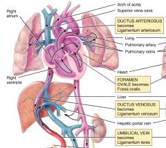
Oxygenated blood enters the fetus via the...
|

the umbilical vein and then the ductus venosus in the liver.
|
|
|
What happens to the blood that enters the umbilical vein?
|

It mixes with deoxygenated blood from the IVC.
|
|
|
After umbilical vein blood mixes with deoxygenated blood from the IVC, what happens next?
|

It enters the right atrium.
|
|
|
Is the blood from the IVC and the umbilical vein the only blood that enters the right atrium?
|
No, you have the SVC too.
|
|
|
Where does the blood in the IVC and SVC come from?
|
As in adults, the IVC drains the lower extremities and the SVC drains the upper extremities.
|
|
|
True or False. The way the valves are placed in the right atrium for the SVC and IVC, cause the two streams of blood to mix.
|
False. The way the valves are placed in the right atrium for the SVC and IVC, the two streams of blood do DO NOT mix that much.
|
|
|
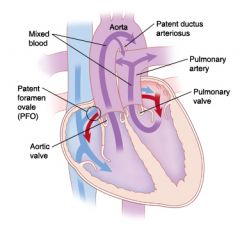
Once in the right atrium the oxygenated blood from the IVC goes where?
|
Foramen ovale ---> Left atrium ---> left ventricle ---> aorta.
|
|
|
Once in the right atrium the de-oxygenated blood from the SVC goes where?
|

Right atrium ---> Right ventricle ---> Pulmonary artery ---> Pulmonary circulation
(minimal blood goes this route) OR Right atrium ---> Right ventricle ---> Pulmonary artery ---> Ductus arteriosis ---> Descending aorta (most blood goes this route) |
|
|
How do the fetal lungs appear in the womb?
|
Collapsed
Fluid filled |
|
|
What can you say about the resistance of fetal pulmonary arteries?
|
High resistance
Constricted |
|
|
How much blood is returned from the pulmonary veins into the left atrium in fetal circ.?
|
Very little
|
|
|
What type of blood does the left atrium in a fetal heart contain? Where does it come from?
|
Oxygenated blood through the foramen ovale.
|
|
|
After the blood moves from the left atrium to the left ventricle, it enters the aorta. Where does it go next?
|
The branches of the aorta supply the:
Heart Head Upper Extremities |
|
|
After the aorta supplies the upper extremeties, what happens to the blood?
|
It blood mixes with the deoxygenated blood from the ductus arteriosus and supplies the lower extremities.
|
|
|
Why is it that the upper extremities are supplied with oxygenated blood first?
|
First priority is to oxygenate the heart and head.
|
|
|
After distributing the blood to the lower extremities what happens to the blood?
|

Enters the umbilical arteries and returns to the placenta for oxygenation.
|
|
|
What happens to the lungs once a baby is born?
|
Inhales ---> Lungs expand ---> Alveoli open ---> Pulmonary vasculature dilates.
|
|
|
What happens to pulmonary resistance after birth?
|
It drops
|
|
|
After the opening of the pulmonary vessels and the cessation of umbilical flow what happens to the pressure in the right atrium?
|
It drops.
|
|
|
What happens to the amount of pulmonary venous return after birth?
|
It increases.
|
|
|
What happens to the pressure in the left atrium after pulmonary venous return increases?
|
It increases.
|
|
|
What causes the foramen ovale to close?
|
The increase in pulmonary venous return which raises left atrium pressure.
|
|
|
What happens to the ductus arteriosis after birth?
|
Shortly after birth the ductus arteriosus constricts.
|
|
|
Sometimes the ductus arteriosus does not close after birth, what type of infants does this occur in?
|
Premature infants
|
|
|
What does the failure of the ductus arteriosis result in?
|
Patent ductus arteriosus (PDA)
|
|
|
What is the ductus arteriosus under the influence of? How does it get a DUI?
|

Prostaglandins
|
|
|
How can the opening and closure of the ductus arteriosus be modulated?
|
With prostaglandins or prostaglandin inhibitors.
|
|
|
What is a type of prostaglandin inhibitor?
|
Indomethacin
|
|
|
Which causes the ductus arteriosus to open? Close?
|
Prostgatlandins open; Prostaglandin inhibitors close
|
|
|
A premature infant would require which type of treatment for a ductus arteriosus?
|
Prostaglandin inhibitors, these close the ductus.
|
|
|
In infants with severe cardiovascular malformations which type of treatment for a ductus arteriosus is used? Why?
|
Prostaglandins to keep the ductus arteriosus open.
A patent ductus arteriosus may actually help them to live until the malformation can be corrected surgically. |
|
|
Which kind of defects do not shunt significant amounts of deoxygenated blood from the right side to the left side?
|
Acyanotic Congenital Heart Defects
|
|
|
Why doesn't cyanosis occur with Acyanotic Congenital Heart Defects?
|
The left ventricle continues ejecting oxygenated blood.
|
|
|
List 4 Acyanotic Congenital Heart Defects.
|
Atrial Septal Defect (ASD)
Ventricular Septal Defect (VSD) Patent Ductus Arteriosus (PDA) Coarctation of the Aorta |
|
|
What percent of all cardiac congenital anomalies are Atrial Septal Defect (ASD)?
|
7%
|
|
|
In general, what is Atrial Septal Defect (ASD) ?
|
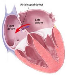
Direct communication between the left and right atria.
|
|
|
When is ASD a significant problem?
|
If large
Accompanied by other defects. |
|
|
What happens when an ASD becomes large?
|
1) Left atrium pushes blood into right atrium
2) Blood goes into the right ventricle 3) Pulmonary circulation does not tolerate the pressure 4) Resistance in the pulmonary vasculature causes blood to flow backwards, right to left though the defect. 5) Deoxygenated blood flows to body, causing systemic hypoxemia. 6) Leads to fatal pulmonary hypertension (after many years) |
|
|
True or False. Most ASDs are medium to large sized.
|
False. Most ASDs are small.
|
|
|
True or False. Most infants with ASD are symptomatic
|
False. ASYMPTOMATIC
|
|
|
How is ASD detected?
|
By murmur.
|
|
|
When is asymptomatic ASD usually detected?
|
School-age.
|
|
|
If an ASD is very small, can the person be asymptomatic for most of his life?
|
Yes.
|
|
|
How many births are Ventricular Septal Defect (VSD)?
|
12 in 10,000 births
|
|
|
In general, what is Ventricular Septal Defect (VSD)?
|
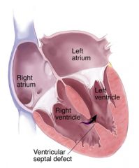
Communication between the right and left ventricles.
|
|
|
What do VSD symptoms depend on? What are some symptoms?
|
The SIZE of the defect and the SEVERITY of the shunt of blood.
A murmur on anterior chest. |
|
|
What happens when an VSD is present?
|
1) Left ventricle pushes blood into the right ventricle.
2) Increased blood enters pulmonary circulation 3) Blood moves into the left atrium. 4) Left atrium DILATES. 5) Left ventricle, takes on extra blood, HYPERTROPHIES. 6) Results in tachypnea due to wet lungs 7) Results in fatigued by feeding. 8) Left-to-right shunt causes pulmonary hypertension. |
|
|
How many births are Patent Ductus Arteriosus (PDA)?
|
8 in 10,000 births
|
|
|
In general, what is Patent Ductus Arteriosus (PDA)?
|
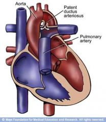
The ductus arteriosus fails to constrict after birth.
|
|
|
When should a PDA normally close by?
|
12 weeks.
|
|
|
What are some diseases associated with PDA?
|
Rubella syndrome or other anomalies.
|
|
|
What does Rubella syndrome cause? Why is this bad news bears?
|
Ductal scarring associated with rubella may prevent constriction and closure.
|
|
|
Why can oxygenated blood go through the PDA from the aorta to the pulmonary artery and lungs? Why is this good?
|
Because aortic pressure is greater than pulmonary arterial pressure.
It's good because PDA can save the life of an infant with cyanotic heart defects and decreased pulmonary blood flow. |
|
|
How many births have Coarctation of the Aorta?
|
3 in 10,000 births
|
|
|
In general, what is Coarctation of the Aorta?
|

Narrowing of the aorta.
|
|
|
There are two types of Coarctation of the Aorta. What are they?
|
Preductal and Postductal
|
|
|
Where is the reference point for preductal and postductal Coarctation of the Aorta?
|
The ductus arteriosus.
|
|
|
What processes occur with preductal Coarctation of the Aorta?
|
1) Left ventricle cannot pump through the aorta
2) Blood flow to the lower half of the body is reduced 3) Backup of blood in the heart 4) Lower extremities are cool, and mottled. 5) The upper half of the body looks normal, because it is being supplied with oxygenated blood. |
|
|
What processes occur with postductal Coarctation of the Aorta?
|
1) Obstruction is not as severe
2) Collateral circulation develops around the narrowed site 3) Collaterals provide perfusion to the lower body |
|
|
True or False. The majority of postductal newborns develop severe circulation problems by school-age.
|
False. The majority of the newborns grow and mature NORMALLY since the obstruction is not as severe.
|
|
|
What are Cyanotic Congenital Heart Defects? What effect does this have on the blood?
|
They shunt deoxygenated blood from the right side to the left.
Causes mixing of the venous and arterial blood. |
|
|
Why is mixing of the blood bad with Cyanotic Congenital Heart Defects?
|
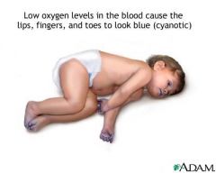
Arterial oxygen level is lower than normal.
Patients look blue (cyantotic). |
|
|
List two types of Cyanotic Congenital Heart Defects. Which is most common?
|
Tetralogy of Fallot (ToF) - MOST COMMON
Transposition of the Great Vessels |
|
|
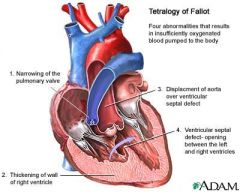
Tetralogy of Fallot (ToF) is a syndrome of four defects. What are they?
|

Ventricular Septal Defect (VSD)
Pulmonic stenosis Anteriorly displaced aorta that receives blood from both ventricles (overriding aorta) Right Ventricular Hypertrophy |
|
|
Why are children with ToF cyanotic?
|
Deoxygenated blood from body is shunted from right ventricle into aorta.
|
|
|
Regarding ToF, why is deoxygenated blood from the body shunted from right ventricle into the aorta?
|

The right ventricle can't pump it through the stenotic pulmonary valve.
|
|
|
Regarding ToF, the right ventricle can't pump it through the stenotic pulmonary valve, where does the blood go instead?
|

Through the VSD.
|
|
|
What are some complications of ToF?
|
Dyspnea on exertion
Murmur from the stenotic pulmonary valve. |
|
|
What shape does the heart take on with ToF? Why?
|
A boot-shaped appearance
Right ventricular hypertrophy |
|
|
What is treatment of ToF?
|
Surgery.
|
|
|
How many births contain Transposition of the Great Vessels?
|
4.8 in 10,000
|
|
|
How common is Transposition of the Great Vessels?
|
This is the second most common cyanotic heart defect.
|
|
|
In general, what is Transposition of the Great Vessels?
|
When the aorta and pulmonary trunk switch positions.
|
|
|
True or False. Newborns with Transposition of the Great Vessels usually survive without surgical intervention.
|
False. Without immediate surgical intervention, it is a lethal defect.
|
|
|
What physiological processes occur with Transposition of the Great Vessels?
|
1) Unoxygenated blood ---> right ventricle ---> aorta ---> systemic circulation.
2) Lungs ---> Oxygenated blood ---> Left ventricle ---> Pulmonary artery ---> Lungs |
|
|
Why was Transposition of the Great Vessels ok in the fetus?
|
the placenta was the primary organ of oxygenation and the pulmonary circulation wasn't all that important.
|
|
|
Regarding Transposition of the Great Vessels, what can help temporarily to permit mixing of venous and arterial blood?
|
A patent ductus arteriosus
|
|
|
From the last test...Chronic diseases are ______ while acute diseases are more _______. Because of this, a chronic patient may develop ________ vessels and be able to withstand an acute attack like MI. In other words, older/chronic patients can withstand these kinds of attacks better than young people.
|
milder; severe; collateral
|
|
|
In adults, systemic circulation moves in what direction? Pulmonary moves in what direction?
|
Systemic: Left ---> Right; Pulmonary: Right ---> Left
|
|
|
In fetal circulation, the placenta plays the role of what three organs?
|
Kidneys, lungs, liver.
|
|
|
True or False. Oxygenation of fetal circulation occurs in the fetal heart with blood mixing.
|
False. Oxygenation occurs on the outside of the fetus. From mother to baby through the placenta.
|
|
|
There are are three natural shunts that develop in the fetus, what are they?
|
1) Ductus Venosus - bypasses the liver to the IVC
2) Foramen Ovale - atrial septum hole 3) Ductus Arteriosus - shunt between pulmonary artery and aorta |
|
|
How many vessels does the umbilical cord hold? What are they?
|
3 Vessels. 2 Arteries, 1 Vein.
|
|
|
True or False. Babies have a the same quality of hemoglobin as adults.
|
False. It's a different quality of hemoglobin.
|
|
|
True or False. Babies have less oxygen concentration in their hemoglobin compared to adults but bring approximately the same amount of oxygen to their tissues.
|
True.
|
|
|
When does fetal circulation switch over to "adult" circulation?
|
At the time of birth, within minutes after being born.
|
|
|
Go through the process of what happens when a baby is born in terms of the change in circulation.
|
1) The umbilical cord is cut
2) The ductus venosus closes (Shunt #1) 3) Baby cries, lungs inflate, pulmonary circulation opens 4) Pressure in left atrium rises, pressure in right atrium lowers 5) Foramen ovale closes (Shunt #2) 6) Ductus arteriosus closes (Shunt #3) |
|
|
The three fetal shunts become something else. What do they become respectively?
|
Ductus venosus ---> Ligamentum venosum
Foramen ovale ---> Fossa ovalis Ductus arteriosus ---> Ligamentum arteriosum |
|
|
What is the MOST COMMON congenital heart pathology?
|
Congenital Heart Disease...about 90% of cases.
|
|
|
The three MOST COMMON classes of CHD's are what? What is the MOST COMMON of these three classes?
|
1) Left ---> Right shunt (MOST COMMON)
2) Right ---> Left shunt 3) Obstructions |
|
|
List 4 left ---> right shunts. Which is MOST COMMON?
|
Atrial septal defect (ASD) (MOST COMMON)
Ventricular septal defect (VSD) (MOST COMMON) Atrial Ventricular Septal Defect (AVSD) Patent Ductal Arteriosis (PDA) |
|
|
What do all 4 left ---> right shunts have in common?
|
There abbreviations have "D" in them.
|
|
|
True or False. Acyanotic heart disease is when right ---> left circulation is affected while cyanotic heart disease is when left ---> right circulation is affected.
|
False. Acyanotic heart disease is when left ---> right circulation is affected while cyanotic heart disease is when right ---> left circulation is affected.
|
|
|
True or False. If systemic circulation is altered only a little bit, oxygen content is significantly diminished.
|
False. If systemic circulation is altered only a little bit, oxygen content is the SAME.
|
|
|
True or False. If systemic circulation is altered, tissues don't suffer.
|
True. This describes an acyanotic heard disease.
|
|
|
True or False. If pulmonary circulation is altered, oxygen content drops, and tissue becomes ischemic.
|
True. This is cyanotic heart disease.
|
|
|
True or False. Any hole in any septum (atrial or ventricular) will give you left to right movement.
|
True. Because the left side of the heart is stronger than the right.
|
|
|
What is a major complication of acyanotic heart disease?
|
Pulmonary Edema
|
|
|
Explain what happens when an ASD or VSD develops.
|
1) Blood moves Left ---> Right
2) Blood moves Right ---> Lungs 3) Blood in lungs causes congestion and edema 4) Activity is limited, including sucking milk. |
|
|
Name three ways and ASD or VSD is diagnosed.
|
Echo
ECD Auscultation - shhhh sound between LUB DUB |
|
|
True or False. VSD is detected post birth, ASD is detected in utero with echo.
|
False. ASD is detected post birth, VSD is detected in utero with echo.
|
|
|
What is another name for Atrial Ventricular Septal Defect (AVSD)?
|
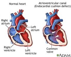
Cushion defect
|
|
|
In general, what is AVSD?
|

When both septum are open
|
|
|
Is AVSD more severe than ASD or VSD alone?
|
Yes-m
|
|
|
So does that mean AVSD results in more severe pulmonary edema than ASD or VSD alone?
|
Why, yes-m.
|
|
|
What is AVSD associated with?
|
Genetics
Trisomy 21 ( Down syndrome) |
|
|
Explain the process of a chronic complication of VSD.
|
1) Lungs overloaded with pressure and pulmonary edema
2) Lung vessels become stronger and develop protective mechanism 3) Pressure in lungs cause Right ventricle to work harder 4) Right ventricle hypertrophies 5) VSD shunt reverses if untreated (Eisenberger phenom) 6) Need surgery due to pulmonary hypertension (irreversible and fatal) |
|
|
List 5 right ---> left shunts. Which is MOST COMMON?
|
Tetrology of Fallot (ToF) (MOST COMMON)
Transposition of Great Vessels (TGV) Tricuspid Valve Atresia (TVA) Persistant Truncus Arteriosum (PTA) Total Anomalous Pulmonary Venous Return (TAPVR) |
|
|
What do all 5 right ---> left shunts have in common?
|
There abbreviations have "T" in them.
|
|
|
True or False. For any right ---> left condition, a PDA is required to keep the child alive.
|
True.
|
|
|
True or False. People will not survive right ---> left shunt without surgery.
|
True.
|
|
|
Explain the process of a ToF.
|

1) Stenosis of the pulmonic valve
2) Stenosis causes RIGHT ventricle hypertrophy 3) RIGHT ventricle hypertrophy causes VSD 4) Overriding aorta |
|
|
True or False. With a ToF, blood is less oxygenated than it should be.
|
True.
|
|
|
With a ToF, what becomes blue?
|
Lips, nose, fingers, toes...nice nursery rhyme.
Also around the eyes. |
|
|
True or False. The "blueness" in ToF and other cyanotic heart diseases becomes less pronounced with exertion.
|
False. The "blueness" in ToF and other cyanotic heart diseases becomes MORE pronounced with exertion.
|
|
|
With a ToF, how does the blood become oxygenated?
|
PDA opens to supply oxygen.
|
|
|
How is ToF diagnosed?
|
Ultrasound.
|
|
|
What is the treatment for ToF?
|
1) Open the stenosis
2) Close the aorta and move it to the left ventricle 3) Close the VSD |
|
|
Before treatment of ToF, what happens to the infant if the PDA closes?
|
They die.
|
|
|
What is the only thing that keeps patients with TGV alive? What is the treatment for TGV?
|
PDA keeps them alive. Treatment is surgery to place vessels in the proper location.
|
|
|
What is the MOST COMMON congenital defect of the tricuspid valve?
|
Tricuspid Valve Atresia (TVA)
|
|
|
What process occurs with a TVA?
|

1) Pressure builds in the Right atrium
2) Right atrium becomes enlarged 3) Right ventricle suffers HYPOplasia 4) PDA develops to connect to pulmonary cirulation |
|
|
What shape does the heart take on with TVA?
|
Boot shaped heart
|
|
|
What is another name for Persistant Truncus Arteriusus (PTA)?
|
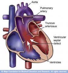
Frog Heart
|
|
|
Explain three characteristics of a PTA.
|

1) Pulmonary artery and aorta don't divide completely
2) Ventricles don't divide completely 3) There is one common valve with 4 cusps. |
|
|
Explain some characteristics of a Total Anomalous Pulmonary Venous Return (TAPVR).
|
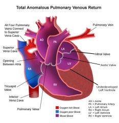
1) Pulmonary veins are not connected to Left atrium
2) To function, blood needs an ASD or VSD to move through. |
|
|
List 3 obstructive defects of the infant heart. Which is MOST COMMON?
|
Congenital Bicuspid Aortic Valve (MOST COMMON)
Aortic Valve Atresia Aortic Coarction |
|
|
What happens to the left ventricle in congenital bicuspid aortic valve?
|
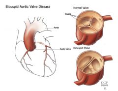
LVH
|
|
|
What risk does congenital bicuspid aortic valve place on patients?
|
The risk of arrhythmia.
|
|
|
Explain the process that occurs with Aortic Valve Atresia.
|
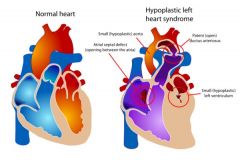
1) Atresia of the aortic valve
2) Left ventricle suffers HYPOplasia 3) Shunt develops from Right ventricle to aorta |
|
|
What are two types of aortic coarction? Which is more severe?
|
Proximal and distal. Proximal is more severe.
|
|
|
What is aortic coarction associated with? Why is this bad?
|
Turner's syndrome; It is the lack of one chromosome.
|
|
|
Explain the process of aortic coarction.
|
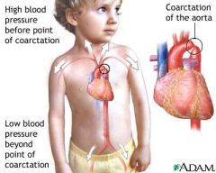
1) Obstruction occurs in the aorta
2) Compromised flow and lower pressure to lower extremities |
|
|
What is a complication of aortic coarction?
|
Rib notches from increased pressure in the mammary arteries. Can be seen in X-Ray.
|