![]()
![]()
![]()
Use LEFT and RIGHT arrow keys to navigate between flashcards;
Use UP and DOWN arrow keys to flip the card;
H to show hint;
A reads text to speech;
76 Cards in this Set
- Front
- Back
|
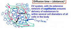
What is the primary purpose of capillaries in the circulatory system? |
|
|
|
How many veins bring blood back to the left atrium? |
Four Pulmonary Veins. |
|
|
Why is it important that the systemic blood vessels are arranged in parallel? |
Delivers blood with the same composition to all the tissues (O2 & CO2) Adequate perfusion pressure for each tissue, while allowing organs and tissues to adjust their own blood flow. |
|
|
What has to be true in steady state blood flow? |
Output from the RV & LV are equal CO=Venous Return |
|
|
What type of tissue is blood? |
Connective Tissue |
|
|
What percent of the body weight is blood? |
8% of body weight. 1 Liter of blood ~ 1 Kg Can estimate Total Blood Volume (TBV) by multiplying weight by 8% |
|
|
What is the composition of Plasma? |
Plasma proteins-7% Other Solutes-1% (electrolytes, organic nutrients and wastes) Contribute to Osmotic Pressure Water-92% |
|
|
What are the four Plasma Proteins? |
Albumin- made in liver, major contributor to Colloid pressure (7%) Globulins-made in liver, transport ions & immune function (35%) Fibrinogens-Made in liver, Clotting (4%) Regulatory Proteins-enzymes, proenzymes, & hormones (<1%) |
|
|
What are the only solutes important in fluid exchange across capillaries? |
Plasma Proteins There is a continuous bulk flow of protein-free ECF across capillaries, proteins exert colloid osmotic pressure. |
|
|
What are the types of WBCs? |
Neutrophils lymphocytes monocytes eosinophils basophils |
|
|
Blood consists of plasma and formed elements, what are the formed elements and their percentages? |
RBCs-99.9% Platelets and WBCs-0.1% |
|
|
What are the characteristics of WBCs? |
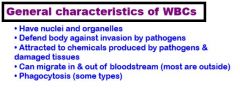
|
|
|
What is the most plentiful cell type in the body? |
RBCs ~25 trillion in the body |
|
|
Describe the structures and functions of the RBCs? |
Biconcave disc providing large membrane surface area (Fick's Law)
No nucleus or Organelles-short lifespan & no O2 consumption
Contains enzyme Carbonic Anhydrase for the transport of CO2 |
|
|
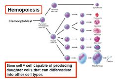
What forms the different formed elements of the blood? |
All elements are derived from the same type of stem cell in the red bone marrow (hemocytoblasts). |
|
|

|
|
|
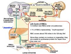
What happens at the end of the RBCs lifespan? |
Macrophages (mostly in spleen) detect worn out membranes and engulfs the RBC (phagocytosis) Globin is digested into component amino acids Iron of heme is recycled (transported by transferrin) Non-iron portion of heme is converted into bilirubin released into blood and cleared by liver |
|

|
|
|
|
What controls the production of RBCs? |
Regulated by erythropoietin-produced in Kidneys Low O2 concentrations cause endocrine cells to release erythropoietin Must have amino acids (globin), iron (heme), B12, folic acid, and other nutrients |
|
|
What is the hematocrit level for: Normal, anemia, polycythemia, and dehydration? |
Normal-45% Anemia<30% Polycythemia-70% Dehydration-70% |
|
|
What are seven types of anemia? |
Nutritional-deficient in Fe, B12, Folic Acid, etc Pernicious-Lacking intrinsic factor in stomach to absorb B12 Renal-insufficient EPO from kidneys Aplastic-failure of hematopoietic stem cells (toxins, radiation, chemo) Hemorrhagic- acute or chronic Hemolytic-rupture of RBCs Sickle Cell |
|
|
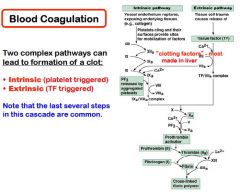
What are the three phases of Hemostasis? |
Vascular spasm Platelet Plug formation Coagulation |
|
|
What are the three layers of the vessels? |
Tunica Interna-single layer of simple squamous endothelium (elastic connective tissue just outside which allows for stretch & recoil) Tunica Media-smooth muscle cells arranged in circle around lumen Tunica Externa-collagen and elastin fibers |
|
|
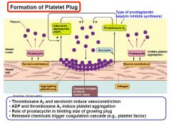
What causes vasospasm in hemostasis? |
Seratonin and Thromboxin A2 released by damaged endothelial cells and platelets causing constriction. May possibly have a sympathetic response to pain |
|
|
What happens in platelet plug formation? |
|
|
|
Describe the intrinsic, extrinsic, and common pathways. |
|
|
|
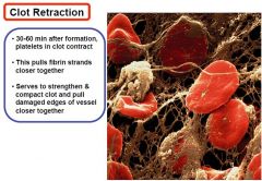
What is Clot retraction? |
|
|
|
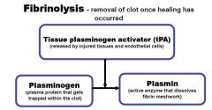
How is a clot brokendown (Fibrinolysis)? |
|
|
|
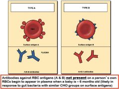
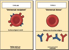
What are blood type antigens and antibodies? |
Antigens are on the surface of the cell and trigger immune responses. (Type A has A antigens, O has no antigens) Antibodies are proteins that bind to foreign antigens. |
|
|
|
|
|
When does a baby develop antigens and what is likely the cause? |
At 6 months of age, likely in response to gut bacteria with similar CHO groups. |
|
|
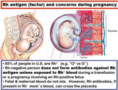
What are the percentages of Rh antigens, and when are they an issue? |
|
|
|
How do you prevent Hemolytic Disease of the Newborn (HDN)? |
Give the mother RhoGam (antibodies that destroy Rh+ fetal RBCs) at 28th week and after delivery. |
|
|
How can a change in the membrane potential be made? |
|
|
|
What are the two general types of Cardiac Action Potentials? |
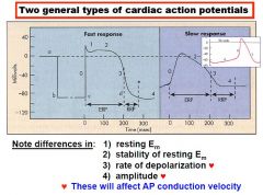
|
|
|
What is happening at each of the phases of the fast action cardiac potentials? |
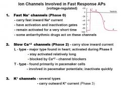
|
|
|
How are slow action potentials different? |
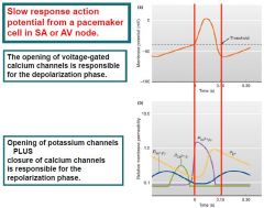
|
|
|
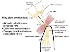
How is the conduction velocity of fast and slow response potentials different (which type of tissue do they conduct through)? |
Fast-purkinje fibers and atrial/ventricular muscle cells Slow-AV and SA nodes Different fiber diameter, rate of activation, and difference in amplitude of AP (greater amplitude the greater spread of Local current) |
|
|
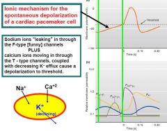
What is automaticity? |
Cells in the heart that have unstable membrane potential causing an AP. Occurs in total absence of any ANS or hormonal stimulation. SA 90-100 bpm AV 40-60 bpm purkinje fibers 14-40 bpm |
|
|
What is the mechanism of the changes in membrane potential that stimulates the AP? |
|
|
|
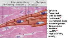
What is the histology of Cardiac Muscle Cells? |
Connected in series Connected end to end by intercalated discs Intercalated discs have gap junctions for quick propagation of APs. |
|
|

|
|
|
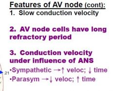
What is the function of the AV node? |
It is the only electrical connection between the atria and the ventricles. |
|
|
How does the AV node slow the conduction of an AP? |
|
|
|
What are other features that affect AV conduction? |
|
|
|
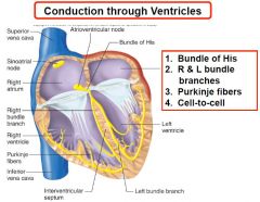
How does an AP conduct through the ventricles? |
|
|
|
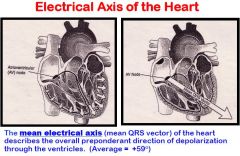
What is the normal axis of the heart conduction? |
|
|
|
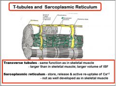
What are specifics of cardiac muscle that is specific to cardiac muscle? |
Has a high capillary density (dependent of aerobic metabolism) Layers that wrap around atria & spiral within the ventricles Many mitochondria |
|

|
|
|
|
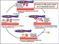
What is the cross-bridge cycle of cardiac muscle? |
|
|
|
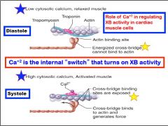
What is responsible for activation in cardiac muscle? |
|
|
|
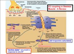
What is different between cardiac muscle when compared to skeletal muscle? |
In skeletal Enough Ca++ is released to bind to every single troponin activating every tropomysin.
In skeletal muscle every Ca++ comes from the sarcoplasmic Reticulum while in Cardiac ECF Ca++ is also involved. |
|

|
|
|
|
How is Ca++ removed after an AP? |
Most reabsorbed by active transport back into the SR. Some Ca++ is transported out of the cell by secondary active transport |
|
|
How is contraction of heart affected by ANS? |
in Absence of ANS stimulation not enough Ca++ is available to activate all cross bridges. ANS affects Ca++ availability. |
|
|
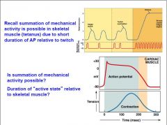
Can you have summation of APs in cardiac muscle? |
|
|
|
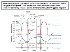
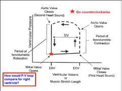
Describe the phases of the Wiggers Diagram. |
|
|
|
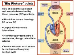
What are the big picture points of blood flow? |
|
|
|
What two factors affect aortic pressure? |
Compliance (constant) Volume of blood (major factor from beat to beat) |
|
|
What causes the a wave on the Wiggers Diagram? |
Atrial Muscle contraction causing a small increase in the left atrial pressure. |
|
|
What causes the c wave in the Wiggers Diagram that occurs during isovolumic contraction? |
The cusps of the semilunar valves bulge into the atria causing a slight increase in atrial pressure. |
|
|
When is the most rapid filling of the ventricle? |
as soon as the AV valves open. |
|
|
What are the volumes of blood during : End Diastolic Volume (EDV) End Systolic Volume (ESV) Stroke Volume (SV) Ejection Fraction (EF) |
EDV-110-120 ml ESV-40-50 ml SV- (EDV-ESV)-70 ml/bt EF- (SV/EDV)-60% |
|
|
What are factors that affect heart volumes? |
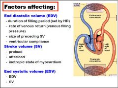
|
|
|
What is charted on the X & Y axis of the pressure volume loop? |
X-volume of blood in the LV y-ventricular pressure |
|

|
|
|
|
What is the cause of heart sounds? |
First-AV valves closing, Ventricular systole Second-Semilunar valves closing, ventricular diastole |
|

|
|
|
|
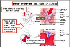
What causes a murmur and what does it symbolize? |
Turbulent blood flow Murmur heard during systole over right AV valve caused by regurgitation Murmur heard over pulmonary semilunar valves caused by pulmonic stenosis |
|
|
When does the heart receive its perfusion? |
Coronary arteries receive perfusion during diastole. |
|
|
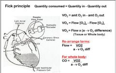
How is cardiac output measured (steady State) using the Fick method? CO is volume of blood pumped by each ventricle per minute (L/min). |
|
|
|
How do you calculate Cardiac Reserve? |
Cardiac Reserve=Max CO-Resting CO |
|
|
What determines Cardiac Output? |
CO=HRxSV Ultimately venous return (VR) determines CO |
|
|
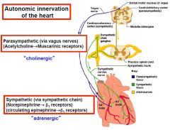
How is the ANS innervating the heart? |
|
|
|
Where does sympathetic nervous system innervation start? |
Originate in the lateral horn from T1-T6 Affects mainly heart rate and force of contraction. Right side SNS mainly pacemaker and conduction system, left to myocardium |
|
|
Where does parasympathetic nervous system innervation start?
|
Vagus Nerve to medulla Right side of PNS innervation goes to the SA node, Left side goes to the AV node Affects mainly heart rate and conduction velocity |