![]()
![]()
![]()
Use LEFT and RIGHT arrow keys to navigate between flashcards;
Use UP and DOWN arrow keys to flip the card;
H to show hint;
A reads text to speech;
16 Cards in this Set
- Front
- Back
- 3rd side (hint)
|
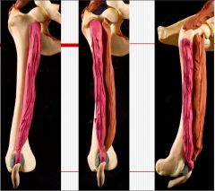
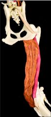
Three femoral heads of the quadriceps group |
Vastus medialis m. O: Proximal end of the craniomedial femur. Vastus intermedius m. O: Lateral part of the proximal 1/4 of the femur. Vastus lateralis m. O: Proximal lateral femur. All 3 femoral heads: I: Tibial tuberosity A: Extend the stifle joint. N: Femoral nerve |
|
|
|
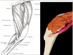
Rectus femoris m. |
O: Ilium, cranial to the acetabulum. I: Tibial tuberosity A: Extend the stifle joint and flex the hip joint (the only head of the quads to do this) N: Femoral nerve |
|
|
|
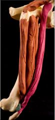
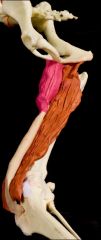
Semimembranosus m. |
O: Ischiatic tuberosity I: Distal medial femur and proximal end of tibia. A: Extend the hip joint. N: Sciatic nerve |
|
|
|
Semimembranosus m. |
A: The part inserting on the tibia can flex or extend the stifle, depending upon the position of the limb. |
|
|
|
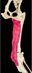
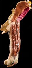
Semitendinosus m. |
O: Ischiatic tuberosity I: Medial surface of the body of the tibia and tuber calcanei via the crural fascia A: Extend the hip joint, flex the stifle joint, extend the tarsal joint. N: Sciatic nerve |
|
|
|
Semitendinosus m. |
A: Extend the hip joint, flex the stifle joint, extend the tarsal joint.
|
|
|
|
Gracilis m. |
O: Pubic symphysis I: Cranial border of the tibia and, with the semitendinosus muscle, the tuber calcaneus. A: Adduct the limb, flex the stifle joint, extend the hip and tarsal joints. |
|
|
|
Tensor fasciae latae |
O: Tuber coxae and adjacent ilium. I: Lateral femoral fascia A: Tense the femoral fascia, flex the hip joint, extend the stifle joint. |
|
|
|
Sartorius m. |
2 heads O: Cranial head: Medial crest of the ilium. Caudal head: Cranial ventral iliac spine. I: Cranial: Patella, with the quadriceps muscles Caudal: Cranial border of the tibia, with the gracilis m. A: Flex the hip (both heads) Cranial: also extends the stifle joint Caudal: also flexes the stifle joint. N: Femoral nerve |
|
|
|
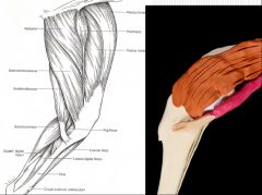
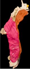
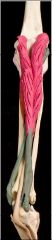
Biceps femoris m. |
O: Sacrotuberous ligament and ischiatic tuberosity I: By means of fascia lata, to patella, patellar ligament and the cranial border of the tibia; by means of crural fascia, the tuber calcanei. A: Extend the hip, stifle and tarsal joints. Caudal part flexes the stifle joint. N: Sciatic nerve |
|
|
|
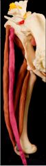
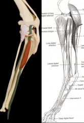
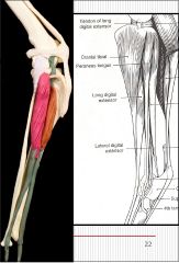
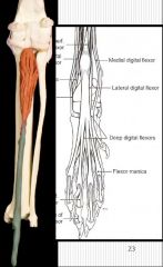
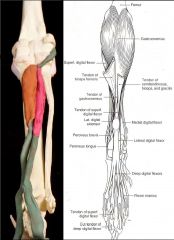
Long digital extensor m. |
O: Extensor fossa of the femur I: Extensor processes of distal phalanges of digits II-V A: Extend digits and flex the tarsal joint. |
|
|
|
Peroneus longus m. |
O: Lateral condyle of tibia, proximal end of fibula. I: 4th tarsal bone, plantar aspect of base of the metatarsals. A: Flex tarsal joint and rotate dorsum of paw medially, so plantar surface faces laterally. |
|
|
|
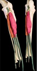
Deep digital flexor m. |
O: Plantar aspect of proximal 2/3 of tibia and proximal 1/2 of fibula. I: Plantar surface of base of each of the distal phalanges. A: Flex digits and extend the tarsal joint. |
|
|
|
Cranial tibial m. |
O: Lateral edge of cranial tibial border. I: Plantar surface of base of metatarsals I and II, with peroneus longus m. A: Flex tarsal joint and rotate dorsum of paw laterally, so plantar aspect of paw faces medially. |
|
|
|
Superficial digital flexor m. |
O: Distal caudal femur I: Tuber calcaneus (with the calcaneal tendon) and bases of middle phalanges of digits II-V A: Flex the proximal two digital joints of the four principal digits; flex the stifle joint, extend the tarsal joint. |
|
|
|
Gastrocnemius m. |
O: Medial and lateral supracondylar tuberosities of femur, over the fabellae. I: Proximal dorsal surface of tuber calcaneus. A: Extend tarsal joint, flex stifle joint. |
|