Reading...
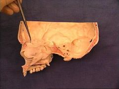
![]()
Play button
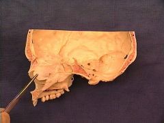
![]()
Play button
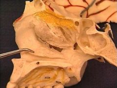
![]()
Use LEFT and RIGHT arrow keys to navigate between flashcards;
Use UP and DOWN arrow keys to flip the card;
H to show hint;
A reads text to speech;
390 Cards in this Set
- Front
- Back
- 3rd side (hint)
|
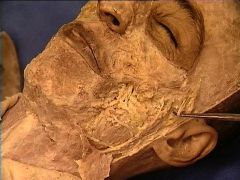
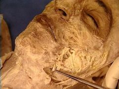
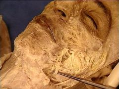
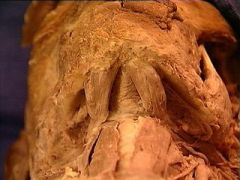
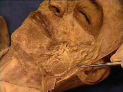
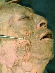
Platysma
|
|
o Origin: superficial fascia over pectoralis major and deltoid
o Insertion: mandible; muscles around mouth o Innervation: CN VII o Action: depresses mandible, draws lower lip downward, tenses skin of neck |
|
|
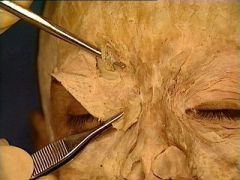
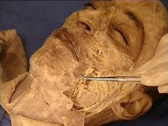
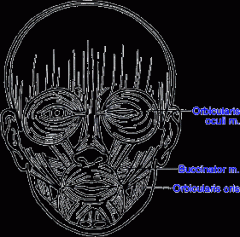
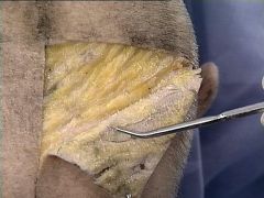
Orbicularis Oculi
|
|
• O: medial orbital margin, medial palpebral ligament, lacrimal bone
• I: skin around margin of orbit, tarsal plate • A: close eyelid |
|
|
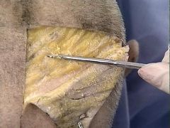
Temporalis
|
|
o Origin: inferior temporal line, temporal fossa, temporalis fascia
o Insertion: coronoid process; anterior border of mandibular ramus o Innervation: deep temporal branches of V3 o Action: anterior fibers-elevate mandible; posterior fibers-retrude mandible |
|
|
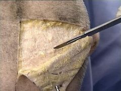
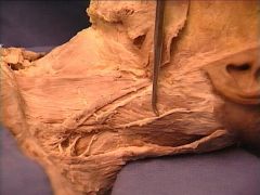
Masseter
|
|
o Origin: zygomatic arch/maxilla
o Insertion: coronoid process/ramus mandible o Innervation: CN V o Action: elevates and retracts the mandible (closes jaw) |
|
|
Masseter
|
|
o Origin: zygomatic arch/maxilla
o Insertion: coronoid process/ramus mandible o Innervation: CN V o Action: elevates and retracts the mandible (closes jaw) |
|
|
Buccinator
|
|
• O: mandible, pterygomandibular raphe, alveolar process of maxilla and mandible
• I: angle of mouth • A: press cheek against molar teeth to keep food between teeth, expel air from oral cavity |
|
|
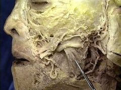
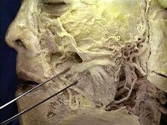
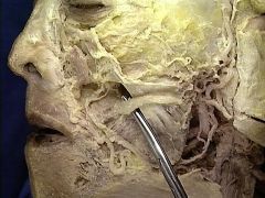
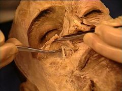
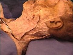
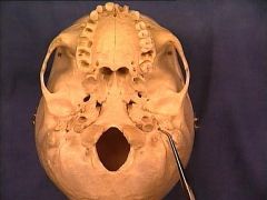
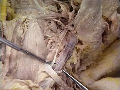
Parotid (Stenson's) Duct
|
|
found along the edge of the parotid gland; exits from the anterior border of the gland and passing about a finger’s breadth below the zygomatic arch over the superficial fibers of masseter, the duct makes a sharp turn over anterior border of masseter to perforate buccinator and enter the oral cavity (around the max 2nd molar)
|
|
|
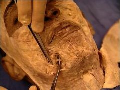
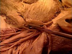
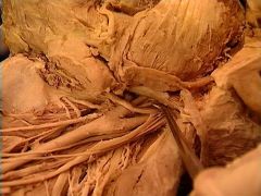
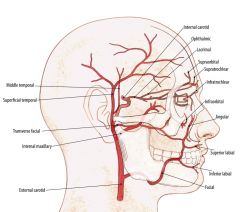
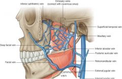
Facial Artery and Vein
|
|
• Facial artery: arises in carotid triangle from ECA, ascends deep to submandibular gland, winds around inferior border of mandible and enters the face (torturous which allows for distention and opening of the jaw); distributes to the muscles of facial expression/face
Facial vein: direct continuation of angular vein past inferior margin of orbit; descends along lateral border of the nose, receiving external nasal and inferior palpebral veins, then obliquely across face to mandible. It receives anterior division of retromandbiular vein, after which it is sometimes called the common facial vein. |
|
|
Facial Nerve - Cervical Branch
|
|
• Cervical:
• Runs forward beneath platysma; one branch descends to join cervical cutaneous nerve from the cervical plexus, which innervates Platysma |
|
|
Facial Nerve - Mandibular Branch
|
|
• Marginal mandibular:
• Innervates muscles of lower lip & chin; [communicates with mental branch of inferior alveolar branch] |
|
|
Facial Nerve - Buccal Branch
|
|
Runs laterally over the Masseter muscle;• Superficial branches innervate procerus; [join with infratrochlear and nasociliary branches of V1]
• Deep branches innervate zygomaticus and levator labii superioris & nasalis; [form infraorbital plexus with infraorbital branch of V1] • Lower deep branches innervate buccinators & orbicularis oris; [join with fibers of buccinator branch of V3] |
|
|
Facial Nerve - Zygomatic Branch
|
|
• Innervates orbicularis oculi
• [Joins with fibers of lacrimal n. and zygomaticofacial branch of V2] |
|
|
Facial Nerve - Temporal Branch
|
|
• Innervates auriculares anterior & superior, [and join with zygomaticotemporal branch of V2 & auriculotemporal branch of V3]
• Anterior branches innervate frontalis, orbicularis oculi, corrugator supercilii, [and join the supraorbital & lacrimal branches of V1] |
|
|
Opthalmic Nerve (V1) -> Frontal Nerve -> Supraorbital Nerve (through Supraorbital Foramen)
|
|
|
|
|
Maxillary Nerve (V2) - Infraorbital Nerve (through Infraorbital Foramen) NVB
|
|
infraorbital NVB exits here (anterior superior and middle superior alveolar nerve/artery branch off before exit)
|
|
|
Mandibular Nerve (V3) -> Inferior Alveolar Nerve -> Mental Nerve (through Mental Foramen)
|
|
|
|
|
Facial Artery
|
|
arises in carotid triangle from ECA, ascends deep to submandibular gland, winds around inferior border of mandible and enters the face (torturous which allows for distention and opening of the jaw); distributes to the muscles of facial expression/face
|
|
|
Facial Vein
|
|
direct continuation of angular vein past inferior margin of orbit; descends along lateral border of the nose, receiving external nasal and inferior palpebral veins, then obliquely across face to mandible. It receives anterior division of retromandbiular vein, after which it is sometimes called the common facial vein.
It terminates at the internal jugular vein, and drains the anterior scalp and forehead, eyelids, external nose, anterior cheek, lips, chin, and submandibular gland. and receives drainage from external palatine vein; joins either the anterior branch of retromandibular vein OR drains directly into internal jugular vein |
|
|
Platysma
|
|
o Origin: superficial fascia over pectoralis major and deltoid
o Insertion: mandible; muscles around mouth o Innervation: CN VII o Action: depresses mandible, draws lower lip downward, tenses skin of neck |
|
|
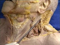
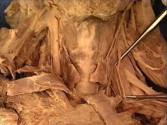
Sternocleidomastoid
|
|
o Origin: sternal head: anterior manubrium; clavicular head: medial surface of clavicle
o Insertion: lateral surface of mastoid process and lateral portion of superior nuchal line o Action: bilaterally – flexion of cervical vertebral column anteriorly, or when supine, raises head; unilaterally – tilt s head toward ipsilateral shoulder and rotates head so that chin faces the opposite side o Innervation: Spinal XI [and C3, C4 ventral rami (sensory)] |
|
|
Trapezius
|
|
|
|
|
Spinal Accessory Nerve
|
|
nerve emerges from the jugular foramen, crosses the internal jugular vein within the posterior triangle and innervates SCM and trapezius
|
|
|
Omohyoid (Inferior Belly)
|
|
O: upper border of scapula and suprascapular lig.
I: lower border body of hyoid A: depresses hyoid bone I: ansa cervicalis (C1-C3) |
|
|
Omohyoid (Superior Belly)
|
|
superior belly: central tendon located deep to SCM; here, superior belly unites with inferior belly
I: lower border body of hyoid A: depresses hyoid bone I: ansa cervicalis (C1-C3) |
|
|
External Jugular Vein
|
|
o Formed by the union of the posterior auricular vein and the posterior branch of the retromandibular vein; passes obliquely downward over SCM to enter the posterior triangle, where it receives three tributaries: transverse cervical vein, suprascapular vein, and anterior jugular vein
o Drains into the subclavian vein. |
|
|
Subclavian Vein
|
|
courses over the anterior border of the anterior scalene and it become the axillary vein once it passes the 1st rib
|
|
|
Scalenus Anterior Muscle (Anterior Scalene)
|
|
(separates subcalvian artery and branchial plexus)
o O: transverse processes of cervical vertebrae 3 thru 6 o I: superior aspect of 1st rib at scalene tubercle o A: bends cervical portion of vertebral column antero-laterally and rotates it towards the opposite side/elevates 1st rib o I: ventral rami of C4 thru C6 |
|
|
Scalenus Medius Muscle (Middle Scalene)
|
|
o O: transverse processes of cervical vertebrae 2 thru 7
o I: superior aspect of 1st rib o A: bends the vertebral column to the same side; elevates 1st rib o I: ventral rami of C3 thru C6 |
|
|
Brachial Plexus Trunks
|
|
roots and trunks of brachial plexus pass through scalene interval anterior to middle scalene
|
|
|
Phrenic Nerve (C3, C4, C5)
|
|
ventral rami of C3-C5 which innervate the diaphragm
|
|
|
Transverse Cervical Artery (Superior) and Suprascapular Artery (Inferior)
|
|
branches off of thyrocervical trunk
• Transverse Cervical • supplies trapezius, serratus anterior, muscles of rotator cuff (teres major, supraspinatus, infraspinatus, subscapular muscles) • Suprascapular • supplies SCM and Supraspinitus |
|
|
Thyrocervical Trunk
|
|
• Inferior thyroid: crosses deep to carotid sheath to reach the thyroid gland
• Dorsal scapular: supplies latissimus dorsi, levator scapulae, trapezius, and rhomboids • Suprascapular: supplies SCM and supraspinitus (travels w/same n.) • Transverse Cervical • supplies trapezius, serratus anterior, muscles of rotator cuff (teres major, supraspinatus, infraspinatus, subscapular muscles) |
|
|
Branches of Facial Nerve
|
|
Two Zebras Bit My Cookies
Temporal, Zygomatic, Buccal, Marginal Mandibular, Cervical (& Posterior Auricular) |
|
|
Zygomatic Arch
|
|
masseter muscle originates here
|
|
|
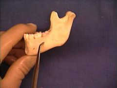
Angle of Mandible
|
|
attachment of stylomandibular ligament, m pterygoid, masseter
|
|
|
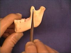
Coronoid Process
|
|
masseter and temporalis insert here
|
|
|
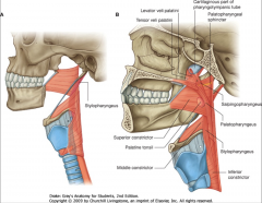
Medial Pterygoid Muscle
|
|
• Origin – deep head (medial side of the lateral pterygoid plate); superficial head (pyramidal process of palatine bone and maxillary tuberosity)
• Insertion – medial surface of the ramus and angle of the mandible • Innervation – nerve to medial pterygoid of V3 • Action – elevates mandible; helps lateral pterygoids in lateral movement |
|
|
Lateral Pterygoid Muscle
|
|
o Origin: greater wing of sphenoid; lateral pterygoid plate
o Insertion: pterygoid fovea; articular disk/meniscus of TMJ (neck of condyle) o Innervation: lateral pterygoid n of V3 o Action: depress mandible; protrude mandible; lateral excursion |
|
|
Lateral Pterygoid Muscle
|
|
o Origin: greater wing of sphenoid; lateral pterygoid plate
o Insertion: pterygoid fovea; articular disk/meniscus of TMJ (neck of condyle) o Innervation: lateral pterygoid n of V3 o Action: depress mandible; protrude mandible; lateral excursion |
|
|
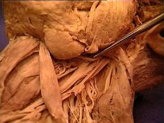
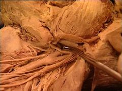
Lingual Nerve
|
|
Between medial and lateral pterygoids. Courses inferiorly on the superficial aspect of medial pterygoid. Adjacent to inferior alveolar nerve (Superior). Innervates anterior 2/3 of tongue; parasympathetic fibers to submandibular ganglion
|
|
|
Lingual Nerve
|
|
Between medial and lateral pterygoids. Courses inferiorly on the superficial aspect of medial pterygoid. Adjacent to inferior alveolar nerve (Superior). Innervates anterior 2/3 of tongue; parasympathetic fibers to submandibular ganglion
|
|
|
Mandibular Nerve (CN V3)
|
|
Branches of Trigeminal nerve; Branches: BAIL (Long Buccal, Auriculotemporal, Inferior Alveolar, Lingual)
|
|
|
Chorda Tympani
|
|
Emerges from petrotympanic fissure, passes anteriorly and joins the Lingual nerve
|
|
|
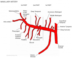
Maxillary Artery
|
|
One of two terminal branches of External Carotid Artery; runs deep to condylar neck; 3 parts: mandibular, pterygoid, pterygopalatine
|
|
|
Maxillary Artery
|
|
One of two terminal branches of External Carotid Artery; runs deep to condylar neck; 3 parts: mandibular, pterygoid, pterygopalatine
|
|
|
Inferior Alveolar Artery (of Maxillary Artery)
|
|
Travels with inferior alveolar nerve and vein to mandibular foramen
|
|
|
Middle Meningeal Artery (of Maxillary Artery)
|
|
1st branch off of maxillary artery (1 of two terminal branches off ECA); passes through foramen spinosum to enter middle cranial fossa; ascends lateral walls of skull and branches to anterior and posterior
branches |
|
|
Middle Meningeal Artery (of Maxillary Artery)
|
|
1st branch off of maxillary artery (1 of two terminal branches off ECA); passes through foramen spinosum to enter middle cranial fossa; ascends lateral walls of skull and branches to anterior and posterior
branches |
|
|
1st part of Maxillary Artery
|

|
Mandibular portion of Maxillary Artery
|
|
|
2nd part of Maxillary Artery
|
|
Pterygoid part of the Maxillary Artery
|
|
|
Posterior Superior Alveolar Artery
|
|
Branch of pterygopalatine segment of Maxillary artery
|
|
|
Left Middle Meningeal Artery
|
|
Supply blood to dura mater and cranial bones
|
|
|
Cephalic Vein
|
|
|
|
|
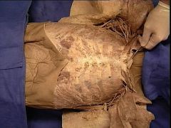
External Intercostal Muscle
|
|
|
|
|
Internal Intercostal Muscle
|
|
|
|
|
Costal Cartilage
|
|
|
|
|
Intercostal NVB (Nerve (shown) + Artery + Vein)
|

|
Located in costal grooves of all ribs; VAN - Vein, Artery, Nerve from superior to inferior
|
|
|
Innermost Intercostal Muscle (deep and lateral to probe)
|
|
Deep to Internal Intercostal Muscle.
|
|
|
Manubrium
|
|
|
|
|
Body of Sternum
|
|
|
|
|
Sternal Angle
|
|
|
|
|
Jugular Notch
|
|
|
|
|
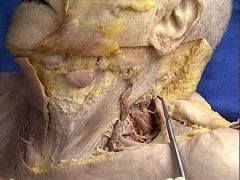
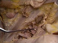
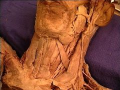
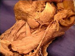
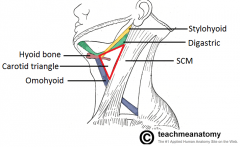
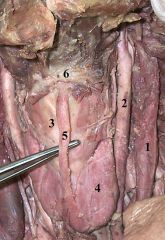
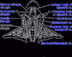
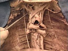
Anterior Cervical Triangle: 1) Inferior Border of Mandible (superior), 2) Medial Border of SCM (posterior), 3) Midline of Neck (anterior)
|
|
|
|
|
Carotid Triangle: 1) Posterior Belly of Digastric (superior), 2) Medial Border of SCM (lateral), 3)
|
|
|
|
|
Submental Triangle: 1) Hyoid Bone (inferior), 2) Midline of Neck (medial), 3) Anterior Belly of Digastric (lateral)
|
|
|
|
|
Submandibular Triangle: 1) Body of Mandible (superior), 2) Anterior Belly of Digastric (anterior), 3) Posterior Belly of Digastric (posterior)
|
|
|
|
|
Posterior Belly of Digastric
|
|
• O: anterior belly: digastric fossa of mandible; posterior belly: mastoid notch of temporal bone
• I: tendinous connection of both bellies through a fascial loop on the hyoid bone • A: depresses mandible/elevates hyoid • I: anterior belly: branch of mylohyoid of V3; posterior belly: branch of CN VII |
|
|
Anterior Belly of Digastric
|
|
• O: anterior belly: digastric fossa of mandible; posterior belly: mastoid notch of temporal bone
• I: tendinous connection of both bellies through a fascial loop on the hyoid bone • A: depresses mandible/elevates hyoid • I: anterior belly: branch of mylohyoid of V3; posterior belly: branch of CN VII |
|
|
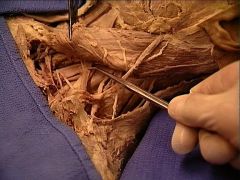
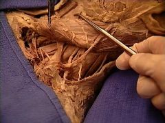
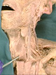
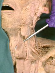
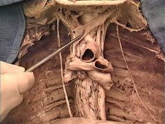
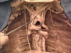
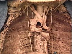
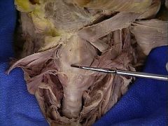
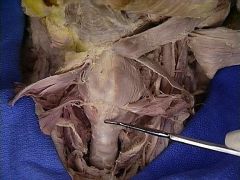
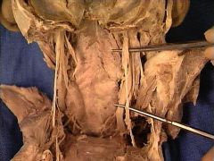
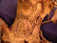
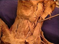
Neck Region: 1) Internal Jugular Vein, 2) Common Carotid Artery, 3) Thyroid Cartilage, 4) Thyroid Gland, 5) Thyroglossal Duct, 6) Hyoid Bone
|
|
|
|
|
Sternohyoid
|
|
O: posterior manubrium, sternoclavicular lig., and medial end of clavicle
I: medial lower body of hyoid A: depresses hyoid bone I: ansa cervicalis (C1-C3) |
|
|
Sternocleidomastoid
|
|
o Origin: sternal head: anterior manubrium; clavicular head: medial surface of clavicle
o Insertion: lateral surface of mastoid process and lateral portion of superior nuchal line o Action: bilaterally – flexion of cervical vertebral column anteriorly, or when supine, raises head; unilaterally – tilt s head toward ipsilateral shoulder and rotates head so that chin faces the opposite side o Innervation: Spinal XI [and C3, C4 ventral rami (sensory)] |
|
|
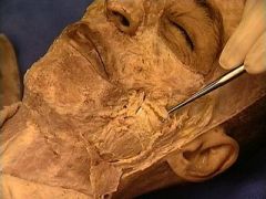
Orbicularis Oris
|
|
• O: some fibers near medial plane of maxilla superiorly and mandible inferiorly, deep surface of skin
• I: mucous membrane of lips • A: compresses and protrudes lips |
|
|
Frontal Bone
|
|
|
|
|
Parietal Bone
|
|
|
|
|
Occipital Bone
|
|
|
|
|
Temporal Bone
|
|
|
|
|
Sphenoid Bone
|
|
|
|
|
Ethmoid Bone
|
|
|
|
|
Zygomatic Bone
|
|
|
|
|
Maxilla Bone
|
|
|
|
|
Nasal Bone
|
|
|
|
|
Lacrimal Bone
|
|
|
|
|
Vomer Bone
|
|
|
|
|
Palatine Bone
|
|
|
|
|
Inferior Nasal Concha
|
|
|
|
|
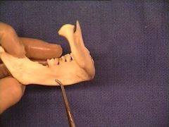
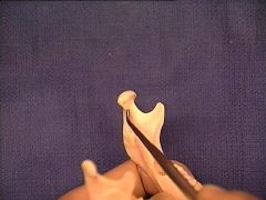
Mandible Bone
|
|
|
|
|
Mental Foramen
|
|
|
|
|
Infraorbital Foramen
|
|
|
|
|
Supraorbital Foramen
|
|
|
|
|
Orbital Plate of Frontal Bone
|
|
|
|
|
Coronal Suture
|
|
|
|
|
Superior Orbital Fissure
|
|
|
|
|
Inferior Orbital Fissure
|
|
|
|
|
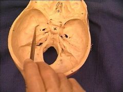
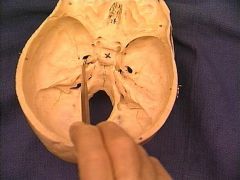
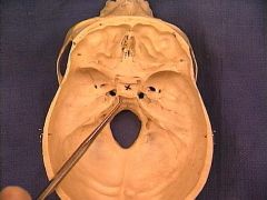
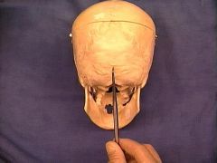
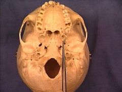
Hypoglossal Canal
|
|
|
|
|
Middle Concha
|
|
|
|
|
Mental Symphysis
|
|
|
|
|
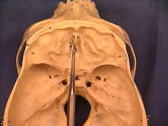
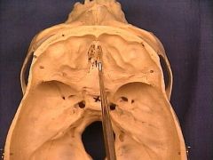
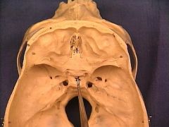
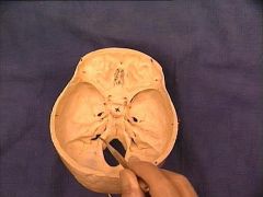
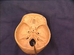
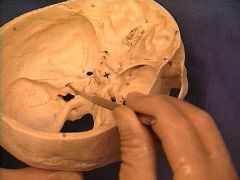
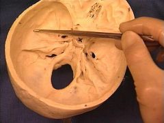
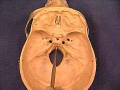
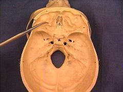
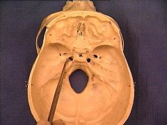
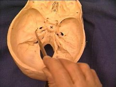
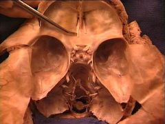
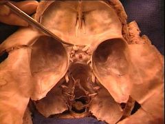
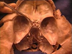
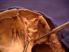
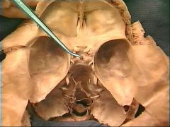
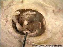
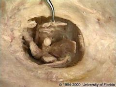
Anterior Cranial Fossa
|
|
|
|
|
Middle Cranial Fossa
|
|
|
|
|
Posterior Cranial Fossa
|

|
|
|
|
Crista Galli
|
|
|
|
|
Cribriform Plate
|
|
part of ethmoid bone; olfactory bulbs sit here - rootlets pass through perforations in plate to reach nasal epithelium in nasal cavity
|
|
|
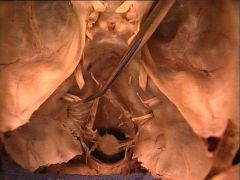
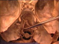
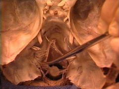
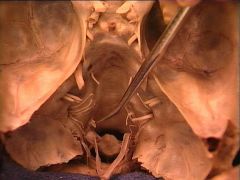
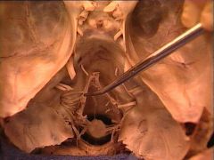
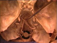
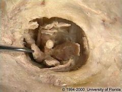
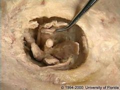
Pituitary/Hypophyseal Fossa (Sella Turcica)
|
|
|
|
|
Groove for Transverse Sinus
|
|

lateral from IOP, Occipital: in tentorium cerebelli; Right: larger, drains superior sagittal; Left: drains straight; drains to internal jugular vein
|
|
|
Groove for Sigmoid Sinus
|

|

continue from transverse sinuses and end at the jugular f.
|
|
|
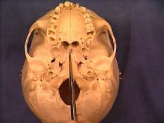
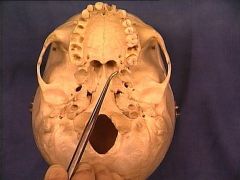
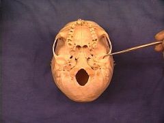
Jugular Foramen
|
|
CN IX, X and XI pass through it
|
|
|
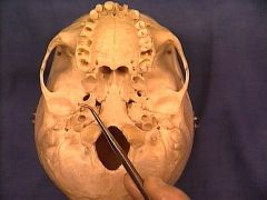
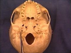
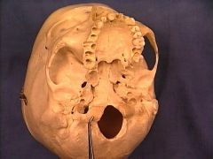
Foramen Magnum
|
|
|
|
|
Internal Acoustic Meatus
|
|
CN VII and VIII pass through it
|
|
|
Petrous Part of Temporal Bone
|

|
|
|
|
Squamous Part of Temporal Bone
|
|
|
|
|
Clivus
|
|
|
|
|
Orbital Plate of Frontal Bone
|
|
|
|
|
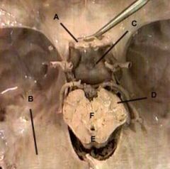
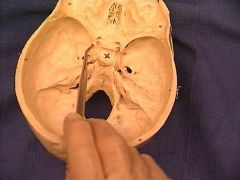
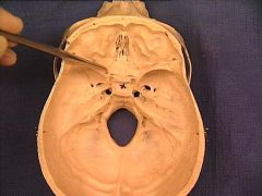
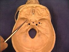
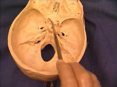
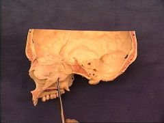
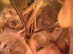
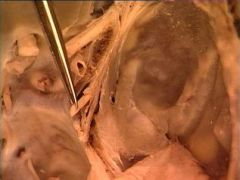
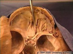
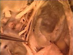
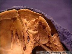
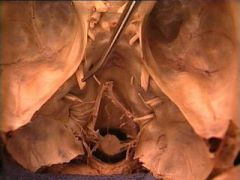
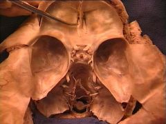
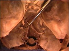
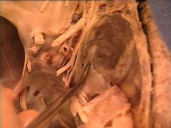
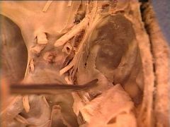
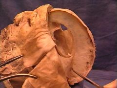
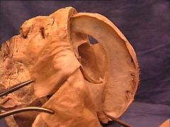
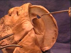
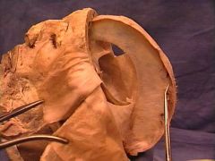
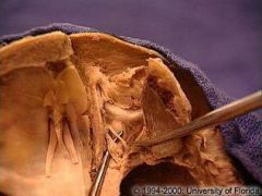
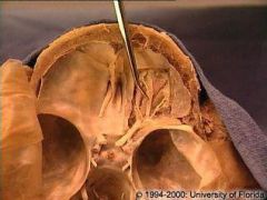
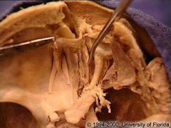
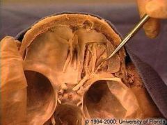
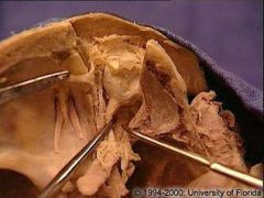
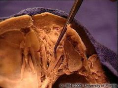
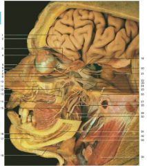
Middle Cranial Fossa Structures
A. Optic nerve B. Tentorium cerebelli C. Diaphragma sellae D. Cerebral peduncles E. Tectum F. Tegmentum |
|
Tentorium cerebelli – horizontal fold across posterior third of skull to separate cerebral hemispheres (occipital lobes) from cerebellum, attaches along transverse sulcus on each side of skull, and attaches along superior petrosal sulcus and ends medially at posterior clinoid process
Diaphragma sellae – dura mater membrane covering sella turcica; stretches from anterior clinoid processes to posterior clinoid processes; pierced by the infundibulum (stalk of pituitary gland) |
|
|
Foramen Lacerum
|
|
Inferior to carotid canal
Greater petrosal nerve-heads to foramen lacerum, enters the pterygoid canal and joins the nerve of the deep petrosal to form the nerve of the pterygoid canal. |
|
|
Foramen Spinosum
|
|
• Middle meningeal artery: passes through foramen spinosum
|
|
|
Groove for Middle Meningeal Artery
|
|
supplies dura of anterior and middle cranial fossae; 1st branch off of maxillary artery (1 of two terminal branches off ECA); passes through foramen spinosum to enter middle cranial fossa; ascends lateral walls of skull and branches to anterior and posterior
branches |
|
|
Foramen Ovale
|
|
V3 passes through
|
|
|
Posterior Clinoid Process
|
|
posterior are lateral ends of dorsum sellae
|
|
|
Foramen Rotundum
|
|
V2 passes through
|
|
|
Anterior Clinoid Process
|
|
anterior are medial ends of lesser
wings of sphenoid bone |
|
|
Lesser Wing of Sphenoid
|
|
|
|
|
Optic Canal
|
|
|
|
|
Lamboidal Suture
|
|
|
|
|
Pterion
|
|
|
|
|
Greater Wing of Sphenoid
|
|
|
|
|
Zygomaticofacial Foramen
|
|
|
|
|
Pterygomaxillary Fissure
|
|
|
|
|
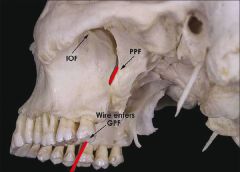
Pterygopalatine Fossa
|
|
small triangular fossa at the angle of the junction between the pterygomaxillary and inferior orbital fissures. It contains the pterygopalatine ganglion, maxillary nerve (V2), and terminal parts of the internal maxillary artery. Six foramina open into it: foramen rotundum, pterygoid canal, pharyngeal canal, sphenopalatine foramen (which transmits the sphenopalatine artery into the nasal cavity), pterygopalatine foramen, and the inferior orbital fissure
|
|
|
Sagittal Suture
|
|
|
|
|
Superior Nuchal Line
|
|
|
|
|
External Occipital Protuberance
|
|
|
|
|
Greater Palatine Foramen
|
|
|
|
|
Lesser Palatine Foramen
|
|
|
|
|
Infratemporal Fossa
|
|
|
|
|
Foramen Ovale
|

|
anterior and medial to foramen spinosum
|
|
|
Foramen Spinosum
|
|
inferior and lateral to foramen ovale
|
|
|
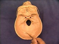
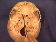
Carotid Canal
|
|
posterior to foramen spinosum; superior to foramen lacerum
|
|
|
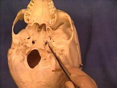
Stylomastoid Foramen
|
|
facial neve (CN VII) exits here
|
|
|
Jugular Foramen
|

|
|
|
|
Occipital Condyle
|
|
|
|
|
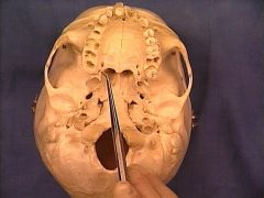
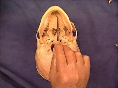
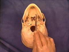
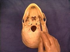
Incisive Foramen
|
|
Nasopalatine nerve enters roof of the mouth through the Incisive foramen
|
|
|
Palatine Plate of Maxilla
|
|
|
|
|
Maxillary Tuberosity
|
|
|
|
|
Vomer
|
|
|
|
|
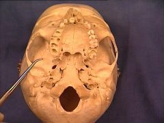
Medial Pterygoid Plate and Hamulus
|
|
|
|
|
Lateral Pterygoid Plate
|
|
|
|
|
Mandibular/Glenoid Fossa
|
|
|
|
|
Pterygoid/Vidian Canal
|
|
|
|
|
Pharyngeal Tubercle
|
|
median pharyngeal raphe attaches superiorly to the pharyngeal tubercles
|
|
|
Spine of Sphenoid Bone
|
|
|
|
|
Petrous Part of Temporal Bone
|
|
|
|
|
Hypoglossal Canal
|
|
|
|
|
Stylomastoid Foramen
|
|
|
|
|
Condylar Process
|
|
|
|
|
Neck of Mandible
|
|
|
|
|
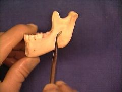
Angle of Mandible
|
|
|
|
|
Body of Mandible
|
|
|
|
|
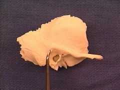
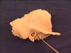
Ramus of Mandible
|
|
|
|
|
Coronoid Process of Mandible
|
|
|
|
|
Mental Foramen
|
|
|
|
|
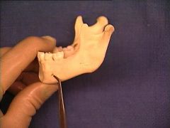
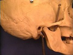
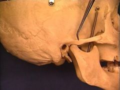
Lingula
|
|
|
|
|
Mylohyoid Line
|
|
|
|
|
Mental Spines/Genial Tubercles
|
|
|
|
|
Mandibular Foramen
|
|
|
|
|
Submandibular Fossa
|
|
|
|
|
Pterygoid Fovea
|
|
|
|
|
Vomer
|
|
|
|
|
Perpendicular Plate of Ethmoid Bone
|
|
|
|
|
Crista Galli
|
|
|
|
|
Nasal Septum
|
|
|
|
|
Inferior Concha
|
|
|
|
|
Middle Concha
|
|
|
|
|
Superior Concha
|
|
|
|
|
Sphenopalatine Foramen
|
|
- Near the posterior aspect of the superior conchae and anterior to the sphenoid sinus; sphenopalatine artery passes through (pterygopalatine portion of maxillary artery)
|
|
|
Pterygoid Hamulus
|
|
tensor veli palatini attaches here
|
|
|
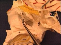
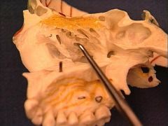
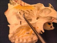
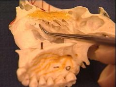
Ostium of Maxillary Sinus
|
|
Below the bulla ethmoidalis, and partly hidden by the inferior end of the uncinate process
|
|
|
Uncinate Process of Ethmoid Bone
|
|
projects downward and backward from this part of the labyrinth; it forms a small part of the medial wall of the maxillary sinus, and articulates with the ethmoidal process of the inferior nasal concha.
|
|
|
Ethmoid Bulla
|
|
|
|
|
Ostium of Middle Ethmoid Air Cells
|
|
|
|
|
Infundibulum (Ethmoidal)
|
|
|
|
|
Pterygoid/Vidian Canal
|
|
|
|
|
Pterygoid Process of Sphenoid Bone
|
|
|
|
|
Semilunar Hiatus
|
|
|
|
|
External Auditory Meatus
|
|
|
|
|
Tympanic Part of Temporal Bone
|
|
|
|
|
Zygomatic Process of Temporal Bone
|
|
|
|
|
Suprameatal Triangle
|
|
|
|
|
Mandibular/Glenoid Fossa
|
|
|
|
|
Parotid Gland
|
|
|
|
|
SCALP - Skin
|
|
contains hair follicles, sweat glands, sebaceous glands
|
|
|
SCALP - Connective Tissue (dense)
|
|
anchored firmly to skin above and to aponeurosis below; carotid arteries (ICA and ECA) anastomose and supply blood here
|
|
|
SCALP - Aponeuroses
|
|
broad, flat tendon for occipitalis and frontalis muscles
|
|
|
SCALP - Loose Connective Tissue
|
|
loose areolar tissue that allows freedom of movement of the superficial three layers
|
|
|
SCALP - Pericranium
|
|
firmly attached to underlying bone
|
|
|
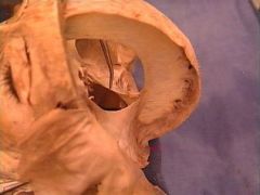
Calvaria
|
|
|
|
|
Arachnoid Granulations
|
|
protrusions in dura that allow filtering of CSF back to circulation
|
|
|
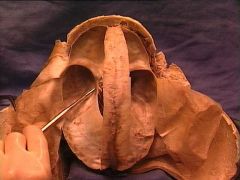
Sagittal Section of Brain:
Falx Cerebri Falx Cerebelli Tentorium Cerebelli |

|
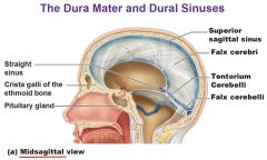
Falx cerebri – separates cerebral hemispheres, attached anteriorly to crista galli, superiorly to lips of superior sagittal sulcus, posteriorly ends at internal occipital protuberance, posterior third of inferior border is attached to tentorium cerebelli, anterior two-thirds of inferior border is free edged
b) Falx cerebelli – smaller fold that also separates cerebral hemispheres but inferior to tentorium cerebelli (from internal occipital crest to foramen magnum) note: impt anastomoses occur btw venous dural sinuses and the internal vertebral venous plexus @f. magnum c) Tentorium cerebelli – horizontal fold across posterior third of skull to separate cerebral hemispheres (occipital lobes) from cerebellum, attaches along transverse sulcus on each side of skull, and attaches along superior petrosal sulcus and ends medially at posterior clinoid process |
|
|
Tentorial Notch
|
|
opening the in the tentorium cerebelli for the brainstem and thru which herniation of the forebrain may cause impt neurological symptoms
|
|
|
CN I - Olfactory Nerve
|
|
not a true nerve actually a tract; tract emerges from brain and ends as the olfactory bulbs
|
|
|
CN II - Optic Nerve
|
|
not a true nerve, outgrowth of the brain; Nerves go through optic foramina (of the sphenoid bone) to enter each orbit, go to posterior aspect of eyeballs; some fibers cross at optic chiasm
|
|
|
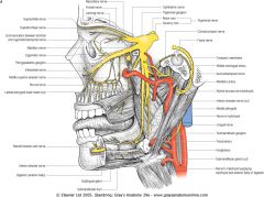
Internal Carotid Artery (next to Optic Nerve)
|
|
enters skull through carotid canal, transverses the petrous portion of the temporal bone, and passes through the cavernous sinus. The two internal carotid arteries join to send communicating branches anteriorly and posteriorly (communicating branches) to join with basilar to form Circle of Willis
|
|
|
Ophthalmic Artery
|
|
First branch from ICA, through optic canal; distal to cavernous sinus; supply all the structures in the orbit as well as some structures in the nose, face and meninges
|
|
|
Infundibulum/Pituitary Stalk
|
|
stalk of the pituitary and the hypophysis: pituitary gland that sits in the sella turcica
|
|
|
CN III - Oculomotor Nerve
|
|
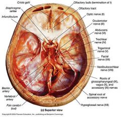
pierces dura and enters cavernous sinus, travels along lateral wall; enters orbit by passing through superior orbital fissure
-Controls eye movement and pupil constriction (superior: levator palpebrae superioris, superior rectus; inferior: medial rectus, inferior rectus, inferior oblique; ciliary muscle |
|
|
CN IV - Trochlear Nerve
|
|
passes through cavernous sinus; exits cranial cavity through superior orbital fissure; innervates superior oblique
|
|
|
CN VI - Abducens Nerve
|
|

pierces dura overlying the clivus and travels a bit (longest intracranial course) and enters cavernous sinus, travels through it most medially of the nerves (inferior to internal carotid artery); exits cranial cavity via superior orbital fissure to enter orbit; innervates lateral rectus
|
|
|
CN V - Trigeminal Nerve
|
|

pierces dura just antero-inferior to the trochlear nerve
|
|
|
Olfactory Bulb
|
|
olfactory bulbs sit on cribriform plates of ethmoid bone; rootlets pass through perforation in the cribriform plates to reach the nasal epithelium in the nasal cavity
|
|
|
CN VII - Facial Nerve
|
|

pierces dura and exits through internal auditory meatus, entering the petrous temporal bone; goes through the facial canal; exits base of skull through stylomastoid foramen; innervates muscles of facial expression (motor trunk divides in parotid gland to temporal, zygomatic, buccal, marginal mandibular, cervical branches); chorda tympani branch of VII eventually joins lingual branch of V3 to hitchhike along it (innervates anterior 2/3 of tongue: taste); Greater petrosal nerve – branches from VII at geniculate ganglion and exits via hiatus of facial nerve, crosses foramen lacerum, and joins deep petrosal nerve to form nerve of the pterygoid canal (Vidian nerve); nerve to the stapedius; PS innervation of submandibular, sublingual, and lacrimal glands
|
|
|
Diaphgrama Sellae
|
|
dura mater membrane covering sella turcica; stretches from anterior clinoid processes to posterior clinoid processes; pierced by the infundibulum (stalk of pituitary gland)
|
|
|
Trigeminal Ganglion
|
|
found in Meckel’s cave – dural projection as the nerve dilates to form the ganglion
|
|
|
CN VIII - Vestibulocochlear Nerve
|
|

arises from brainstem lateral to facial nerve; exits cranial cavity via the internal auditory meatus to supply the inner ear
|
|
|
CN V1 - Ophthalmic Division of Trigeminal
|
|
enters cavernous sinus and travels along lateral wall and enters orbit via superior orbital fissure; supplies forehead, eyes, nose, temples, meninges, paranasal sinuses, part of nasal mucosa; NFL: Nasociliary, Frontal -supraorbital, supratrochlear, Lacrimal
|
|
|
CN V2 - Maxillary Division of Trigeminal
|
|
also enters cavernous sinus and exits cranial cavity via foramen rotundum and enters pterygopalatine fossa; supplies max and mand dentition, lower eyelid, part of nose, upper lip, cheeks, hard palate, maxillary sinuses; ZIP: Zygomatic-zygomaticotemporal, zygomaticoorbital, zygomaticofacial, Infraorbital- superior labial, nasal, inferior palpebral, Palatine-greater and lesser palatine nerves, nasopalatine- incisive canal
|
|
|
CN V3 - Mandibular Division of Trigeminal
|
|
does not go through cavernous sinus; instead, drops straight down through foramen ovale to infratemporal region; contains motor portion of the nerve; supplies muscles of mastication, tensor tympani, tensor
veli palatini, anterior digastric, mylohyoid; BAIL: Long Buccal, Auriculotemporal, Inferior alveolar and Lingual |
|
|
CN IX - Glossopharyngeal
|
|

exits cranial cavity via jugular foramen; motor innervation to stylopharyngeus muscle, taste and sensation for posterior 1/3 of tongue, soft palate, pharynx, tonsils, auditory tube, tympanic cavity; afferent limb of gag reflex; PS innervation to parotid gland (via otic ganglion)
|
|
|
CN X - Vagus Nerve
|
|

nerve exits at jugular foramen; supplies muscles of larynx, pharynx, palate, and upper esophagus; taste from epiglottis and valleculae
|
|
|
CN XI - Spinal Accessory Nerve
|
|

cranial portion arises from converging rootlets below X; spinal portion arises from motor spinal contributions of C1-C6 that entered the cranium through foramen magnum to travel with the cranial portion; nerve exits through jugular foramen; innervates the SCM and trap
|
|
|
CN XII - Hypoglossal Nerve
|
|
nerve rootlets combine and exit via hypoglossal canal; innervates the intrinsic and extrinsic muscles of the tongue (expt palatoglossus)
|
|
|
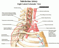
Vertebral Arteries
|
|
first branches from subclavian arteries; ascends in neck to enter transverse foramen of C6; enters skull via foramen magnum; right and left vertebral arteries join to form basilar artery (which goes on to supply the circle of Willis)
|
|
|
Basilar Artery
|
|
right and left vertebral arteries join to form basilar artery (which goes on to supply the circle of Willis)
|
|
|
Straight Sinus
|
|

base of falx tentorium cerebelli; drainage: inferior sagittal and great cerebral vein to confluence of sinuses
|
|
|
Cavernous Sinus
|
|
paired, 1cm wide, R and L of sphenoid bone; transversed by network of fibrous filaments (unusual): serve to slow flow of venous blood through these structures; allows pathogens to colonize within these regions
- extends 2cm from superior orbital fissure anteriorly to apex of petrous temporal bone posteriorly; anterior and posterior intercavernous sinuses (forms circular sinus) connects both L and R cavernous sinus; receives blood from: Pterygoid plexus via inferior ophthalmic vein, emissary veins, and deep facial vein drain into cavernous thru sphenoid foramen |
|
|
Confluence of Sinuses
|
|
sinus junction at internal occipital protuberance; drainage from: Superior Sagittal, Straight, and Occipital Sinuses; drainage to: transverse sinuses
|
|
|
Inferior Sagittal Sinus
|
|
inferior to falx cerebri; anterior: crista galli; posterior: joins Great Cerebral Vein; drainage: small cerebral veins
|
|
|
Superior Sagittal Sinus
|
|
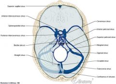
largest dural sinus; cranial falx cerebri; anterior: foramen cecum; posterior: internal occipital protuberance; drains: cerebral veins, emissary veins, diploic veins
|
|
|
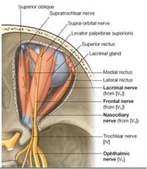
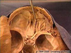
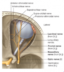
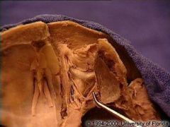
Nasociliary Nerve (CN V1)
|
|
ciliary ganglion, long ciliary nerves, anterior and posterior ethmoidal nerves, infratrochlear nerve; runs obliquely toward medial wall of the orbit, passes b/w superior oblique and medial rectus; travels w/ the ophthalmic artery
|
|
|
Frontal Nerve (CN V1)
|
|
supraorbital nerve, supratrochlear nerve; provides sensory info for forehead skin, frontal sinus mucosa, and upper eyelid; above levator palpebrae superioris
|
|
|
Lacrimal Nerve (CN V1)
|
|
innervates lacrimal gland (smallest of three branches)
|
|
|
Ciliary Ganglion
|
|
sensory root from nasociliary nerve of CN V1; parasympathetic root from inferior branch of CN 3 (pupillary constriction, ciliary muscle→ accommodation- narrows pupil and increases lens convexity); sympathetic root from internal carotid plexus (pupillary dilator, ophthalmic artery, tarsal muscle and levator palpebrae superioris); gives off short ciliary nerves
|
|
|
Supraorbital Nerve
|
|
branch of Frontal Nerve (CN V1)
|
|
|
Supratrochlear Nerve
|
|
branch of Frontal Nerve (CN V1)
|
|
|
Lateral Rectus
|
|
abducts; innervated by CN VI
|
|
|
Medial Rectus
|
|
adducts; inferior division of CN III
|
|
|
Superior Rectus
|
|
elevates, adducts, intorts; innervated by CN III
|
|
|
Inferior Rectus
|
|
depresses, adducts, extorts; innervated by CN III
|
|
|
Levator Palpebrae Superioris
|
|
elevates upper eyelid; innervated by CN III
|
|
|
Superior Oblique
|
|
depresses, abducts, extorts; arises from superomedial margin of the optic foramen, runs forward forming a tendon passing though the trochlea; innervated by CN IV
|
|
|
Inferior Oblique
|
|
o action: depresses, adducts, extorts (3 axes movement)
o course: arise from annular tendon to attach to anterior sclera o innervation: inferior division of CN 3 |
|
|
Trochlea
|
|
|
|
|
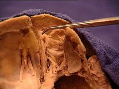
Lacrimal Gland
|
|
• located in the lacrimal fossa of the frontal bone
• sensory innervation from lacrimal nerve (V1) • secretomotor nerves piggyback on zygomatic nerve (V2) o parasympathetic innervation from greater petrosal nerve of CN 7 o sympathetic innervation from deep petrosal nerve from internal carotid plexus o greater petrosal and deep petrosal → Vidian nerve → pterygopalatine ganglion → infraorbital nerve (V2) → zygomatic branch of infraorbital → lacrimal gland • arterial supply from lacrimal artery (branch of ophthalmic artery) |
|
|
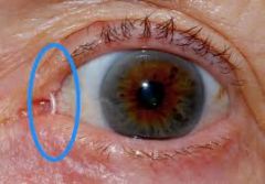
Lacrimal Sac & Nasolacrimal Duct
|
|
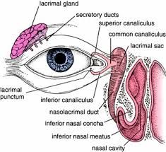
• tears are secreted by the lacrimal gland (at superolateral part of eye) to lubricate the eyes
• tears pool in the lacrimal caruncle/lake (at medial part of eye) • tears drain into the puncta lacrimalis and then through the lacrimal canaliculi to the lacrimal sac • lacrimal sac (above medial palpebral ligament) drains into the nasolacrimal duct which opens at the inferior nasal meatus (of the same side) • remember tears are disseminated by actions of the orbicularis oculi and related papebral musculature |
|
|
Zygomaticus Major
|
|
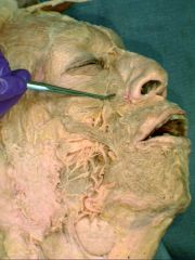
Innervated by buccal and zygomatic branches of facial nerve; extends from zygomatic arch to corner of the mouth; supplied by facial artery
|
|
|
Lesser Occipital Nerve (C2) - cutaneous branch of cervical plexus
|
|
(C2) - innervates lateral scalp behind ear
o [Auricular branch: innervates skin along the side of the head behind the ear; joins with greater occipital n, great auricular n. & posterior auricular branch of CN VII] Superficial Nerve of Cervical Plexus |
|
|
Ansa Cervicalis (Superior and Inferior Roots): C1-C3 - muscular branch of cervical plexus
|
|
innervates all infrahyoid muscles except thyrohyoid; Superior Root: ventral ramus of C1 spinal root (descendans hypoglossi)
• These nerve fibers travel w/ hypoglossal n. before branching to form the superior root; hypoglossal n. will always be found looping around (90 degree turn) the occipital branch of the external carotid A en route to the floor of the mouth (XII also courses btw the mylohyoid and hyoglossus) • Inferior Root: ventral rami of C2 and C3 Deep Nerve of Cervical Plexus |
|
|
Phrenic Nerve (C3-C5) - muscular branch of cervical plexus
|
|
important nerve that lies on the anterior surface of the anterior scalene; descends from its origins in the cervical plexus along the anterior surface of the anterior scalene; deep nerve of cervical plexus
|
|
|
Hypoglossal Nerve
|
|
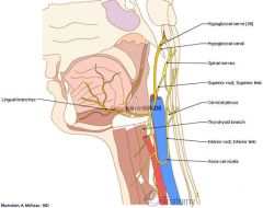
courses between the mylohyoid and hyoglossus muscles
|
|
|
Superior Thyroid Artery (ECA)
|
|
• Infrahyoid: GT
• Sternocleidomastoid: GT • Superior Laryngeal: GT (upper larynx) • Cricothyroid: cricothyroid ligament |
|
|
Ascending Pharyngeal Artery (ECA)
|
|
• Pharyngeal: middle constrictors
• Palatine: soft palate & tonsil • Prevertebral: longus capitis & colli • Inferior tympanic: tympanic cavity • Posterior meningeal: dura mater |
|
|
Lingual Artery (ECA)
|

|
• Suprahyoid: GT
• Dorsal lingual: post. part of dorsum of tongue, tonsil, soft palate & epiglottis • Sublingual: sublingual gland & mylohyoid m. • Deep lingual: underside of tongue, genioglossus |
|
|
Facial Artery (ECA)
|
|
• Cervical branches: ATGS
• Ascending palatine: soft palate, palatine glands • Tonsilar: tonsil & tongue • Glandular: submandibular gland • Submental: chin & lip • Facial branches: ISLAM • Inferior labial: GT • Superior labial: GT • Lateral nasal: GT • Angular (terminal branch): lacrimal sac; anastomoses with V1 • Muscular: neck, face |
|
|
Occipital Artery (ECA)
|
|
supplies scalp, mastoid process, SCM & Trapezius
• Muscular: digastric, stylohyoid • SCM: GT • Auricular: mastoid air cells • Meningeal: dura of PC fossa • Terminal: occipital bone Hypoglossal Nerve passes over it |
|
|
Transverse Facial Artery
Labial Arteries Angular Arteries Superficial Temporal Arteries |
|
|
|
|
Levator Labii Superioris
|
|
• O: frontal process of maxilla and infraorbital region
• I: skin of upper lip and alar cartilage of nose • A: elevates lip, dilate nostril, raise angle of mouth |
|
|
Deep Facial Vein
Angular Vein Posterior Auricular Vein |
|
|
|
|
Mylohyoid
|
|
• O: mylohyoid line of mandible
• I: median raphe from chin to hyoid bone and onto hyoid (mylohyoid raphe) • A: elevates floor of mouth & hyoid bone/depresses mandible (raises tongue in early stage of swallowing) • I: mylohyoid branch of inferior alveolar branch of V3 |
|
|
Geniohyoid
|
|
• O: inferior genial tubercle
• I: anterior border of hyoid • A: elevates the hyoid and draws it forward/depresses mandible • I: C1 through the hypoglossal n. |
|
|
Genioglossus
|
|
Origin – superior genial tubercle/superior part of mental spine of mandible
Insertion – dorsum of tongue and body of hyoid Innervation – CN XII Action – depresses tongue, posterior part pulls tongue anteriorly for protrusion |
|
|
Stylohyoid
|
|
• O: posterior border of styloid process of temporal bone
• I: hyoid bone, at junction of the body and greater cornu • A: elevates hyoid & draws it posteriorly • I: stylohyoid branch of VII |
|
|
Stylopharyngeus
|
|
o Only muscle derived from pharyngeal arch 3
o Origin: styloid process o Insertion: thyroid cartilage (pharynx) o Innervation: only muscle innervated by CN 9; CN 9 accompanies stylopharyngeus thru the gap between the superior and middle constrictors o Action: elevates the pharynx and larynx |
|
|
Hyoglossus
|
|
Origin – body and greater horn of hyoid
- Insertion – side and inferior aspect of tongue - Innervation – CN XII - Action – depresses and retracts tongue |
|
|
Sternothyroid
|
|
deep to sternohyoid; is raised and stretched by the mass of the underlying thyroid gland;
O: posterior manubrium, deep to sternohyoid; 1st costal cartilage I: oblique line on lamina of thyroid cartilage A: depresses larynx I: ansa cervicalis (C1-C3) |
|
|
Thyrohyoid
|
|
(deep to the sternohyoid)
O: oblique line on thyroid cartilage I: lower border of body and greater cornu of hyoid A: depresses hyoid bone/elevates larynx I: thyrohyoid branch of C1 through the hypoglossal |
|
|
Ansa Cervicalis (Superior Root)
|
|
Superior Root: ventral ramus of C1 spinal root (descendans hypoglossi)
• These nerve fibers travel w/ hypoglossal n. before branching to form the superior root; hypoglossal n. will always be found looping around (90 degree turn) the occipital branch of the external carotid A en route to the floor of the mouth (XII also courses btw the mylohyoid and hyoglossus) |
|
|
Levator Anguli Oris
|
|
immediately below the infraorbital foramen; innervated by buccal branches of the facial nerve
|
|
|
Vagus Nerve (in neck)
|
|
internal jugular vein courses with the nerve laterally, common carotid medially
|
|
|
Carotid Sinus
|
|
dilation of the ICA near the bifurcation of the common carotid a. containing baroreceptors
• Convey info abt changes in BP; innervated by IX to medulla |
|
|
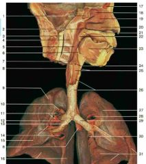
Common Carotid Artery
|
|
Part of the carotid sheath (with internal jugular vein and vagus nerve); bifurcates into internal and external branches
|
|
|
External Carotid Artery
|
|
More superficial branch of the common carotid artery; branches into SALFOPSM
|
|
|
Internal Carotid Artery
|
|
More deep branch of the common carotid artery; supplies the brain
|
|
|
Brachiocephalic Artery
|
|
supplies blood to the R arm, head and neck; divides into the R subclavian and right common carotid artery, only on the right side of body
|
|
|
Left Common Carotid Artery
|
|
Part of the carotid sheath (with internal jugular vein and vagus nerve); bifurcates into internal and external branches; adjacent to left subclavian artery
|
|
|
Right Vagus Nerve
|
|
clearly visible b/w IJV and carotid artery throughout the neck in the carotid sheath; passes through jugular foramen and carotid sheath, and then courses posterior to the root of the lungs and branches to form the esophageal plexus (along w/ direct visceral branches arising from the sympathetic turns bilaterally) on the anterior surface of the of esophagus
|
|
|
Left Brachiocephalic Vein
|
|
Drains left and right inferior thyroid veins; Internal jugular vein and Subclavian vein join to form brachiocephalic; union of the two brachiocephalics forms SVC; conduct the venous outflow of the head (IJV) and upper extremities (subclavian vein) to the SVC (the left one is particularly long, crossing the midline almost horizontally and receiving the drainage of the inferior thyroid vein from the neck en route to the SVC)
|
|
|
Superior Vena Cava
|
|
Carries deoxy blood from the upper half of the body into the RA and is formed by the union of the L+R brachiocephalic veins; the azygos vein joins it just before it enters the RA
|
|
|
Depressor Labii Inferioris
|
|
helps lower bottom lip; innervated by mandibular division of facial nerve
|
|
|
Left Vagus Nerve
|
|
internal jugular vein courses with the nerve laterally, common carotid medially; L vagus nerve descends from the neck toward the posterior mediastinum (posterior to root of the lung) along the L side of the aortic arch and it ramifies extensively here, providing visceral efferent (PS) fibers to both the pulmonary and cardiac plexuses.
|
|
|
Internal Branch of Superior Laryngeal Nerve
|
|

Superior Laryngeal: branch of the vagus nerve. It arises from the middle of the ganglion nodosum and in its course receives a branch from the superior cervical ganglion of the sympathetic.
It descends, by the side of the pharynx, behind the internal carotid artery, and divides into two branches Internal Branch: goes through the thyrohyoid membrane (accompanied by superior laryngeal a. and v.); innervates mucous membrane superior to vocal folds |
|
|
External Branch of Superior Laryngeal Nerve
|
|

Superior Laryngeal: branch of the vagus nerve. It arises from the middle of the ganglion nodosum and in its course receives a branch from the superior cervical ganglion of the sympathetic.
It descends, by the side of the pharynx, behind the internal carotid artery, and divides into two branches; External Branch: goes to cricothyroid muscle (only laryngeal muscle not innervated by recurrent laryngeal nerve) |
|
|
Thyroid Gland (Isthmus)
|
|
covered by a pretracheal layer of deep cervical fascia; two lobes and an isthmus); look for pyramidal lobe or partially obliterated thyroglossal duct; supplied by superior and inferior thyroid arteries; drains to superior, middle and inferior thyroid veins
|
|
|
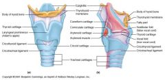
Cricothyroid Muscle
|
|
• O: lateral surface of cricoid cartilage
• I: (lower fibers) anterior margin of inferior horn of thyroid C; (upper fibers) lower border of lamina of thyroid C • I: external laryngeal nerve (from superior laryngeal branch of X) • A: tenses vocal folds (raise pitch of speech) Cricothyroid joint: Movement at this joint changes length and tension of the vocal ligaments, determining pitch of speech |
|
|
Thyroid Cartilage
|
|
contains the larynx; laryngeal prominence in front is palpable, superior and inferior thyroid notches; posteriorly, superior horns of the thyroid come close to greater horns of hyoid; many muscles originate and insert here; sternothyroid inserts, thyrohyoid originates, inferior pharyngeal constrictor inserts, stylopharyngeus inserts, palatopharyngeus inserts
|
|
|
Cricoid Cartilage (Signet Ring)
|
|
only complete ring around the trachea; disparity in anterior arch thickness (band) and posterior lamina thickness (slightly broader)… like a signet ring; opposite the sixth cervical vertebra
|
|
|
Hyoid Bone
|
|
floating bone just below the mandible with lesser and greater horns
|
|
|
Right Brachiocephalic Vein
|
|
Internal jugular vein and Subclavian vein join to form brachiocephalic; union of the two brachiocephalics forms SVC; conduct the venous outflow of the head (IJV) and upper extremities (subclavian vein) to the SVC
|
|
|
Greater Petrosal Nerve
|
|
branches from VII at geniculate ganglion and exits via hiatus of facial nerve, crosses foramen lacerum, and joins deep petrosal nerve to form nerve of the pterygoid canal
|
|
|
Depressor Anguli Oris
|
|
frowning; from mandible to angle of the mouth; innervated by mandibular branch of facial nerve
|
|
|
Thoracic Duct (& Right Lymphatic Duct)
|
|
On the left side of the body;
Arises from: cysterna chyli in abdomen (small sac) o Enters thorax through aortic opening in diaphragm o Ascends within thorax in between the azygos vein & aorta o Drains: lower extremities, pelvis, abdomen, upper left quadrant of body o Picks up bronchomediastinal trunk in thorax • Contains visceral lymphatic drainage o As duct approaches root of neck, swings to left, arches around L IJV o Empties: confluence of the left IJV and subclavian veins Right lymphatic duct: receives drainage from the right upper quadrant and drains into the right subclavian v.; only segment of the body not drained by thoracic duct |
|
|
Deep Temporal Arteries (Pterygoid Portion of Maxillary Artery)
|

#18
|

|
|
|
Inferior Alveolar Artery (Mandibular Portion of Maxillary Artery)
|
#29
|
Runs with inferior alveolar nerve
|
|
|
Middle Meningeal Artery (Mandibular Portion of Maxillary Artery) w/ Auriculotemporal Nerve
|
#41
|
Mandibular portion of Maxillary Artery; passes through foramen spinosum; surrounded by two branches of auriculotemporal nerve
|
|
|
Buccal Artery (Pterygoid Portion of Maxillary Artery) (& Nerve)
|

#23
|

Runs with buccal nerve
|
|
|
Sphenopalatine Artery (Pterygopalatine Portion of Maxillary Artery)
|

#20
|
Near the posterior aspect of the superior conchae and anterior to the sphenoid sinus is the sphenopalatine foramen thru which courses the sphenopalatine artery
|
|
|
Submandibular Ganglion
|
#10
|
lingual nerve and chorda tympani carry taste sensations (anterior 2/3 of tongue) and PS fibers to submandibular ganglion;
Attached to the inferior loop of the lingual nerve o Responsible for the innervation of the submandibular and sublingual glands o Hangs via two filaments from the lingual nerve (branch of V3) o Preganglionic parasympathetic fibers synapse from the medulla’s superior salivatory nucleus |
|
|
Pterygopalatine Ganglion
|
#12
|
Part of CN V2; Vidian nerve passes through foramen lacerum and synapses here; contained in the pterygopalatine fossa
|
|
|
23: Inferior Thyroid Artery
21: Ascending Cervical Artery 19: Superior Thyroid Artery |
#23 + #21 + #19
|
inferior thyroid artery (supplies trachea, esophagus, larynx, thyroid gland) is a branch off the thyrocervical trunk, ascending cervical artery (supplies vertebrae and neck muscles) branches off inferior thyroid; superior thyroid is a branch off ECA; both thyroid arteries supply the thyroid gland
|
|
|
Vertebral Artery
|
#47
|
branch off Subclavian artery; ascends and courses posteriorly toward the cervical spine where it normally ascends thru the transverse foramina of the upper six vertebrae
• Supplies blood to the posterior part of circle of Willis and anastomoses with blood supplied to the anterior part of the circle of Willis from the carotid |
|
|
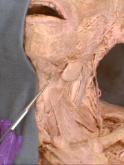
Submandibular Gland
|
|
mixed gland found below the mandible and superior to the digastric muscles; located inferior to mylohyoid muscle and close to medial surface of the body of the mandible
|
|
|
Right Internal Thoracic Artery
|

#24
|
Branch off subclavian artery; supplies the anterior chest wall and the breasts
• Anterior intercostal arteries (supplies ribs) • Superior epigastric A • Descends inferiorly to anastomose with inferior epigastric (from external iliac) |
|
|
Costocervical Trunk
|
|
Branch off subclavian artery; Arteries that supply back of neck
• Posterior Intercostal arteries • supplies first two intercostals spaces]] branches into deep cervical artery and superior intercostal artery |
|
|
Inferior Thyroid Veins (& Left Brachiocephalic)
|
#21
|
left brachiocephalic vein drains both inferior thyroid veins
|
|
|
Superior Thyroid Veins (& Middle)
|
#15
|
Both drain to IJV
• Superior thyroid vein; begins in substance of and on top of the thyroid gland and also drains in IJV • Middle thyroid vein: drains blood from the lower part of the thyroid gland to the IJV |
|
|
Retropharyngeal Space
|
|

bound by the buccopharyngeal fascia anteriorly and the anterior lamina (alar fascia) of the prevertebral fascia posteriorly (btw the pharynx and prevertebral fascia); b/c serious infections of teeth can spread down this space into the posterior mediastinum, it is can be designated as the danger space
|
|
|
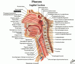
Nasopharynx
|

#13
|
o Communicates anteriorly with nasal cavity
o Nasal cavity is split into left and right by the nasal septum o Continuous with nasal cavity at the choanae or posterior nasal aperture o Nasopharynx and oropharynx separated by soft palate musculature |
|
|
Oropharynx
|
#15
|
o Communicates anteriorly with oral cavity
o Separated from oral cavity by the fauces or pillars o Nasopharynx and oropharynx separated by soft palate musculature |
|
|
Laryngopharynx
|

#17
|
Below the oropharynx to the esophagus
|
|
|
Cervical Sympathetic Trunk & Superior Cervical Ganglion
|
#22
|

• Cervical sympathetic trunk located behind the carotid sheath as it rises from the thorax towards the base of the skull embedded in prevertebral fascia
• Superior cervical ganglion: superior extent of trunk |
|
|
Inferior Cervical Ganglion
|
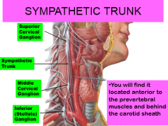
|
• Inferior cervical ganglion: opposite the transverse process of the 7th cervical vertebrae where it is often continuous with the first thoracic ganglion to form the stellate ganglion
|
|
|
Anterior Jugular Vein
|
|
drains anterior aspect of neck and descends on either side of the midline to point above the jugular notch of the manubrium and empties into external jugular; connects with internal jugular vein
|
|
|
Superior and Middle Cervical Ganglia
|
|
• Superior cervical ganglion: superior extent of trunk
• Middle cervical ganglion: foten diminutive |
|
|
Internal Jugular Vein
|
|
formed by joining of the inferior petrosal sinus and the sigmoid sinus; runs down side of the neck vertically (lateral to ICA and then to common carotid) joins with subclavian vein to form brachiocephalic vein
|
|
|
Common Facial Vein
|
|
o Union of facial vein and anterior retromandibular; anterior branch of retromandibular vein and facial vein join to form the common facial vein --> IJV
|
|
|
Submandibular/Wharton's Duct
|
|
runs with the lingual nerve in the floor of the mouth; crosses superior to the lingual nerve as the duct courses toward its opening on the sublingual caruncle
|
|
|
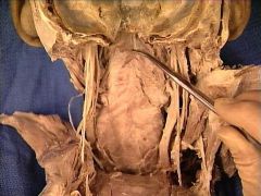
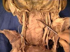
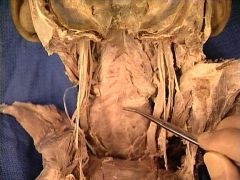
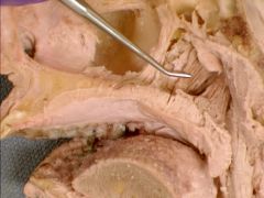
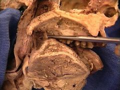
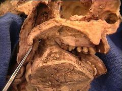
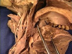
Hard Palate
|

#2
|
Oral and nasal cavity separated by hard palate
|
|
|
Soft Palate & Uvula
|

#14
|
soft palate divides nasopharynx from oropharynx; o Soft palate muscles attach to a fibrous aponeurosis
o Soft palate is elevated and tensed to limit reflux of fluid into nasopharynx during swallowing and to enable certain plosive speech sounds o Musculus uvulae, levator veli palatini, tensor veli palatini |
|
|
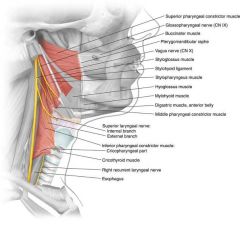
Superior Pharyngeal Constrictor
|
|
o Attaches to lower 1/3 of posterior of medial pterygoid, pterygomandibular raphe, and alveolar process of mandible
o Passavant’s ridge: a prominence seen during swallowing on the nasopharyngeal wall by contraction of the superior constrictors; when the palate is not functioning during swallowing, the deficiency is compensated by a greater convergence of Passavant’s ridge |
|
|
Middle Pharyngeal Constrictor
|
|

o Attaches to both horns of hyoid bone and stylohyoid ligament
|
|
|
Inferior Pharyngeal Constrictor
|
|

|
|
|
Median Pharyngeal Raphe
|
|

fibrous median septum to which the pharyngeal constrictor muscles attach posteriorly; located in the midline on the posterior wall of the pharynx and attaches superiorly to the pharyngeal tubercle of the occipital bone
|
|
|
Thryopharyngeal portion of Inferior Pharyngeal Constrictor
|
#16
|
o Upper part: thyropharyngeal part (from oblique thyroid line)
|
|
|
Cricopharyngeal portion of Inferior Pharyngeal Constrictor
|

#18
|
cricopharyngeal part (from cricoid cartilage btw the cricothyroid and the inferior part of the thyroid cartilage)
|
|
|
Pterygomandibular raphe
|

#2
|
o Tendinous band of buccalpharyngeal fascia
o Attached to pterygoid hamulus of the medial pteryogid and the posterior end of the mylohyoid line of the mandible o Attached to the superior middle constrictor and buccinators |
|
|
Styloid Process
|
|
Its proximal part (tympanohyal) is ensheathed by the vaginal process of the tympanic portion.
Its distal part (stylohyal) gives attachment to the following: stylohyoid ligament stylomandibular ligament styloglossus muscle (innervated by the hypoglossal nerve) stylohyoid muscle (innervated by the facial nerve) stylopharyngeus muscle (innervated by the glossopharyngeal nerve) |
|
|
Salpingopharyngeus/Salpingopharyngeal Fold
|

#24
|
• Salpingopharyngeus (covered by mucosal fold)
o Origin: inferior part of the cartilage of the pharyngotympanic tube (Eustachian tube) in the nasal cavity o Insertion: palatopharyngeus muscle o Innervation: pharyngeal plexus of CN 10 o Action: raises the nasopharynx during swallowing and laterally draws the pharyngeal walls up |
|
|
Palatopharyngeus/Palatopharyngeal Fold
|
#19
|
o Origin: palatine aponeurosis and hard palate
o Insertion: thyroid cartilage o Innervation: vagus and cranial accessory nerve o Action: pulls pharynx and larynx upward |
|
|
Choana
|

#16
|
opening of the nasal cavity into the nasopharynx; aka posterior nares/nasal aperture
|
|
|
Torus Tubarius
|
|
broad cartilaginous end of the auditory/eustachian/pharyngotympanic tube; tubal elevation of the base of the cartilaginous portion of the Eustachian tube above the orifice of the tube
|
|
|
Auditory/Eustachian/Pharyngotympanic Tube
|
#27
|
extends from middle ear to lateral wall of nasopharynx at inferior nasal concha level; opening of the auditory tube is found below the broad cartilaginous end of the tube know as the torus tubarius
|
|
|
Pharyngeal Recess
|

#8
|
fossa of Rosenmuller; posterior to the opening of the auditory tube and the torus tubarius; at the superior end, the pharyngeal tonsils/adenoids will occupy this area
|
|
|
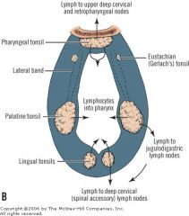
Pharyngeal Tonsil/Adenoids
|

#5
|
occupy superior end of pharyngeal recess; when enlarged, called adenoid hypertrophy or adenoid facies
|
|
|
Maxillary Vein
|
|
o Short trunk joining pterygoid plexus to retromandibular
|
|
|
Levator Veli Palatini
|
|
soft palate muscle;
o Origin: petrous temporal + cartilaginous auditory tube (torus tubarius) o Insertion: contralateral muscle in the velum palatinum (soft palate) • Review: torus tubarius is ridge in the nasopharyngeal wall posterior to the opening of the auditory tube, caused by the projection of the cartilaginous portion of this tube. The salpingopharyngeal fold also descends from this torus tubarius. o Action: elevates the soft palate, pulling posteriorly and narrowing the walls of the nasopharynx (drawing medially) o Innervation: pharyngeal plexus of CNX. |
|
|
Tensor Veli Palatini
|
|
soft palate muscle;
o Origin: scaphoid fossa, spina angularis, and cartilaginous part of the auditory tube o Insertion: velum palatinum after being redirected by the tendinous portion of the pterygoid hamulus. o Action: tenses the soft palate and opens the cartilaginous auditory tube. In other words, it elevates the lateral edges, providing a good seal of the soft palate from the nasopharynx. o Innervation: tensor veli palatini branch of CNV3 |
|
|
Lingual Tonsil (& Root of Tongue)
|

#16
|
lymph tissue covering the base of the tongue
|
|
|
Palatoglossus
|
|
Origin – palatine aponeurosis of soft palate
- Insertion – side of tongue - Inneration – cranial root of CN XI vial pharyngeal branch of CN X and pharyngeal plexus - Action – elevates posterior part of tongue |
|
|
Palatopharyngeus
|
|
(covered by a mucosal fold)
o Origin: palatine aponeurosis and hard palate o Insertion: thyroid cartilage o Innervation: vagus and cranial accessory nerve o Action: pulls pharynx and larynx upward |
|
|
Oropharyngeal Isthmus/Oropharynx
|
#5
|
• Boundaries of oropharynx: laryngopharynx inferior, oral cavity anterior (by the oropharyngeal isthmus or the isthmus of the fauces= bounded superiorly by soft palate, inferiorly by root of tongue (where lingual tonsils lie), laterally by palatoglossal and paaltopharyngeal arches/folds), nasopharynx superior (by the soft palate muscles), pharyngeal wall posteriorly (sup constrictor)
|
|
|
Palatine Tonsils/Tonsilar Fossa
|
|
palatoglossal fold and palatopharyngeal fold (aka anterior and posterior pillars of the fauces); palatine tonsils lie btw the pillars of the fauces on the loose connective tissue covering the superior pharyngeal constrictor
|
|
|
Waldeyer's Tonsillar Ring
|
|
annular arrangement of lymphoid tissue in the pharynx. Waldeyer's ring circumscribes the naso- and oropharynx, with some of its tonsillar tissue located above and some below the soft palate (and to the back of the oral cavity)
Pharyngeal tonsils, tubal tonsils, palatine tonsils, lingual tonsils |
|
|
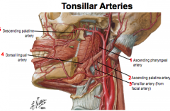
Tonsillar & Ascending Palartine Branches of Facial Artery
|
|
The palatine tonsil receives its BS from a tonsillar branch of the facial artery and the ascending palatine arteries
|
|
|
Epiglottis
|
|
o Lid-like flap of cartilage guarding the entrance of the laryngeal inlet
o During swallowing, elevation of the hyoid draws the epiglottis down to direct food to esophagus o Upper epiglottis: CN 9 fibers contribute to afferent gag reflex o Lower epiglottis: CN 10 fibers contribute to afferent cough reflex |
|
|
Glossoepiglottic Folds
|
Walls of this depression/opening
|
o Median glossoepiglottic fold: middle fold on epiglottis
o Lateral glossoepiglottic folds: lateral folds on epiglottis attached to pharyngeal wall |
|
|
Piriform Recess
|

#11
|
on either side of the laryngeal orifice; bounded medially by the aryepiglottic folds and bounded laterally by the thyroid cartilage and thyrohyoid membrane
o Often pills or capsules can get stuck here |
|
|
Valleculae
|

|
depression between the median and lateral glossoepiglottic folds; two small depressions created by the three glossoepiglottic folds
(run anteriorly from epiglottis to base of posterior 1/3 of tongue – one median glossoepiglottic fold and two lateral glossoepiglottic folds); the depressions lie on either side of the median fold |
|
|
Ary-epiglottic fold
|

#9
|
stretched between the sides of the epiglottis and the apex of the arytenoid cartilages→contain the aryepiglottic muscles
|
|
|
Vestibule of Larynx/Laryngeal Inlet
|
#50
|
opening connecting pharynx and larynx, formed by epiglottis edges, aryepiglottic folds, arytenoids, and interarytenoid notch
|
|
|
Vestibular/False Vocal Fold
|
|
• Contain no muscle and are found superior to the true vocal folds; Vestibule: portion of the larynx above the vocal folds
|
|
|
True Vocal Fold
|

|
contain: vocal ligament, vocalis muscle, thyroarytenoid muscle; Ventricle: fossa between the vocal folds; inferior to false vocal folds
|
|
|
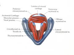
Vocal Ligament
|
#19
|
core of each vocal cord, run from vocal process of arytenoids to inner surface of thyroid lamina; position of these ligaments determines quantity of air flow
o Vocal process of arytenoid cartilage: attaches vocalis muscle and vocal ligament o Movement at the cricothyroid joint changes length and tension of the vocal ligaments, determining pitch of speech |
|
|
Ventricle of larynx
|
#24
|
fossa between the vocal folds
|
|
|
Rima Glottidis
|
|
opening between the left and right true vocal cords, closed by the arytenoid muscles, opened only by posterior cricoarytenoid muscles; Valsalva manaveur: straining maneuver to raise abdominal pressure, maximum adduction of rima glottidis
|
|
|
Vocalis Muscle (part of Thyro-arytenoid muscle)
|

#20
|
Vocal process of arytenoid cartilage: attaches vocalis muscle and vocal ligament; fine adjustments of vocal cords
|
|
|
Vocal Processes of Arytenoid Cartilage
|
|
anterior angle of the base of the arytenoid cartilage projects horizontally forward; it gives attachment to the vocal ligament
|
|
|
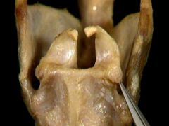
Arytenoid Cartilage
|

#14
|

triangular cartilages articulating inferiorly with cricoid lamina
o Vocal process: attaches vocalis muscle and vocal ligament o Muscular process: attached thyroarytenoid muscles |
|
|
Corniculate Cartilage
|
#13
|

articulate with the summits of the arytenoid cartilages and serve to prolong them posteriorly and medially
|
|
|
Cuneiform Cartilage
|

#24
|
paired cartilages that sit on top of and move with the arytenoids; located above and in front of the corniculate cartilages, and the presence of these two pairs of cartilages result in small bulges on the surface of the mucous membrane; covered by ary-epiglottic folds
|
|
|
Inferior Laryngeal (Branch of Recurrent Laryngeal)
|

#16
|
Recurrent laryngeal of CNX: all the intrinsic muscles of the larynx except cricothyroid
|
|
|
Posterior Cricoarytenoid
|

#8
|
only muscle to abduct the vocal cords/open rima glottidis
|
|
|
Transverse Arytenoid
|

#7
|
adducts vocal cords
|
|
|
Oblique Arytenoid
|
|

adduct vocal cords; continuation of aryepiglottic muscles
|
|
|
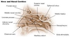
Superior Nasal Concha
|

#13
|

protect olfactory bulb, part of ethmoid bone
|
|
|
Middle Nasal Concha
|

#14
|

protect sinuses from coming into direct contact with nasal airflow, part of ethmoid bone
|
|
|
Inferior Nasal Concha
|

#15
|

responsible for airflow, humidification, filtering; are separate bones (superior and middle conchae are parts of other bones)
|
|
|
Superior Nasal Meatus
|

#2
|
sphenopalatine foramen opens into it posteriorly, posterior ethmoidal cells anteriorly (drain here)
|
|
|
Middle Nasal Meatus
|

#3
|
bulla ethmoidalis(=elevation containing the middle ethmoidal cells (drain here)), hiatus semilunaris (anterior ethmoidal cells (drain here), frontonasal duct, ostium of maxillary sinus…. Sooooo frontal, middle ethmoid, and maxillary drain here!!!), uncinate process
|
|
|
Inferior Nasal Meatus
|
#17
|
ostium of nasolacrimal duct anteriorly (draining tears form the lacrimal sac into the inferior meatus
|
|
|
Spheno-ethmoidal recess
|

#12
|
sphenoethmoidal recess behind the superior concha
|
|
|
Nasolacrimal Duct Opening
|

#19
|
ostium of nasolacrimal duct anteriorly (draining tears form the lacrimal sac into the inferior meatus
|
|
|
Semilunar Hiatus
|

#16
|
hiatus semilunaris (anterior ethmoidal cells (drain here); on lateral wall of middle nasal meatus
|
|
|
Bulla ethmoidalis (& Superior Nasal Meatus)
|
#15
|
bulla ethmoidalis(=elevation containing the middle ethmoidal cells (drain here) in the middle nasal meatus
|
|
|
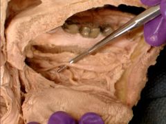
Greater Palatine (Pterygopalatine) Canal
|
|
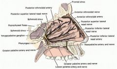
passage in the skull that transmits the descending palatine artery, vein, and greater and lesser palatine nerves between the pterygopalatine fossa and the oral cavity
|
|
|
Greater Palatine Artery and Nerve
|
#14
|

Maxillary lingual gingivae of posterior teeth supplied by gingival branches of greater palatine nerve
Nerve travels through the pterygopalatine canal and greater palatine foramina and supplies the mucosa and glands of the hard palate, as well as the anterior soft palate; Artery is branch of pterygopalatine part of Maxillary artery |
|
|
Lesser Palatine Artery and Nerves
|

#15
|

usually two lesser palatine nerves which emerge from the lesser palatine foramina and supply the mucosa and glands of the soft palate and uvula. The posterior superior lateral nasal branches reach this aspect of the nasal cavity and also supply the superior and middle nasal concha/meatuses and posterior ethmoidal sinuses
|
|
|
Posterior Superior Nasal/Alveolar Nerve (of Pterygopalatine Ganglion, CN V2 - ZIPS)
|

#13
|
Travel as NVB; reach this aspect of the nasal cavity and also supply the superior and middle nasal concha/meatuses and posterior ethmoidal sinuses. (The posterior superior medial nasal branches supply the posterior superior nasal septum
|
|
|
Anterior Superior Alveolar (of Infraorbital Branch of CN V2 - ZIPS)
|

#53
|
Travels with NVB; Maxillary vestibular gingivae of anterior teeth supplied by anterior and
middle superior alveolar and infraorbital; run along the lateral walls of the sinus btw the mucosa and bone from the infraorbital nerve |
|
|
Middle Superior Alveolar (of Infraorbital Branch of CN V2 - ZIPS)
|
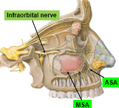
|
Maxillary vestibular gingivae of anterior teeth supplied by anterior and middle superior alveolar and infraorbital; run along the lateral walls of the sinus btw the mucosa and bone from the infraorbital nerve
|
|
|
Infraorbital Canal
|
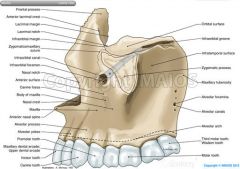
|
opens just below the margin of the orbit; transmits the infraorbital nerve as well as infraorbital artery, both of which enter this canal at the infraorbital groove and after coursing through the maxillary sinus exit via the infraorbital foramen
|
|
|
Zygomaticofacial Nerve (Zygomatic of CN V2 - ZIPS)
|
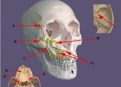
B
|
lateral to infraorbital canal
|
|
|
Nasopalatine Nerve
|
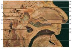
#22
|
nerve lies in the pterygopalatine fossa as a branch of CNV2. It enters the nasal cavity through the sphenopalatine foramen to reach the septum, then descends to the roof of the mouth through the incisive foramen, giving rise to the incisal branch and communicating with the corresponding nerve of the opposite side along with the greater palatine nerve. It supplies palatal structure around the maxillary central and lateral incisors and the nasal septum
|
|
|
Lingual Branch of Glossopharyngeal Nerve
|
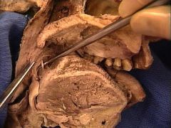
|
posterior to palatoglossal fold, runs medially and on posterior border of stylopharyngeus muscle for a short distance before it passes deep to hyoglossus muscle to enter the deep portion of the tongue
|
|
|
Lingual Frenulum
|
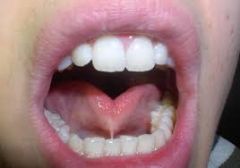
|
midline fold of mucous membrane running from lingual gingiva
behind mandibular central incisors posteriorly to undersurface of tongue |
|
|
Sublingual folds
|
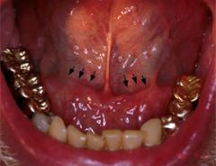
|
produced by underlying sublingual salivary glands running
posteriorly and laterally from the lingual frenulum – horse-shoe shaped |
|
|
Sublingual gland
|
|
primarily mucous-secreting salivary gland
- Located in floor of mouth, nestles laterally in sublingual fossa of mandible and medially against base of tongue - It is elongated – L and R meet anteriorly →horseshoe shape of sublingual folds - Covered by a thin lining of mucosa to separate from oral cavity |
|
|
Sublingual caruncle
|
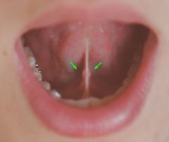
|
eminence on either side of the frenulum of the tongue, on which the major sublingual and the submandibular duct open
|
|
|
Median Lingual Sulcus
|

|
runs posteriorly along the dorsum from the apex of the
tongue – divides the tongue into two lateral halves |
|
|
Foramen cecum
|
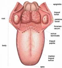
|
small pit at apex of sulcus terminalis – vestige of proximal
end of thyroglossal duct |
|
|
Fungiform papillae
|
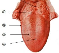
#2
|
found on anterior 2/3 tongue, globular, bright red, mainly
at tip and lateral margins of tongue, contain few taste buds |
|
|
Filiform papillae
|

#4
|
most numerous papillae on anterior 2/3 tongue, arranged in
-shaped rows parallel to sulcus terminalis, give tongue rough texture (helps in handling food), contain sensory nerve endings (very sensitive touch corpuscles) |
|
|
Parotid Papilla
|
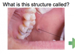
|
where orifice/opening of parotid duct into oral cavity is located – at
occlusal level of maxillary 2nd molar |