Reading...
![]()
Play button
![]()
Play button
![]()
Use LEFT and RIGHT arrow keys to navigate between flashcards;
Use UP and DOWN arrow keys to flip the card;
H to show hint;
A reads text to speech;
56 Cards in this Set
- Front
- Back
|
What are the immunobullous skin disorders?
|
Pemphigus foliaceus
Pemphigus vulgaris Bullous pemphigoid Epidermolysis bullosa acquisita |
|
|
Where does pemphigus foliaceous occur?
|
Within the granular layer
|
|
|
Where does pemphigus vulgaris occur?
|
Spinous layer
|
|
|
Where does bullous pemphigoid occur?
|
Below the basal layer but above the basement membrane
|
|
|
Where does epidermolysis bullosa occur?
|
Below the basal layer; high dermis
|
|
|
What is the function of the deesmosomes?
|
Hold the cells together in the epidermis
|
|
|
What is the function of the hemidesmosome?
|
Hold the epidermis to the dermis, like an anchor
|
|
|
What proteins are in the desmosomes?
|
DSG 1
DSG 3 |
|
|
What are the proteins in the hemidesmosome?
|
Bpag1/2
Laminin 5 Alpha6/Beta4 integrin |
|
|
What are tests that you should order up if you suspect an autoimmune blistering disease?
|
Direct immunofluoresence (in the skin)
Biopsy Indirect immunoflouresence (in the blood) |
|
|
What is the target for pemphigus foliaceus?
|
DSG1
|
|
|
What do the lesions look like in pemphigus foliaceus?
|
Skin lesions:
Scaly Superficial Crusted erosions Looks like crusty corn flakes |
|
|
Where does pemphigus foliaceus localize?
|
Trunk, extremities
The V's |
|
|
What does p. foliaceous look like under the scope?
|
LIght microscopy: a splitting under the epidermis; immune deposits in the granular layer
|
|
|
Where is desmoglein 1 expression the highest?
|
Inside the granular layer, decreases as you go down into the skin
|
|
|
What is the treatment for foliaceus?
|
Corticosteroids
-Locally if in one place -Systemically if you have serious disease |
|
|
Where do blisters take place in pemphigus vulgaris?
|
Within the spinous layer
|
|
|
What kinds of autoantibodies are there in p. vulgaris?
|
DSG 3/DSG 1 autoantibodies
|
|
|
What's the visual difference between pemphigus vulgaris and foliaceus?
|
IN vulgaris you can actually see the blisters
Also, in foliaceous you don't have blisters in the mouth, in vulgaris you do. |
|
|
What do the lesions look like in pemphigus vulgaris?
|
Flaccid blisters on normal looking skin
|
|
|
What do you see in light microscopy in pemphigus vulgaris?
|
Tombstones.
|
|
|
What's the difference between p. vulgaris and foilaceous on direct IF?
|
You can't tell the difference!
|
|
|
What proteins are mutated in pemphigus vulgaris?
|
Desmoglein 3, 1
Desmoglein 3 in the oral mucosa |
|
|
What's the treatment for pemphigus vulgaris?
|
Corticosteroids
Plasmapheresis-->cyclophosphamide Rituxan |
|
|
What are the proteins mutated in bullous pemphigoid?
|
BPag1/BPag2
|
|
|
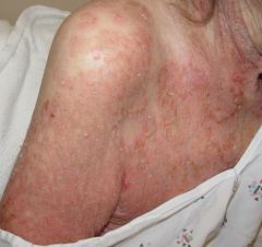
What do the lesions look like in bullous pemphigoid?
|
Large, tense bullae on an erythematous base; very puritic
YOU SEE TSNES BLISTERS!!! |
|
|
What's the distribution of bullous pemphigoid?
|
Trunk
Extremities Mucosal involvementis rare |
|
|
When do people get bullous pemphigoid?
|
When they're old
|
|
|
What does a light microscopy slide look likein bullous pemphigoid?
|
You see a split below the epidermis that's just above the BM
Eosinophils (IT"S WHAT CAUSES THE ITCH!) |
|
|
What kinds of proteins deposit in bullous pemphigoid?
|
IgG
C3 |
|
|
What's the prognosis for bullous pemphigoid?
|
Self-limited; may last 3 years
Some do last longer, though |
|
|
What's the treatment for bullous pemphigoid?
|
Mild disease: topical steroids
Extensive disease: prednisone, tetracyclines, |
|
|
What is cictrical pemphigoid?
|
Disease of the mucous membranes
You have ulcers and erosions |
|
|
Where does cicatrical pemphigoid distribute?
|
Mucus membranes:
Eyes (especially concerning!) Oropharynx Nasal cavity Larynx Esophagus |
|
|
What are the antibodies that are mutated in cicatrical pemphigoid?
|
Laminin 5
Alpha6/beta4 integrin BP antigen 2 |
|
|
What are the major concerns with cicatrical pemphigoid?
|
Eye pain/problems
Laryngeal/esophagical strictures, scarring |
|
|
What's the treatment for cicatrical pemphigoid?
|
Cortisteroids
Immunosuppressives |
|
|
What protein is mutated in epidermolysis bullosa?
|
Collagen 7
|
|
|
Where does epidermolysis bullosa happen?
|
Within the dermis
|
|
|
What are the kinds of skin lesions that happen with epidermiolysis bullosa?
|
Blisters on non-erythematous skin that heals with formation of milia
|
|
|
What does a light slide of epidermolysis bullos look like?
|
Complete shearing of the basement membrane
|
|
|
What's the course for epidermolysis?
|
Frequently self-limited
|
|
|
What's the treatment for epidermolysis?
|
We can't treat this.
|
|
|
What are the different kinds of inherited mechanobullous diseases?
|
Epidermolysis bullosa:
-Simplex -Junctional -Dystrophic |
|
|
What proteins are mutated in epidermolysis bullosa simplex?
|
Keratins 5, 14
Right near the basal cell. |
|
|
What proteins are mutated in junctional EB?
|
Laminin 5
Above lamina densa. Cell is sheared off, lamina densa is still intact |
|
|
What proteins are mutated in dystrophic EB?
|
Collagen VIII
|
|
|
What's the inheritance of epidermolysis bullosa simplex?
|
Autosomal dominant
|
|
|
What do the lesions look like in epidermolysis bullosa simplex?
|
Mild: blisters in traumatized areas in adults
Severe: generalized blisters in neonates |
|
|
What's the treatment for epidermolysis bullosa?
|
Can't.
|
|
|
What's the inheritance of junctional epidermolysis bullosa?
|
AR
|
|
|
What do the lesions look like in junctional epidermolysis bullosa?
|
Blisters and erosions at birth
MUCOSA ALL OVER THE PLACE! |
|
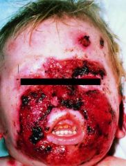
What disease is this?
|
This is a perioral granulation tissue
Junctional epidermolysis bullosa |
|
|
What's the gene mutated in junctional epidermolysis bullosa?
|
Laminin 5
|
|
|
Where does dystrophic epidermolysis bullosa distribute?
|
Generalized to all mucosla surfaces?
|
|
|
What's a common cancer in people with dystrophic epidermolysis bullosa?
|
SCC
|